Maxillary space closure using a digital manufactured Mesialslider in a single appointment workflow
- Affiliations
-
- 1Department of Orthodontics, HeinrichHeine-University, Düsseldorf, Germany
- 2Private Practice, Duisburg, Germany
- 3Department of Orthodontics, University of Sydney, Sydney, Australia
- 4Private Practice, Sydney, Australia
- KMID: 2529946
- DOI: http://doi.org/10.4041/kjod21.203
Abstract
- New digital technologies, many involving three-dimensional printing, bring benefits for clinical applications. This article reports on the clinical procedure and fabrication of a skeletally anchored mesialization appliance (Mesialslider) using computer-aided design/computer-aided manufacturing (CAD/CAM) for space closure of a congenitally missing lateral incisor in a 12-year-old female patient. The insertion of the mini-implants and appliance was performed in a single appointment. Bodily movement of the molars was achieved using the Mesialslider. Anchorage loss, such as deviation of the anterior midline or palatal tilting of the anterior teeth, was completely avoided. CAD/CAM facilitates safe and precise insertion of mini-implants. Further, mini-implants can improve patient comfort by reducing the number of office visits and eliminating the need for orthodontic bands and physical impressions.
Keyword
Figure
-
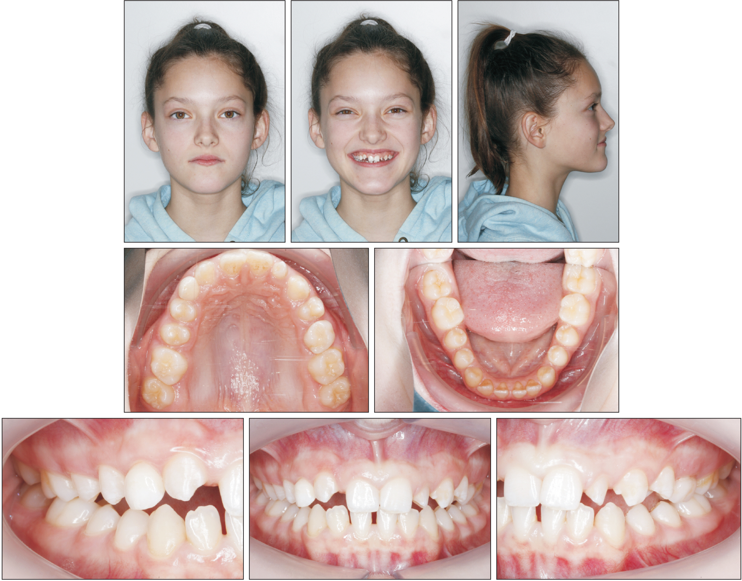
Figure 1 Pretreatment photographs. The maxillary right lateral incisor was congenitally missing.
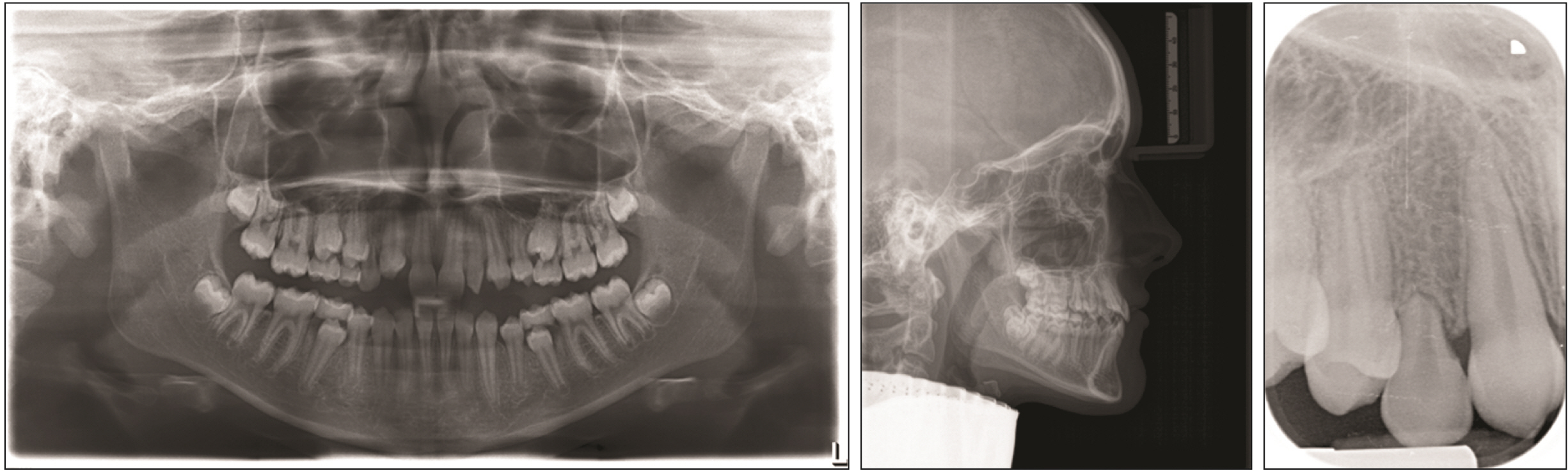
Figure 2 Pretreatment radiographs. Panoramic radiograph was taken two months ahead.
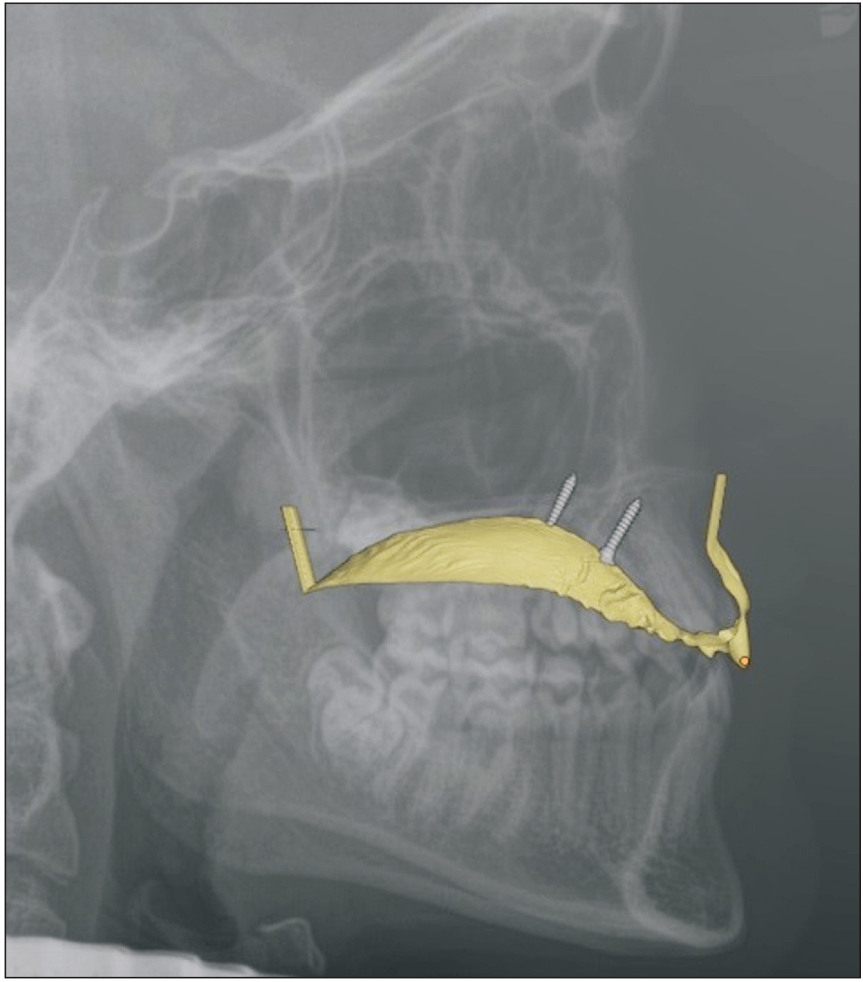
Figure 3 Merging of the stereolithography (STL) file with the lateral cephalometric radiograph for implant positioning.

Figure 4 Insertion guide placed on the three-dimensional printed study model (left) and the diamond cylinder used to reduce resistance to sliding (right).
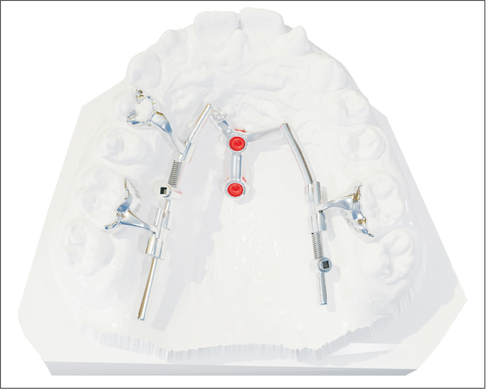
Figure 5 Final three-dimensional design of the Mesialslider.

Figure 6 Three-dimensional design of the Versalock tube (TADMAN, Gunningen, Germany).
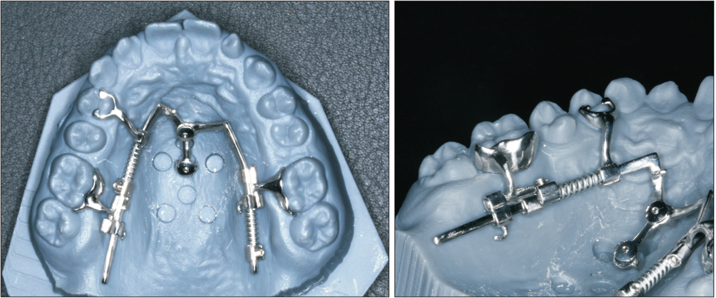
Figure 7 Finalized computer-aided design/computer-aided manufacturing fabricated Mesialslider on printed study model.

Figure 8 Insertion and activation of the Mesialslider right before extraction of the deciduous canine.

Figure 9 Intraoral photographs after 5 months of treatment. Spaces were almost closed in the anterior region.
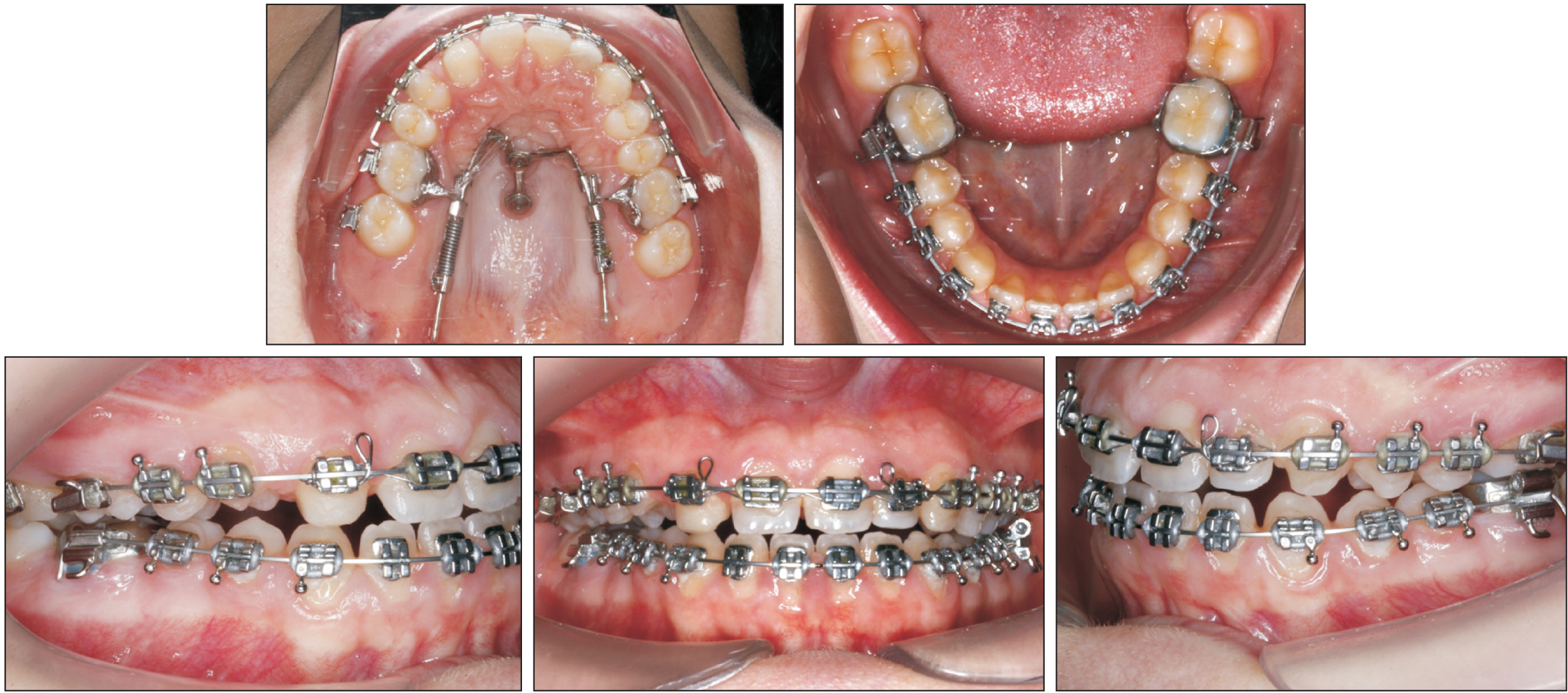
Figure 10 Intraoral photographs after 14 months of treatment.
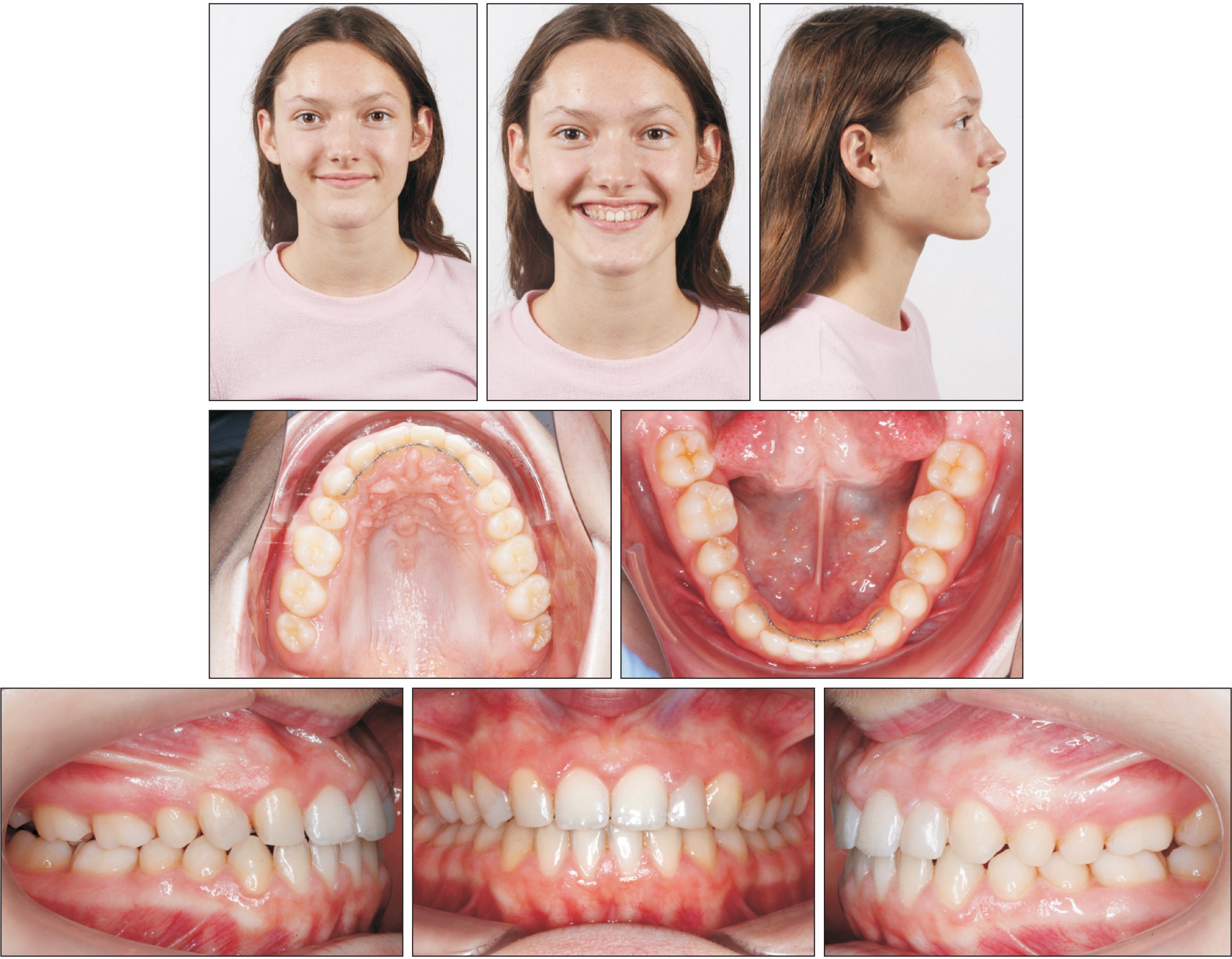
Figure 11 Post-treatment photographs. The treatment duration was less than 24 months.

Figure 12 Post-treatment radiographs.

Figure 13 Three-dimensional comparative evaluation of the clinical outcome: analysis of the pre-treatment (blue) and post-treatment (white) conditions shows a so-called reverse anchorage loss.
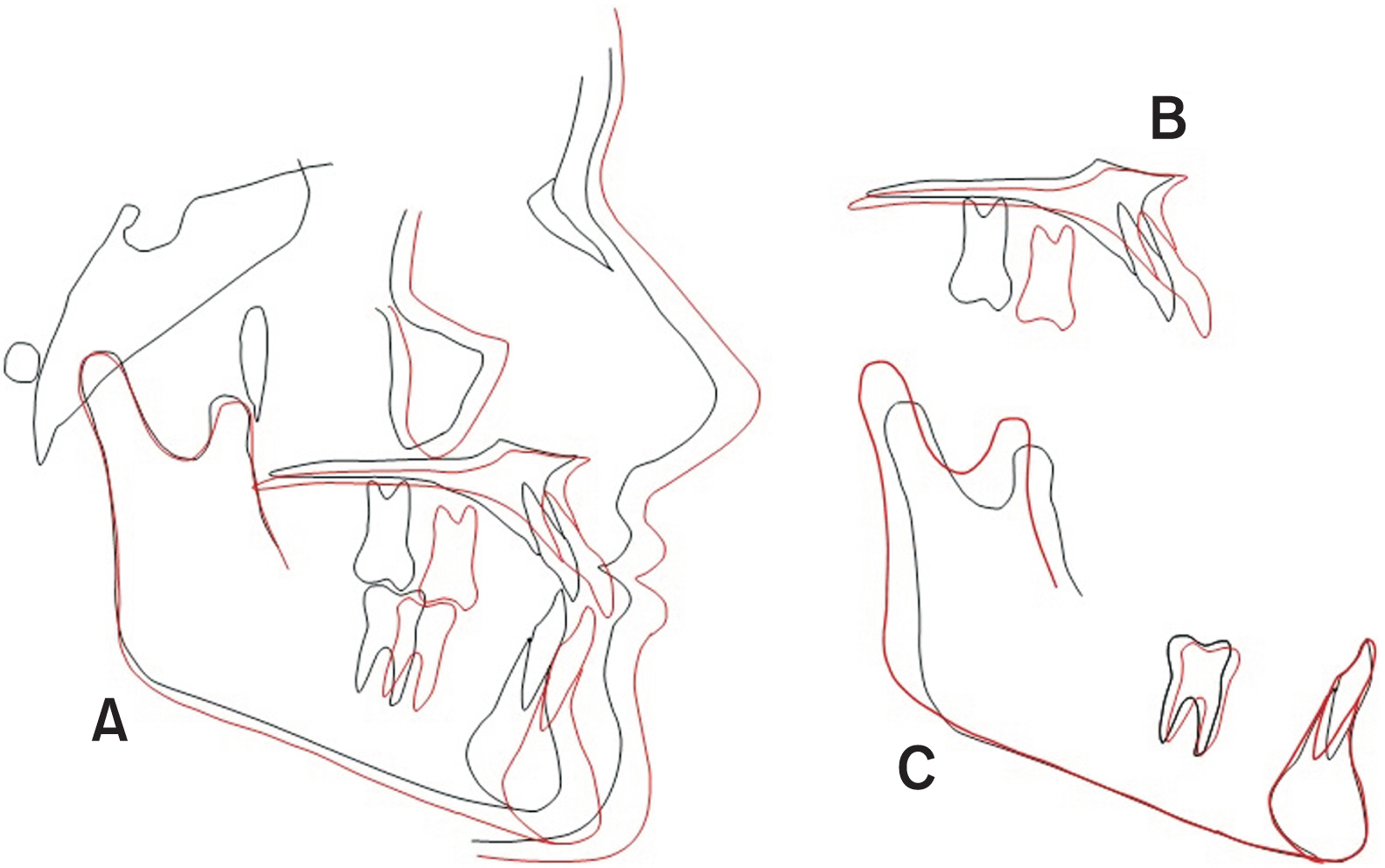
Figure 14 Skeletal growth superimposition and analysis of vertical changes. A, Cranial base superimposition. B, Maxillary superimposition. C, Mandibular superimposition. Black line, pretreatment; Red line, posttreatment.
Reference
-
1. Agarwal N, Kumar D, Anand A, Bahetwar SK. 2016; Dental implants in children: a multidisciplinary perspective for long-term success. Natl J Maxillofac Surg. 7:122–6. DOI: 10.4103/0975-5950.201362. PMID: 28356682. PMCID: PMC5357921.
Article2. Robertsson S, Mohlin B. 2000; The congenitally missing upper lateral incisor. A retrospective study of orthodontic space closure versus restorative treatment. Eur J Orthod. 22:697–710. DOI: 10.1093/ejo/22.6.697. PMID: 11212605.
Article3. Zachrisson BU. 1978; Improving orthodontic results in cases with maxillary incisors missing. Am J Orthod. 73:274–89. DOI: 10.1016/0002-9416(78)90134-3. PMID: 274073.
Article4. Zachrisson BU, Rosa M, Toreskog S. 2011; Congenitally missing maxillary lateral incisors: canine substitution. Point. Am J Orthod Dentofacial Orthop. 139:434. 436. 438 passim. DOI: 10.1016/j.ajodo.2011.02.003. PMID: 21457853.5. Zitzmann NU, Arnold D, Ball J, Brusco D, Triaca A, Verna C. 2015; Treatment strategies for infraoccluded dental implants. J Prosthet Dent. 113:169–74. DOI: 10.1016/j.prosdent.2014.08.012. PMID: 25444288.
Article6. Oesterle LJ, Cronin RJ Jr, Ranly DM. 1993; Maxillary implants and the growing patient. Int J Oral Maxillofac Implants. 8:377–87. PMID: 8270306.7. Zachrisson BU, Stenvik A. 2004; Single implants-optimal therapy for missing lateral incisors? Am J Orthod Dentofacial Orthop. 126:A13–5. DOI: 10.1016/j.ajodo.2001.01.001. PMID: 15384198.8. Rosa M, Lucchi P, Ferrari S, Zachrisson BU, Caprioglio A. 2016; Congenitally missing maxillary lateral incisors: long-term periodontal and functional evaluation after orthodontic space closure with first premolar intrusion and canine extrusion. Am J Orthod Dentofacial Orthop. 149:339–48. DOI: 10.1016/j.ajodo.2015.08.016. PMID: 26926021.
Article9. Johal A, Katsaros C, Kuijpers-Jagtman AM. Angle Society of Europe membership. 2013; State of the science on controversial topics: missing maxillary lateral incisors--a report of the Angle Society of Europe 2012 meeting. Prog Orthod. 14:20. DOI: 10.1186/2196-1042-14-20. PMID: 24325884. PMCID: PMC4384898.10. Ludwig B, Zachrisson BU, Rosa M. 2013; Non-compliance space closure in patients with missing lateral incisors. J Clin Orthod. 47:180–7. PMID: 23660791.11. Wehrbein H, Glatzmaier J, Mundwiller U, Diedrich P. 1996; The Orthosystem--a new implant system for orthodontic anchorage in the palate. J Orofac Orthop. 57:142–53. DOI: 10.1007/BF02191878. PMID: 8655109.12. Wilmes B, Drescher D. 2008; A miniscrew system with interchangeable abutments. J Clin Orthod. 42:574–80. quiz 595PMID: 19075372.13. Wilmes B, Drescher D, Nienkemper M. 2009; A miniplate system for improved stability of skeletal anchorage. J Clin Orthod. 43:494–501. PMID: 19904040.14. Wilmes B, Nienkemper M, Nanda R, Lübberink G, Drescher D. 2013; Palatally anchored maxillary molar mesialization using the mesialslider. J Clin Orthod. 47:172–9. PMID: 23660790.15. Wilmes B, Katyal V, Willmann J, Stocker B, Drescher D. 2015; Mini-implant-anchored Mesialslider for simultaneous mesialisation and intrusion of upper molars in an anterior open bite case: a three-year follow-up. Aust Orthod J. 31:87–97. DOI: 10.21307/aoj-2020-144. PMID: 26219151.
Article16. Wilmes B, Beykirch S, Ludwig B, Becker K, Willmann J, Drescher D. 2018; The B-Mesialslider for non-compliance space closure in cases with missing upper laterals. Semin Orthod. 24:66–82. DOI: 10.1053/j.sodo.2018.01.007.
Article17. Wilmes B, Vasudavan S, Drescher D. 2019; Maxillary molar mesialization with the use of palatal mini-implants for direct anchorage in an adolescent patient. Am J Orthod Dentofacial Orthop. 155:725–32. DOI: 10.1016/j.ajodo.2019.01.011. PMID: 31053288.
Article18. Becker K, Wilmes B, Grandjean C, Vasudavan S, Drescher D. 2018; Skeletally anchored mesialization of molars using digitized casts and two surface-matching approaches: analysis of treatment effects. J Orofac Orthop. 79:11–8. DOI: 10.1007/s00056-017-0108-y. PMID: 29134232.
Article19. Wilmes B, Ludwig B, Vasudavan S, Nienkemper M, Drescher D. 2016; The T-zone: median vs. paramedian insertion of palatal mini-implants. J Clin Orthod. 50:543–51. PMID: 27809213.20. Graf S, Vasudavan S, Wilmes B. 2018; CAD-CAM design and 3-dimensional printing of mini-implant retained orthodontic appliances. Am J Orthod Dentofacial Orthop. 154:877–82. DOI: 10.1016/j.ajodo.2018.07.013. PMID: 30477785.
Article21. Willmann JH, Chhatwani S, Drescher D. 2018; [Blender- freeware als dentales CAD-programm]. Kieferorthopädie. 32:161–5. German.22. Graf S, Cornelis MA, Hauber Gameiro G, Cattaneo PM. 2017; Computer-aided design and manufacture of hyrax devices: can we really go digital? Am J Orthod Dentofacial Orthop. 152:870–4. DOI: 10.1016/j.ajodo.2017.06.016. PMID: 29173866.
Article23. Rosa M, Zachrisson BU. 2001; Integrating esthetic dentistry and space closure in patients with missing maxillary lateral incisors. J Clin Orthod. 35:221–34. PMID: 11345569.24. Willmann JH, Wilmes B, Drescher D. 2019; [Digitale mini-implantat getragene Suprakonstruktionen - design und workflows]. J Compr Dentof Orthod Orthop. 3-4:16–20. German.25. Jamilian A, Perillo L, Rosa M. 2015; Missing upper incisors: a retrospective study of orthodontic space closure versus implant. Prog Orthod. 16:2. DOI: 10.1186/s40510-015-0072-2. PMID: 25769117. PMCID: PMC4385022.
Article26. Rosa M, Zachrisson BU. 2010; The space-closure alternative for missing maxillary lateral incisors: an update. J Clin Orthod. 44:540–9. quiz 561PMID: 21280545.27. Re S, Cardaropoli D, Corrente G, Abundo R. 2001; Bodily tooth movement through the maxillary sinus with implant anchorage for single tooth replacement. Clin Orthod Res. 4:177–81. DOI: 10.1034/j.1600-0544.2001.040308.x. PMID: 11553102.
Article28. Svejda M, Strobl N, Bantleon HP. 2008; [Tooth movement through the maxillary sinus - case report]. Inf Orthod Kieferorthop. 40:249–54. German. DOI: 10.1055/s-2008-1076969.29. Wehrbein H, Riess H, Meyer R, Schneider B, Diedrich P. 1990; [Bodily movement of teeth in atrophic jaw segments]. Dtsch Zahnarztl Z. 45:168–71. German. PMID: 2257823.30. Lindskog-Stokland B, Hansen K, Ekestubbe A, Wennström JL. 2013; Orthodontic tooth movement into edentulous ridge areas--a case series. Eur J Orthod. 35:277–85. DOI: 10.1093/ejo/cjr029. PMID: 21367817.
Article31. Lindskog-Stokland B, Wennström JL, Nyman S, Thilander B. 1993; Orthodontic tooth movement into edentulous areas with reduced bone height. An experimental study in the dog. Eur J Orthod. 15:89–96. DOI: 10.1093/ejo/15.2.89. PMID: 8500541.
Article32. Hom BM, Turley PK. 1984; The effects of space closure of the mandibular first molar area in adults. Am J Orthod. 85:457–69. DOI: 10.1016/0002-9416(84)90085-X. PMID: 6587780.
Article33. Stepovich ML. 1979; A clinical study on closing edentulous spaces in the mandible. Angle Orthod. 49:227–33. DOI: 10.1043/0003-3219(1979)049<0227:ACSOCE>2.0.CO;2. PMID: 292345.34. Ghislanzoni LH, Berardinelli F, Ludwig B, Lucchese A. 2016; Considerations involved in placing miniscrews near the nasopalatine bundle. J Clin Orthod. 50:321–8. PMID: 27323275.35. Züger J, Pandis N, Wallkamm B, Grossen J, Katsaros C. 2014; Success rate of paramedian palatal implants in adolescent and adult orthodontic patients: a retrospective cohort study. Eur J Orthod. 36:22–5. DOI: 10.1093/ejo/cjt003. PMID: 23525601.
Article36. Mraiwa N, Jacobs R, Van Cleynenbreugel J, Sanderink G, Schutyser F, Suetens P, et al. 2004; The nasopalatine canal revisited using 2D and 3D CT imaging. Dentomaxillofac Radiol. 33:396–402. DOI: 10.1259/dmfr/53801969. PMID: 15665234.
Article37. Fukuda M, Matsunaga S, Odaka K, Oomine Y, Kasahara M, Yamamoto M, et al. 2015; Three-dimensional analysis of incisive canals in human dentulous and edentulous maxillary bones. Int J Implant Dent. 1:12. DOI: 10.1186/s40729-015-0012-4. PMID: 27747634. PMCID: PMC5005666.
Article38. Song WC, Jo DI, Lee JY, Kim JN, Hur MS, Hu KS, et al. 2009; Microanatomy of the incisive canal using three-dimensional reconstruction of microCT images: an ex vivo study. Oral Surg Oral Med Oral Pathol Oral Radiol Endod. 108:583–90. DOI: 10.1016/j.tripleo.2009.06.036. PMID: 19778745.
Article
- Full Text Links
-
- Actions
-
Cited
- CITED
-
- Close
- Share
-
- Similar articles
-
- Comparative evaluation of two sets of complete dentures fabricated by digital and conventional workflow in a single patient: A case report
- Ceramic molar crown reproducibility by digital workflow manufacturing: An in vitro study
- CAD-CAM technique based digital diagnosis and fixed partial denture treatment on maxillary congenital missing teeth with skeletal class Ⅲ tendency patient: A case report
- Integration of Intraoral Scanner-based Mandibular Movement Data for An Anterior Single-implant Restoration
- Spatial changes of the upper dentition following en-masse space closure: A comparison between first and second premolar extraction
