Three dimensional finite element analysis of continuous and segmented arches with use of orthodontic miniscrews
- Affiliations
-
- 1Department of Dentistry, College of Medicine, Inha University, Korea.
- 2Department of Orthodontics, College of Dentisrty, Yonsei University, Korea. ypark@yuhs.ac
- KMID: 2274259
- DOI: http://doi.org/10.4041/kjod.2011.41.4.237
Abstract
OBJECTIVE
The purpose of this study was to compare the displacement patterns shown by finite element analysis when the maxillary anterior segment was retracted from different orthodontic miniscrew positions and different lengths of lever arms in lingual continuous and segmented arch techniques.
METHODS
A three dimensional model was produced, the translation of teeth in both models was measured and individual displacement was calculated.
RESULTS
When traction was carried out from miniscrews in the palatal slope, lingual tipping of crowns and extrusion of the maxillary anterior segment were found in both continuous and segmented arches as the lever arms were made shorter. With miniscrews in the midpalatal suture area, the displacement patterns were similar to the palatal slope, but bodily movement of the upper incisors was observed in both continuous and segmented arches with the lever arm at 20 mm. When lever arms were longer, there was less extrusion of the incisors and more buccal displacement of the canines. Such displacement was shown less in the continuous arch than the segmented arch. The second premolar showed crown mesial tipping and intrusion, and the molars showed distal tipping in the continuous arch. The posterior segment was displaced three dimensionally in the segmented arch, but the amount of displacement was less than the continuous arch.
CONCLUSIONS
It is recommended that lever arms of 20 mm in length be used for bodily movement of the anterior segment. Use of continuous or segmented arches affect the displacement patterns and induce differences in the amount of displacement.
MeSH Terms
Figure
-
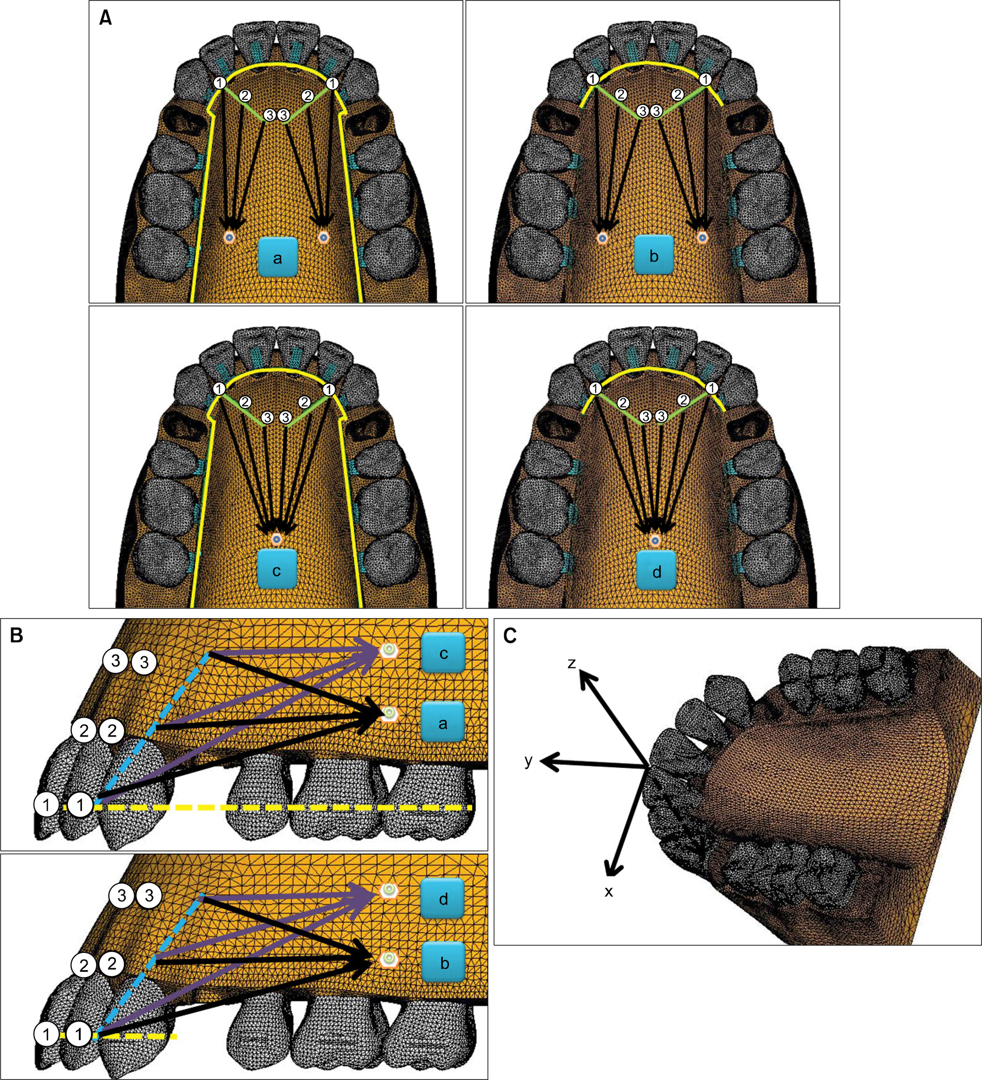
Fig 1. A Occlusal view of experimental groups. a, Continuous arch with lever arms and miniscrews in the palatal slope (condition 1A, lever arm length 0 mm; condition 2A, lever arm length 10 mm; condition 3A, lever arm length 20 mm); b, segmented arch with lever arms and miniscrews in the palatal slope (condition 1B, lever arm length 0 mm; condition 2B, lever arm length 10 mm; condition 3B, lever arm length 20 mm); c, continuous arch with lever arms and miniscrew in the midpalatal suture area (condition 1C, lever arm length 0 mm; condition 2C, lever arm length 10 mm; condition 3C, lever arm length 20 mm); d, segmented arch with lever arms and miniscrew in the midpalatal suture area (condition 1D, lever arm length 0 mm; condition 2D, lever arm length 10 mm; condition 3D, lever arm length 20 mm). B, Lateral view of experimental groups. a, Continuous arch with lever arms and miniscrews in the palatal slope; b, segmented arch with lever arms and miniscrews in the palatal slope; c, continuous arch with lever arms and miniscrew in the midpalatal suture area; d, segmented arch with lever arms and miniscrew in the midpalatal suture area. C, The coordinate system (X, medio-lateral direction (+), medial (-), lateral; Y, anterio-posterior direction (+), anterior (-), posterior; Z, superio-inferior direction (+), superior (-), inferior.
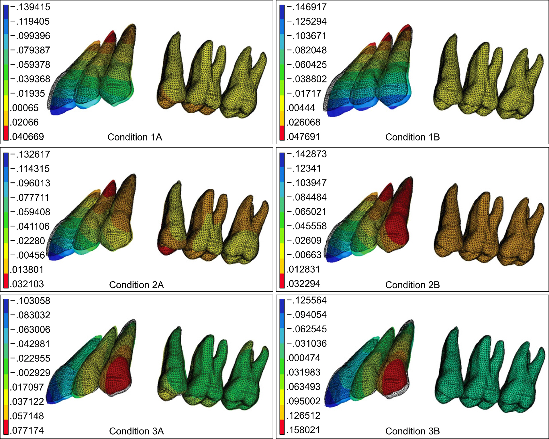
Fig 2. Contour plot of antero-posterior displacement of the continuous and segmented arches with miniscrews in the palatal slope. Condition 1A, Lever arm length of 0 mm with continuous arch; condition 2A, lever arm length of 10 mm with continuous arch; condition 3A, lever arm length of 20 mm with continuous arch; condition 1B, lever arm length of 0 mm with segmented arch; condition 2B, lever arm length of 10 mm with segmented arch; condition 3B, lever arm length of 20 mm with segmented arch.
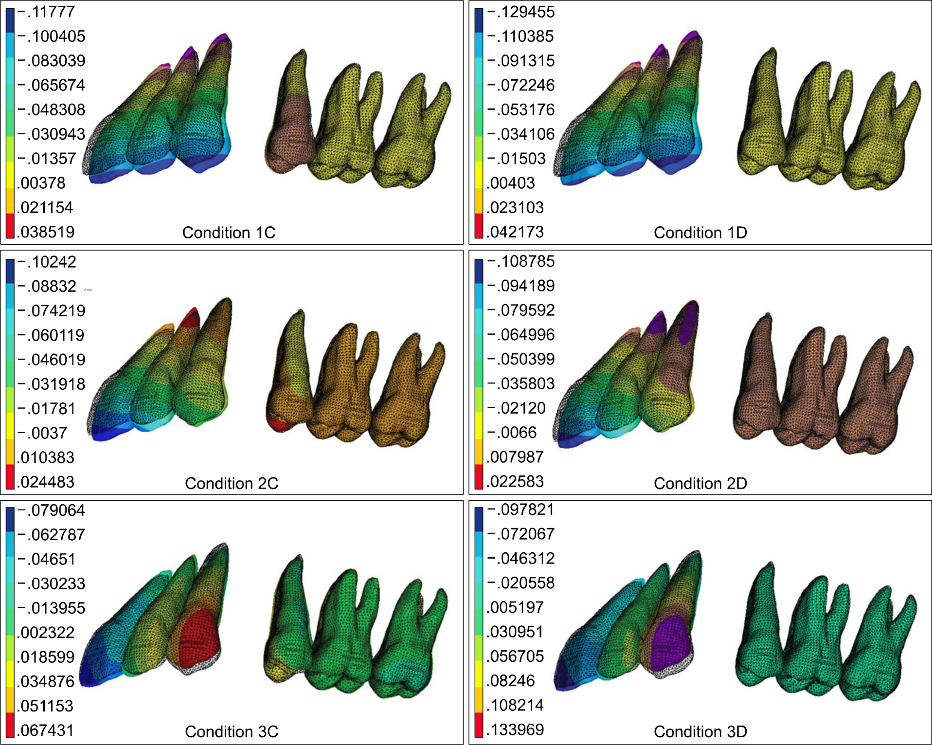
Fig 3. Contour plot of antero-posterior displacement of continuous and segmented arches with miniscrew in the mid-palatal suture area. Condition 1A, Lever arm length of 0 mm with continuous arch; condition 2A, lever arm length of 10 mm with continuous arch; condition 3A, lever arm length of 20 mm with continuous arch; condition 1B, lever arm length of 0 mm with segmented arch; condition 2B, lever arm length of 10 mm with segmented arch; condition 3B, lever arm length of 20 mm with segmented arch.
Fig 4. Contour plot of vertical displacement of the continuous and segmented arches with miniscrews in the palatal slope. Condition 1C, Lever arm length of 0 mm with contitnuous arch; condition 2C, lever arm length of 10 mm with continuous arch; condition 3C, lever arm length of 20 mm with continuous arch; condition 1D, lever arm length of 0 mm with segmented arch; condition 2D, lever arm length of 10 mm with segmented arch; condition 3D, lever arm length of 20 mm with segmented arch.

Fig 5. Contour plot of vertical displacement of continuous and segmented arches with miniscrew in the midpalatal suture area. Condition 1C, Lever arm length of 0 mm with contitnuous arch; condition 2C, lever arm length of 10 mm with continuous arch; condition 3C, lever arm length of 20 mm with continuous arch; condition 1D, lever arm length of 0 mm with segmented arch; condition 2D, lever arm length of 10 mm with segmented arch; condition 3D, lever arm length of 20 mm with segmented arch.
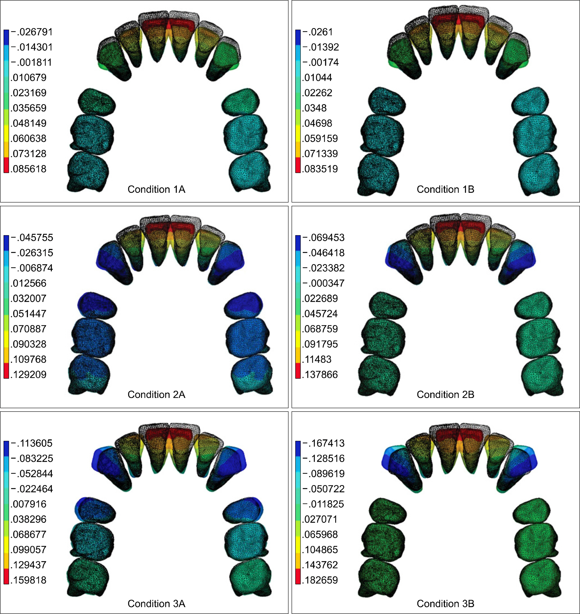
Fig 6. Contour plot of lateral displacement of continuous and segmented arches with miniscrews in the palatal slope. Condition 1C, Lever arm length of 0 mm with contitnuous arch; condition 2C, lever arm length of 10 mm with continuous arch; condition 3C, lever arm length of 20 mm with continuous arch; condition 1D, lever arm length of 0 mm with segmented arch; condition 2D, lever arm length of 10 mm with segmented arch; condition 3D, lever arm length of 20 mm with segmented arch.
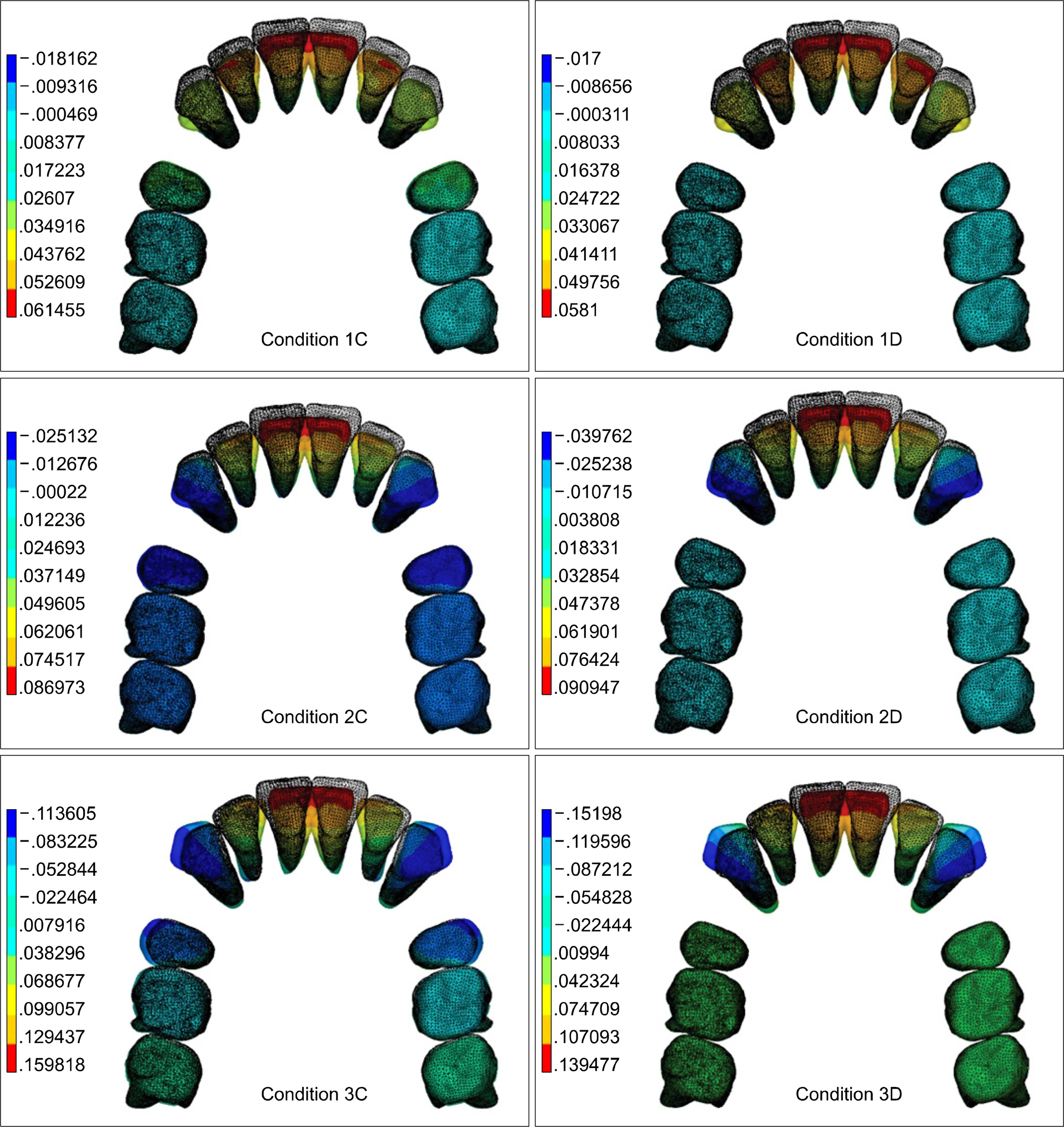
Fig 7. Contour plot of lateral displacement of continuous and segmented arches with miniscrew in the midpalatal suture area. Condition 1C, Lever arm length of 0 mm with contitnuous arch; condition 2C, lever arm length of 10 mm with continuous arch; condition 3C, lever arm length of 20 mm with continuous arch; condition 1D, lever arm length of 0 mm with segmented arch; condition 2D, lever arm length of 10 mm with segmented arch; condition 3D, lever arm length of 20 mm with segmented arch.
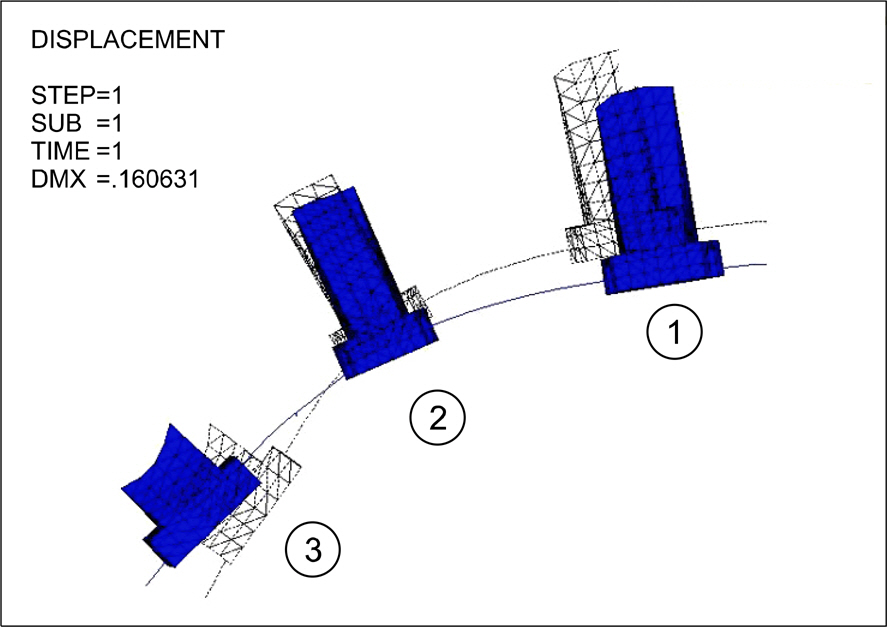
Fig 8. Deformation of main arch wire (meshed, before deformation; colored, after deformation; 1, central incisor; 2, lateral incisor; 3, canine).
Cited by 3 articles
-
Displacement pattern of the anterior segment using antero-posterior lingual retractor combined with a palatal plate
Kyung-Won Seo, Soon-Yong Kwon, Kyung A Kim, Ki-Ho Park, Seong-Hun Kim, Hyo-Won Ahn, Gerald Nelson
Korean J Orthod. 2015;45(6):289-298. doi: 10.4041/kjod.2015.45.6.289.The effects of alveolar bone loss and miniscrew position on initial tooth displacement during intrusion of the maxillary anterior teeth: Finite element analysis
Sun-Mi Cho, Sung-Hwan Choi, Sang-Jin Sung, Hyung-Seog Yu, Chung-Ju Hwang
Korean J Orthod. 2016;46(5):310-322. doi: 10.4041/kjod.2016.46.5.310.Palatal en-masse retraction of segmented maxillary anterior teeth: A finite element study
Jae Hyun Park, Yoon-Ah Kook, Yukio Kojima, Sunock Yun, Jong-Moon Chae
Korean J Orthod. 2019;49(3):188-193. doi: 10.4041/kjod.2019.49.3.188.
Reference
-
1.Sung SJ., Baik HS., Moon YS., Yu HS., Cho YS. A comparative evaluation of different compensating curves in the lingual and labial techniques using 3D FEM. Am J Orthod Dentofacial Orthop. 2003. 123:441–50.
Article2.Vásquez M., Calao E., Becerra F., Ossa J., Enríquez C., Fresneda E. Initial stress differences between sliding and sectional mechanics with an endosseous implant as anchorage: a 3-dimensional finite element analysis. Angle Orthod. 2001. 71:247–56.3.Sia S., Shibazaki T., Koga Y., Yoshida N. Experimental determination of optimal force system required for control of anterior tooth movement in sliding mechanics. Am J Orthod Dentofacial Orthop. 2009. 135:36–41.
Article4.Chung AJ., Kim US., Lee SH., Kang SS., Choi HI., Jo JH, et al. The pattern of movement and stress distribution during retraction of maxillary incisors using a 3-D finite element method. Korean J Orthod. 2007. 37:98–113.5.Jang HJ., Roh WJ., Joo BH., Park KH., Kim SJ., Park YG. Locating the center of resistance of maxillary anterior teeth retracted by Double J Retractor with palatal miniscrews. Angle Orthod. 2010. 80:1023–8.
Article6.Kucher G., Weiland FJ., Bantleon HP. Modified lingual lever arm technique. J Clin Orthod. 1993. 27:18–22.7.Park YC., Choy K., Lee JS., Kim TK. Lever-arm mechanics in lingual orthodontics. J Clin Orthod. 2000. 34:601–5.8.Nägerl H., Kubein-Meesenburg D. A FEM (Finite-Element-Measurement) study for the biomechanical comparison of labial and palatal force application on the upper incisors. Fortschr Kieferorthop. 1993. 54:229–30.9.Vanden Bulcke MM., Burstone CJ., Sachdeva RC., Dermaut LR. Location of the centers of resistance for anterior teeth during retraction using the laser reflection technique. Am J Orthod Dentofacial Orthop. 1987. 91:375–84.
Article10.Sia S., Koga Y., Yoshida N. Determining the center of resistance of maxillary anterior teeth subjected to retraction forces in sliding mechanics. An in vivo study. Angle Orthod. 2007. 77:999–1003.11.Jeong GM., Sung SJ., Lee KJ., Chun YS., Mo SS. Finite-element investigation of the center of resistance of the maxillary dentition. Korean J Orthod. 2009. 39:83–94.
Article12.Sung SJ., Kim IT., Kook YA., Chun YS., Kim SH., Mo SS. Finite-element analysis of the shift in center of resistance of the maxillary dentition in relation to alveolar bone loss. Korean J Orthod. 2009. 39:278–88.
Article13.Creekmore TD., Eklund MK. The possibility of skeletal anchorage. J Clin Orthod. 1983. 17:266–9.14.Costa A., Raffainl M., Melsen B. Miniscrews as orthodontic anchorage: a preliminary report. Int J Adult Orthodon Orthog-nath Surg. 1998. 13:201–9.15.Linkow LI. The endosseous blade implant and its use in orthodontics. Int J Orthod. 1969. 7:149–54.16.Kanomi R. Mini-implant for orthodontic anchorage. J Clin Orthod. 1997. 31:763–7.17.Sugawara J. Dr. Junji Sugawara on the skeletal anchorage system. Interview by Dr. Larry W. White. J Clin Orthod. 1999. 33:689–96.18.Chung KR., Kook YA., Kim SH., Mo SS., Jung JA. Class II malocclusion treated by combining a lingual retractor and a palatal plate. Am J Orthod Dentofacial Orthop. 2008. 133:112–23.
Article19.Hong RK., Heo JM., Ha YK. Lever-arm and mini-implant system for anterior torque control during retraction in lingual orthodontic treatment. Angle Orthod. 2005. 75:129–41.20.Burstone CJ. Rationale of the segmented arch. Am J Orthod. 1962. 48:805–22.
Article21.Burstone CJ. The segmented arch approach to space closure. Am J Orthod. 1982. 82:361–78.
Article22.Row J., Ryu YK. Three dimentional force analysis of force system in continuous archwire by finite element method. Koren J Orthod. 1996. 26:17–32.23.Melsen B. Tissue reaction to orthodontic tooth movement--a new paradigm. Eur J Orthod. 2001. 23:671–81.
Article24.Tanne K., Nagataki T., Inoue Y., Sakuda M., Burstone CJ. Patterns of initial tooth displacements associated with various root lengths and alveolar bone heights. Am J Orthod Dentofacial Orthop. 1991. 100:66–71.
Article25.Rudolph DJ., Willes PMG., Sameshima GT. A finite element model of apical force distribution from orthodontic tooth movement. Angle Orthod. 2001. 71:127–31.26.Tahk SG. A study on craniofacial growth analysis of Korean children by the finite element method. Korean J Orthod. 1988. 18:343–66.27.Ricketts RM., Bench RW., Gugino CF., Hilgers JJ., Schulhof RJ. Bioprogressive therapy. Denver: Rocky Mountain Orthodontics;1979.28.Lim SJ., Kwak BM., Lee JS. Introduction to finite element analysis. Seoul: Dongmyung Publishing;2003.29.Kim KH. A finite element analysis of effectiveness of lever arm in lingual sliding mechanics (thesis). Seoul: Yonsei University. 2009.30.Andrews LF. The six keys to normal occlusion. Am J Orthod. 1972. 62:296–309.
Article31.Kronfeld R. Histologic study of the influence of function on the human periodontal membrane. J Am Dent Assoc. 1931. 18:1242–74.32.Coolidge E. The thickness of the human periodontal membrane. J Am Dent Assoc. 1937. 24:1260–5.
Article33.Kim SH., Park SB., Yang HC. Three-dimensional finite element analysis of the bracket positioning plane in lingual orthodontics. Korean J Orthod. 2006. 36:30–44.34.Tanne K., Sakuda M., Burstone CJ. Three-dimensional finite element analysis for stress in the periodontal tissue by orthodontic forces. Am J Orthod Dentofacial Orthop. 1987. 92:499–505.
Article35.McLaughlin RP., Bennett JC. Anchorage control during leveling and aligning with a preadjusted appliance system. J Clin Orthod. 1991. 25:687–96.36.Mo SS., Kim SH., Sung SJ., Chung KR., Chun YS., Kook YA, et al. Factors controlling anterior torque with C-implants depend on en-masse retraction without posterior appliances: bio-creative therapy type II technique. Am J Orthod Dentofacial Orthop. 2011. 139:e183–91.
Article37.Jeong HS., Sung SJ., Moon YS., Cho YS., Lim SM. Factors influencing the axes of anterior teeth during SWA en masse sliding retraction with orthodontic mini-implant anchorage: a finite element study. Korean J Orthod. 2006. 36:339–48.38.Upadhyay M., Yadav S., Nagaraj K., Patil S. Treatment effects of mini-implants for en-masse retraction of anterior teeth in bialveolar dental protrusion patients: a randomized controlled trial. Am J Orthod Dentofacial Orthop. 2008. 134:18–29. .e1.
Article39.Liang W., Rong Q., Lin J., Xu B. Torque control of the maxillary incisors in lingual and labial orthodontics: a 3-dimensional finite element analysis. Am J Orthod Dentofacial Orthop. 2009. 135:316–22.
Article40.Lee HK., Chung KR. The vertical location of the center of resistance for maxillary six anterior teeth during retraction using three dimensional finite element analysis. Korean J Orthod. 2001. 31:425–38.41.Alexander CM., Alexander RG., Gorman JC., Hilgers JJ., Kurz C., Scholz RP, et al. Lingual orthodontics: a status report. Part 5. Lingual mechanotherapy. J Clin Orthod. 1983. 17:99–115.42.Sung SJ., Jang GW., Chun YS., Moon YS. Effective en-masse retraction design with orthodontic mini-implant anchorage: a finite element analysis. Am J Orthod Dentofacial Orthop. 2010. 137:648–57.
Article43.Tominaga JY., Tanaka M., Koga Y., Gonzales C., Kobayashi M., Yoshida N. Optimal loading conditions for controlled movement of anterior teeth in sliding mechanics. Angle Orthod. 2009. 79:1102–7.
Article44.Yoon HJ., Lim YK., Lee DY., Jo YS. Three-dimensional finite element analysis on the effect of maxillary incisor torque. Korean J Orthod. 2005. 35:137–47.45.Kim CH., Sung JH., Kyung HM. Three-dimensional finite element analysis of initial tooth displacement according to force application point during maxillary six anterior teeth retraction using skeletal anchorage. Korean J Orthod. 2003. 33:339–50.
- Full Text Links
-
- Actions
-
Cited
- CITED
-
- Close
- Share
-
- Similar articles
-
- Three dimentional force analysis of force system in continuous archwire by finite element method
- Stress distributions in peri-miniscrew areas from cylindrical and tapered miniscrews inserted at different angles
- Effect of archwire stiffness and friction on maxillary posterior segment displacement during anterior segment retraction: A three-dimensional finite element analysis
- 3-D FEA on the intrusion of mandibular anterior segment using orthodontic miniscrews
- Influence of changing various parameters in miniscrew-assisted rapid palatal expansion: A three-dimensional finite element analysis
