Influence of changing various parameters in miniscrew-assisted rapid palatal expansion: A three-dimensional finite element analysis
- Affiliations
-
- 1Department of Orthodontics, Graduate School of Clinical Dentistry, Korea University, Seoul, Korea.
- 2Department of Orthodontics, Korea University Guro Hospital, Seoul, Korea.
- 3Department of Orthodontics, Korea University Ansan Hospital, Ansan, Korea. jgosggg@gmail.com
- KMID: 2450224
- DOI: http://doi.org/10.4041/kjod.2019.49.3.150
Abstract
OBJECTIVE
This study aimed to analyze the effect of changing various parameters of the bone-borne rapid palatal expander (RPE) using the finite element method (FEM).
METHODS
In eight experimental groups, we investigated the effect of the number, position, and length of miniscrews; positional changes of the expander; and changes in the hook length on maxillary expansion. In finite element analysis, we compared the magnitude and distribution of stress, and the displacement changes following expansion of the bone-borne RPE.
RESULTS
When we compared the number and position of miniscrews, placing miniscrews in the anterior and posterior sides was advantageous for maxillary expansion in terms of stress distribution and displacement changes. Miniscrew length did not significantly affect stress distribution and displacement changes. Furthermore, anteroposterior displacement of the expander did not significantly affect transverse maxillary expansion but had various effects on vertical changes of the maxilla. The maxilla rotated clockwise when the miniscrews were placed in the anterior region. The hook length of the expander did not show consistent results in terms of changes in stress distribution and magnitude or in displacement changes.
CONCLUSIONS
The findings of this study suggest that changes in the location and length of the miniscrews and displacement of the bone-borne RPE could affect the pattern of the maxillary expansion, depending on the combination of these factors.
Keyword
Figure
-
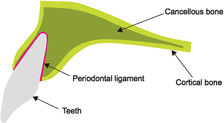
Figure 1 Cross-sectional image used in this study.
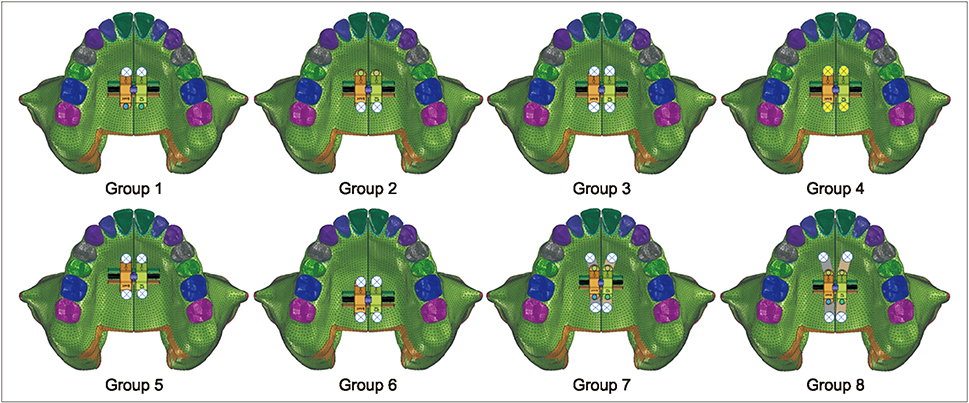
Figure 2 Groups used in this study. Group 1, Miniscrew front 8 mm; Group 2, miniscrew rear 6 mm; Group 3, miniscrew front 8 mm and rear 6 mm; Group 4, miniscrew front 16 mm and rear 10 mm; Group 5, miniscrew front 8 mm, rear 6 mm, expander moved +3 mm; Group 6, miniscrew front 8 mm, rear 6 mm, expander moved −3 mm; Group 7, miniscrew front 8 mm, rear 6 mm, hook extended +3 mm; Group 8, miniscrew front 8 mm, rear 6 mm, hook extended +6 mm.

Figure 3 Landmarks used in this study. A, Incisal point of the maxillary teeth (U1–U7); B, six points that divide the midpalatal suture into five equal parts (PM1–PM6); C, skeletal points of the skull. N, N point; ZMS, zygomaxillary suture; MT, maxillary tuberosity; INB, nasal bone; NB, nasal base; ANS, anterior nasal spine.
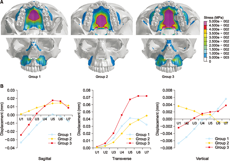
Figure 4 A, Stress distribution at the palatal and frontal skull: Group 1, Group 2, and Group 3, respectively. B, Sagittal, transverse, and vertical displacement of the maxillary teeth: Group 1, Group 2, and Group 3.
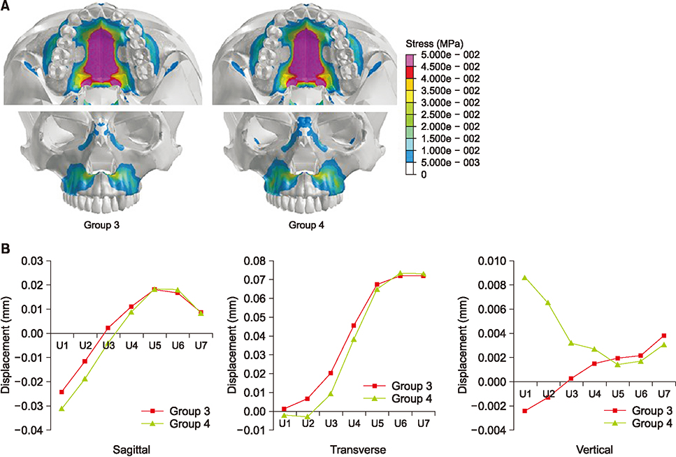
Figure 5 A, Stress distribution at the palatal and frontal skull: Group 3 and Group 4, respectively. B, Sagittal, transverse, and vertical displacement of the maxillary teeth: Group 3 and Group 4.
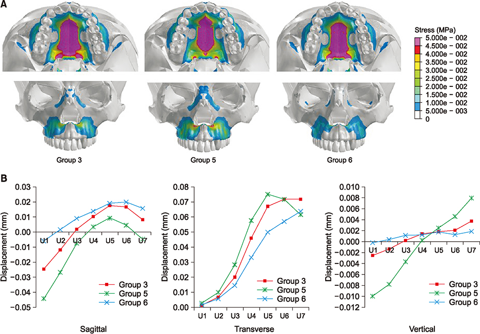
Figure 6 A, Stress distribution at the palatal and frontal skull: Group 3, Group 5, and Group 6, respectively. B, Sagittal, transverse, and vertical displacement of the maxillary teeth: Group 3, Group 5, and Group 6.
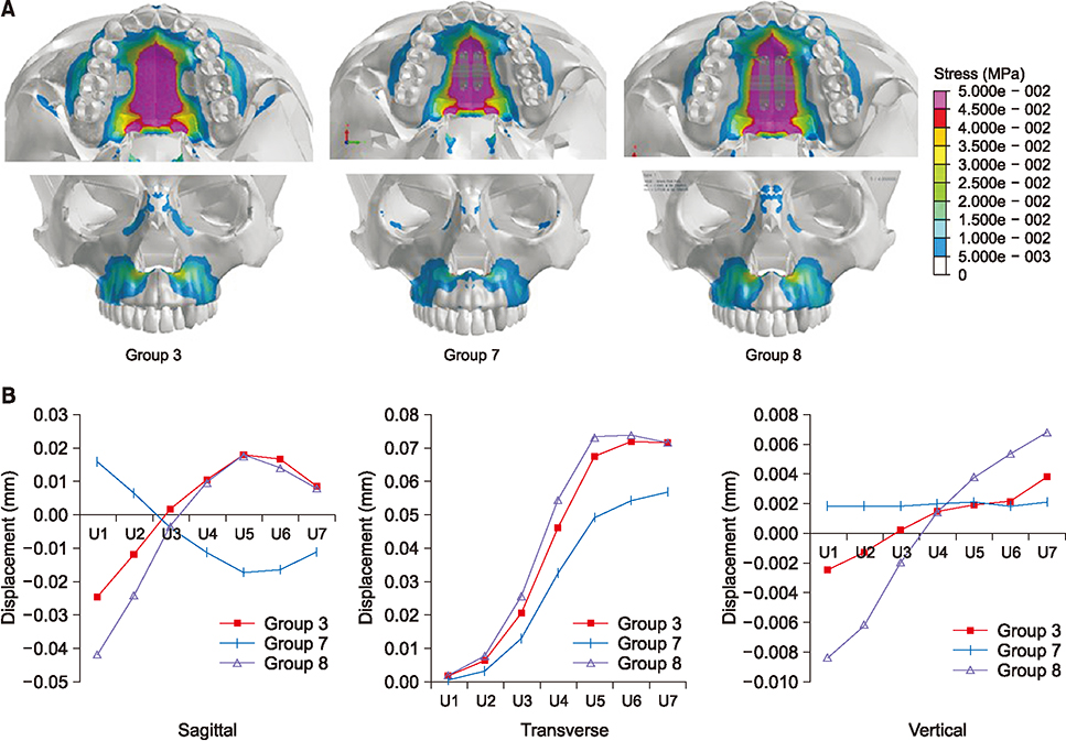
Figure 7 A, Stress distribution at the palatal and frontal skull: Group 3, Group 7, and Group 8, respectively. B, Sagittal, transverse, and vertical displacement of the maxillary teeth: Group 3, Group 7, and Group 8.
Cited by 2 articles
-
Reader's Forum
Mohammed Alfaifi
Korean J Orthod. 2019;49(5):277-278. doi: 10.4041/kjod.2019.49.5.277.Complications reported with the use of orthodontic miniscrews: A systematic review
Antonino Lo Giudice, Lorenzo Rustico, Miriam Longo, Giacomo Oteri, Moschos A. Papadopoulos, Riccardo Nucera
Korean J Orthod. 2021;51(3):199-216. doi: 10.4041/kjod.2021.51.3.199.
Reference
-
1. Angell EH. Treatment of irregularities of the permanent or adult teeth. Dent Cosm. 1860; 1:540–544. 599–600.2. Lux CJ, Dücker B, Pritsch M, Komposch G, Niekusch U. Occlusal status and prevalence of occlusal malocclusion traits among 9-year-old schoolchildren. Eur J Orthod. 2009; 31:294–299.
Article3. Will LA. Transverse maxillary deformities: diagnosis & treatment. Sel Read Oral Maxillofac Surg. 1996; 5:1–28.4. Haas AJ. The treatment of maxillary deficiency by opening the midpalatal suture. Angle Orthod. 1965; 35:200–217.5. Adkins MD, Nanda RS, Currier GF. Arch perimeter changes on rapid palatal expansion. Am J Orthod Dentofacial Orthop. 1990; 97:194–199.
Article6. Baccetti T, Franchi L, Cameron CG, McNamara JA Jr. Treatment timing for rapid maxillary expansion. Angle Orthod. 2001; 71:343–350.7. Chung CH, Font B. Skeletal and dental changes in the sagittal, vertical, and transverse dimensions after rapid palatal expansion. Am J Orthod Dentofacial Orthop. 2004; 126:569–575.
Article8. Persson M, Thilander B. Palatal suture closure in man from 15 to 35 years of age. Am J Orthod. 1977; 72:42–52.
Article9. Brossman RE, Bennett CG, Merow WW. Facioskeletal remodelling resulting from rapid palatal expansion in the monkey (Macaca cynomolgus). Arch Oral Biol. 1973; 18:987–994.
Article10. Vassar JW, Karydis A, Trojan T, Fisher J. Dentoskeletal effects of a temporary skeletal anchorage device-supported rapid maxillary expansion appliance (TSADRME): a pilot study. Angle Orthod. 2016; 86:241–249.
Article11. Lin L, Ahn HW, Kim SJ, Moon SC, Kim SH, Nelson G. Tooth-borne vs bone-borne rapid maxillary expanders in late adolescence. Angle Orthod. 2015; 85:253–262.
Article12. Kim KB, Helmkamp ME. Miniscrew implant-supported rapid maxillary expansion. J Clin Orthod. 2012; 46:608–612. quiz 631.13. Zandi M, Miresmaeili A, Heidari A. Short-term skeletal and dental changes following bone-borne versus tooth-borne surgically assisted rapid maxillary expansion: a randomized clinical trial study. J Craniomaxillofac Surg. 2014; 42:1190–1195.
Article14. Lee HK, Bayome M, Ahn CS, Kim SH, Kim KB, Mo SS, et al. Stress distribution and displacement by different bone-borne palatal expanders with micro-implants: a three-dimensional finite-element analysis. Eur J Orthod. 2014; 36:531–540.
Article15. Tanne K, Sakuda M, Burstone CJ. Three-dimensional finite element analysis for stress in the periodontal tissue by orthodontic forces. Am J Orthod Dentofacial Orthop. 1987; 92:499–505.
Article16. Rees JS, Jacobsen PH. Elastic modulus of the periodontal ligament. Biomaterials. 1997; 18:995–999.
Article17. Provatidis CG, Georgiopoulos B, Kotinas A, McDonald JP. Evaluation of craniofacial effects during rapid maxillary expansion through combined in vivo/in vitro and finite element studies. Eur J Orthod. 2008; 30:437–448.
Article18. Tanne K, Hiraga J, Kakiuchi K, Yamagata Y, Sakuda M. Biomechanical effect of anteriorly directed extraoral forces on the craniofacial complex: a study using the finite element method. Am J Orthod Dentofacial Orthop. 1989; 95:200–207.
Article19. Kyung SH. A study on the bone thickness of midpalatal suture area for miniscrew insertion. Korean J Orthod. 2004; 34:63–70.20. Pirelli P, Ragazzoni E, Botti F, Arcuri C, Cocchia D. A comparative light microscopic study of human midpalatal suture and periodontal ligament. Minerva Stomatol. 1997; 46:429–433.21. Lee H, Ting K, Nelson M, Sun N, Sung SJ. Maxillary expansion in customized finite element method models. Am J Orthod Dentofacial Orthop. 2009; 136:367–374.
Article22. Lee SC, Park JH, Bayome M, Kim KB, Araujo EA, Kook YA. Effect of bone-borne rapid maxillary expanders with and without surgical assistance on the craniofacial structures using finite element analysis. Am J Orthod Dentofacial Orthop. 2014; 145:638–648.
Article23. Liu S, Xu T, Zou W. Effects of rapid maxillary expansion on the midpalatal suture: a systematic review. Eur J Orthod. 2015; 37:651–655.
Article24. MacGinnis M, Chu H, Youssef G, Wu KW, Machado AW, Moon W. The effects of micro-implant assisted rapid palatal expansion (MARPE) on the nasomaxillary complex--a finite element method (FEM) analysis. Prog Orthod. 2014; 15:52.
Article25. Lee RJ, Moon W, Hong C. Effects of monocortical and bicortical mini-implant anchorage on bone-borne palatal expansion using finite element analysis. Am J Orthod Dentofacial Orthop. 2017; 151:887–897.
Article26. Seong EH, Choi SH, Kim HJ, Yu HS, Park YC, Lee KJ. Evaluation of the effects of miniscrew incorporation in palatal expanders for young adults using finite element analysis. Korean J Orthod. 2018; 48:81–89.
Article
- Full Text Links
-
- Actions
-
Cited
- CITED
-
- Close
- Share
-
- Similar articles
-
- Evaluation of the effects of miniscrew incorporation in palatal expanders for young adults using finite element analysis
- Three dimensional finite element method for stress distribution on the length and diameter of orthodontic miniscrew and cortical bone thickness
- A finite element analysis of the stress distribution and displacement in human maxilla to rapid palatal expansion
- Effectiveness of miniscrew assisted rapid palatal expansion using cone beam computed tomography: A systematic review and meta-analysis
- Displacement and stress distribution of the maxillofacial complex during maxillary protraction using palatal plates: A three-dimensional finite element analysis
