Korean J Orthod.
2018 Sep;48(5):304-315. 10.4041/kjod.2018.48.5.304.
Displacement and stress distribution of the maxillofacial complex during maxillary protraction using palatal plates: A three-dimensional finite element analysis
- Affiliations
-
- 1Private Practice, Gimpo, Korea.
- 2Department of Dentistry, College of Medicine, The Catholic University of Korea, Seoul, Korea.
- 3Department of Postgraduate Studies, the Universidad Autonóma del Paraguay, Asunción, Paraguay.
- 4Postgraduate Orthodontic Program, Arizona School of Dentistry & Oral Health, A.T. Still University, Mesa, AZ, USA.
- 5Graduate School of Dentistry, Kyung Hee University, Seoul, Korea.
- 6Department of Orthodontics, Seoul St. Mary's Hospital, College of Medicine, The Catholic University of Korea, Seoul, Korea.
- 7Division of Orthodontics, Department of Dentistry, St. Vincent's Hospital, College of Medicine, The Catholic University of Korea, Seoul, Korea. seonghh@hotmail.com
- KMID: 2421315
- DOI: http://doi.org/10.4041/kjod.2018.48.5.304
Abstract
OBJECTIVE
The purpose of this study was to analyze initial displacement and stress distribution of the maxillofacial complex during dentoskeletal maxillary protraction with various appliance designs placed on the palatal region by using three-dimensional finite element analysis.
METHODS
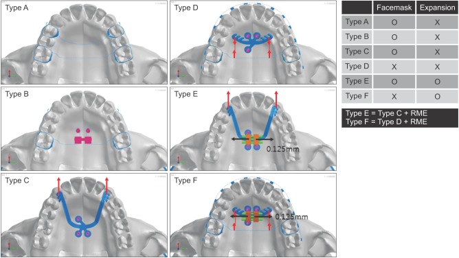
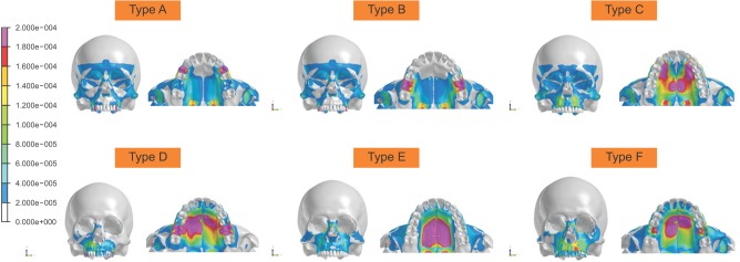
Six models of maxillary protraction were developed: conventional facemask (Type A), facemask with dentoskeletal hybrid anchorage (Type B), facemask with a palatal plate (Type C), intraoral traction using a Class III palatal plate (Type D), facemask with a palatal plate combined with rapid maxillary expansion (RME; Type E), and Class III palatal plate intraoral traction with RME (Type F). In Types A, B, C, and D, maxillary protraction alone was performed, whereas in Types E and F, transverse expansion was performed simultaneously with maxillary protraction.
RESULTS
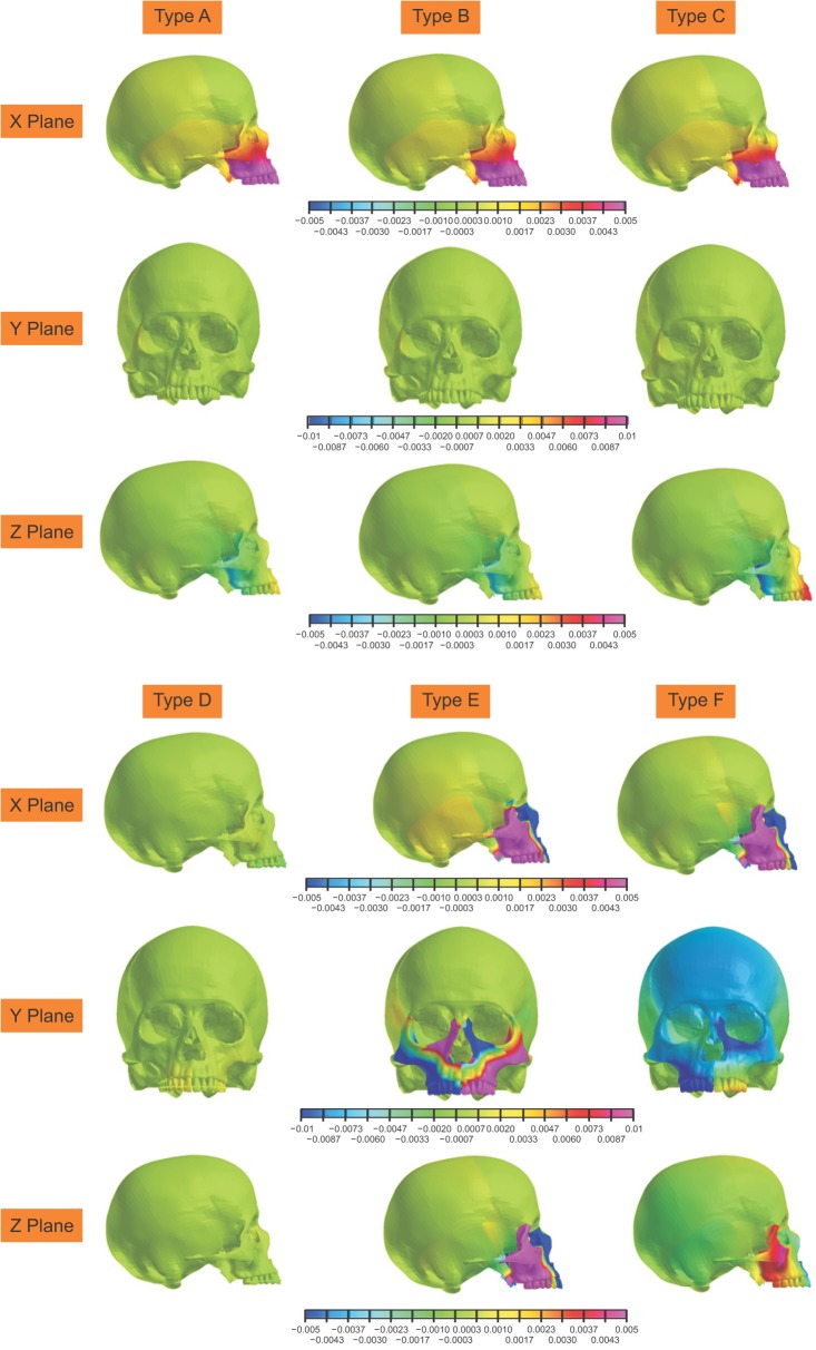
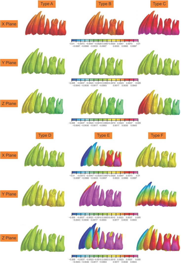
Type C displayed the greatest amount of anterior dentoskeletal displacement in the sagittal plane. Types A and B resulted in similar amounts of anterior displacement of all the maxillofacial landmarks. Type D showed little movement, but Type E with expansion and the palatal plate displayed a larger range of movement of the maxillofacial landmarks in all directions.
CONCLUSIONS
The palatal plate served as an effective skeletal anchor for use with the facemask in maxillary protraction. In contrast, the intraoral use of Class III palatal plates showed minimal skeletal and dental effects in maxillary protraction. In addition, palatal expansion with the protraction force showed minimal effect on the forward movement of the maxillary complex.
Keyword
Figure
Reference
-
1. Baik HS. Clinical results of the maxillary protraction in Korean children. Am J Orthod Dentofacial Orthop. 1995; 108:583–592. PMID: 7503035.
Article2. Kim JH, Viana MA, Graber TM, Omerza FF, BeGole EA. The effectiveness of protraction face mask therapy: a meta-analysis. Am J Orthod Dentofacial Orthop. 1999; 115:675–685. PMID: 10358251.
Article3. Singer SL, Henry PJ, Rosenberg I. Osseointegrated implants as an adjunct to facemask therapy: a case report. Angle Orthod. 2000; 70:253–262. PMID: 10926436.4. Hong H, Ngan P, Han G, Qi LG, Wei SH. Use of onplants as stable anchorage for facemask treatment: a case report. Angle Orthod. 2005; 75:453–460. PMID: 15898388.5. Kircelli BH, Pektas ZO. Midfacial protraction with skeletally anchored face mask therapy: a novel approach and preliminary results. Am J Orthod Dentofacial Orthop. 2008; 133:440–449. PMID: 18331946.
Article6. Baek SH, Kim KW, Choi JY. New treatment modality for maxillary hypoplasia in cleft patients. Protraction facemask with miniplate anchorage. Angle Orthod. 2010; 80:783–791. PMID: 20482368.7. Lee NK, Baek SH. Stress and displacement between maxillary protraction with miniplates placed at the infrazygomatic crest and the lateral nasal wall: a 3-dimensional finite element analysis. Am J Orthod Dentofacial Orthop. 2012; 141:345–351. PMID: 22381495.
Article8. Han S, Bayome M, Lee J, Lee YJ, Song HH, Kook YA. Evaluation of palatal bone density in adults and adolescents for application of skeletal anchorage devices. Angle Orthod. 2012; 82:625–631. PMID: 22077190.
Article9. Ryu JH, Park JH, Vu Thi, Bayome M, Kim Y, Kook YA. Palatal bone thickness compared with cone-beam computed tomography in adolescents and adults for mini-implant placement. Am J Orthod Dentofacial Orthop. 2012; 142:207–212. PMID: 22858330.
Article10. Vu T, Bayome M, Kook YA, Han SH. Evaluation of the palatal soft tissue thickness by cone-beam computed tomography. Korean J Orthod. 2012; 42:291–296. PMID: 23323243.
Article11. Lee SM, Park JH, Bayome M, Kim HS, Mo SS, Kook YA. Palatal soft tissue thickness at different ages using an ultrasonic device. J Clin Pediatr Dent. 2012; 36:405–409. PMID: 23019841.
Article12. Kook YA, Bayome M, Park JH, Kim KB, Kim SH, Chung KR. New approach of maxillary protraction using modified C-palatal plates in Class III patients. Korean J Orthod. 2015; 45:209–214. PMID: 26258067.
Article13. Kim KY, Bayome M, Park JH, Kim KB, Mo SS, Kook YA. Displacement and stress distribution of the maxillofacial complex during maxillary protraction with buccal versus palatal plates: finite element analysis. Eur J Orthod. 2015; 37:275–283. PMID: 25090997.
Article14. Kook YA, Park JH, Kim Y, Ahn CS, Bayome M. Sagittal correction of adolescent patients with modified palatal anchorage plate appliances. Am J Orthod Dentofacial Orthop. 2015; 148:674–684. PMID: 26432323.
Article15. Yu HS, Baik HS, Sung SJ, Kim KD, Cho YS. Three-dimensional finite-element analysis of maxillary protraction with and without rapid palatal expansion. Eur J Orthod. 2007; 29:118–125. PMID: 17218719.
Article16. Park JH, Bayome M, Zahrowski JJ, Kook YA. Displacement and stress distribution by different bone-borne palatal expanders with facemask: A 3-dimensional finite element analysis. Am J Orthod Dentofacial Orthop. 2017; 151:105–117. PMID: 28024761.
Article17. Erkmen E, Simşek B, Yücel E, Kurt A. Three-dimensional finite element analysis used to compare methods of fixation after sagittal split ramus osteotomy: setback surgery-posterior loading. Br J Oral Maxillofac Surg. 2005; 43:97–104. PMID: 15749208.
Article18. Mahoney E, Holt A, Swain M, Kilpatrick N. The hardness and modulus of elasticity of primary molar teeth: an ultra-micro-indentation study. J Dent. 2000; 28:589–594. PMID: 11082528.19. Rees JS, Jacobsen PH. Elastic modulus of the periodontal ligament. Biomaterials. 1997; 18:995–999. PMID: 9212195.
Article20. Farnsworth D, Rossouw PE, Ceen RF, Buschang PH. Cortical bone thickness at common miniscrew implant placement sites. Am J Orthod Dentofacial Orthop. 2011; 139:495–503. PMID: 21457860.
Article21. Kronfeld R. Histologic study of the influence of function on the human periodontal membrane. J Am Dent Assoc. 1931; 18:1242–1274.22. Fricke-Zech S, Gruber RM, Dullin C, Zapf A, Kramer FJ, Kubein-Meesenburg D, et al. Measurement of the midpalatal suture width. Angle Orthod. 2012; 82:145–150. PMID: 21812573.
Article23. Kook YA, Lee DH, Kim SH, Chung KR. Design improvements in the modified C-palatal plate for molar distalization. J Clin Orthod. 2013; 47:241–248. PMID: 23660818.24. Gautam P, Valiathan A, Adhikari R. Stress and displacement patterns in the craniofacial skeleton with rapid maxillary expansion: a finite element method study. Am J Orthod Dentofacial Orthop. 2007; 132:5.e1–5.e11.
Article25. Ngan PW, Hagg U, Yiu C, Wei SH. Treatment response and long-term dentofacial adaptations to maxillary expansion and protraction. Semin Orthod. 1997; 3:255–264. PMID: 9573887.
Article26. Vaughn GA, Mason B, Moon HB, Turley PK. The effects of maxillary protraction therapy with or without rapid palatal expansion: a prospective, randomized clinical trial. Am J Orthod Dentofacial Orthop. 2005; 128:299–309. PMID: 16168327.
Article27. Cevidanes L, Baccetti T, Franchi L, McNamara JA Jr, De Clerck H. Comparison of two protocols for maxillary protraction: bone anchors versus face mask with rapid maxillary expansion. Angle Orthod. 2010; 80:799–806. PMID: 20578848.
Article28. Hino CT, Cevidanes LH, Nguyen TT, De Clerck HJ, Franchi L, McNamara JA Jr. Three-dimensional analysis of maxillary changes associated with facemask and rapid maxillary expansion compared with bone anchored maxillary protraction. Am J Orthod Dentofacial Orthop. 2013; 144:705–714. PMID: 24182587.
Article29. Elnagar MH, Elshourbagy E, Ghobashy S, Khedr M, Evans CA. Comparative evaluation of 2 skeletally anchored maxillary protraction protocols. Am J Orthod Dentofacial Orthop. 2016; 150:751–762. PMID: 27871701.
Article
- Full Text Links
- Actions
-
Cited
- CITED
-
- Close
- Share
- Similar articles
-
- Three-dimensional finite element analysis on the effects of maxillary protraction with an individual titanium plate at multiple directions and locations
- A finite element analysis on the effect of the reverse headgear to the maxillary complex
- Influence of changing various parameters in miniscrew-assisted rapid palatal expansion: A three-dimensional finite element analysis
- Effects of maxillary protraction on the displacement of the maxilla
- A three-dimensional finite element analysis of obturator prosthesis for edentulous maxilla

