Stress distributions in peri-miniscrew areas from cylindrical and tapered miniscrews inserted at different angles
- Affiliations
-
- 1Department of Orthodontics, The Institute of Cranial-Facial Deformity, College of Dentistry, Yonsei University, Seoul, Korea. hwang@yuhs.ac
- 2Private Practice, Gwangju, Korea.
- 3Division of Orthodontics, Department of Dentistry, University of Ulsan College of Medicine, Seoul, Korea.
- 4Department of Orthodontics, Graduate School of Clinical Dentistry, Ewha Womans University, Seoul, Korea.
- KMID: 2392199
- DOI: http://doi.org/10.4041/kjod.2016.46.4.189
Abstract
OBJECTIVE
The purpose of this study was to analyze stress distributions in the roots, periodontal ligaments (PDLs), and bones around cylindrical and tapered miniscrews inserted at different angles using a finite element analysis.
METHODS
We created a three-dimensional (3D) maxilla model of a dentition with extracted first premolars and used 2 types of miniscrews (tapered and cylindrical) with 1.45-mm diameters and 8-mm lengths. The miniscrews were inserted at 30°, 60°, and 90° angles with respect to the bone surface. A simulated horizontal orthodontic force of 2 N was applied to the miniscrew heads. Then, the stress distributions, magnitudes during miniscrew placement, and force applications were analyzed with a 3D finite element analysis.
RESULTS
Stresses were primarily absorbed by cortical bone. Moreover, very little stress was transmitted to the roots, PDLs, and cancellous bone. During cylindrical miniscrew insertion, the maximum von Mises stress increased as insertion angle decreased. Tapered miniscrews exhibited greater maximum von Mises stress than cylindrical miniscrews. During force application, maximum von Mises stresses increased in both groups as insertion angles decreased.
CONCLUSIONS
For both cylindrical and tapered miniscrew designs, placement as perpendicular to the bone surface as possible is recommended to reduce stress in the surrounding bone.
Figure
-
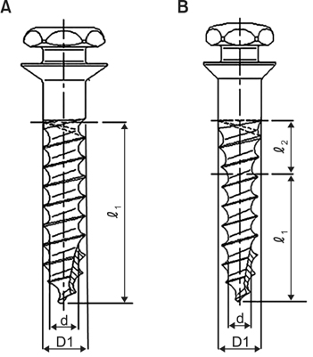
Figure 1 Schematic diagrams of the 1508C and 1508T miniscrews. A, Cylindrical type. B, Taper type. D1, External diameter; d, internal diameter; ℓ1, length of threaded part; ℓ2, length of tapered part.
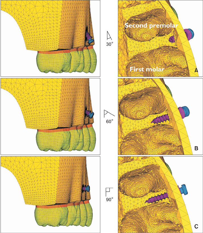
Figure 2 Three miniscrew insertion angles are shown; the insertion angles were measured between the miniscrew and bone surface. A, 30°; B, 60°; C, 90°.
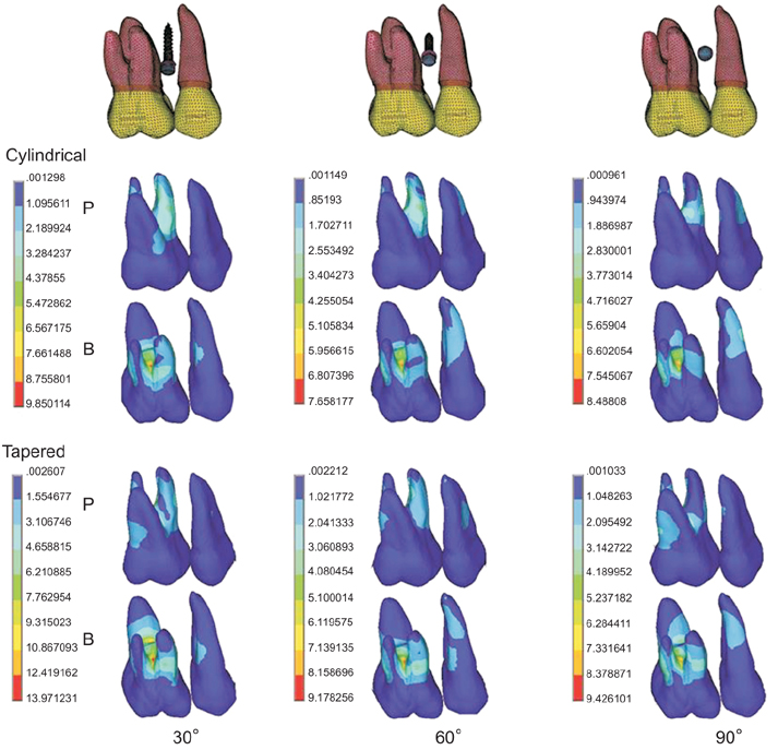
Figure 3 The von Mises stress distributions during miniscrew placement at the roots. P, Palatal view; B, buccal view; left, first molar; right, second premolar.
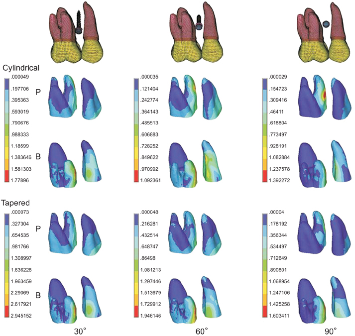
Figure 4 The von Mises stress distributions during miniscrew placement at the periodontal ligaments. P, Palatal view; B, buccal view; left, first molar; right, second premolar.
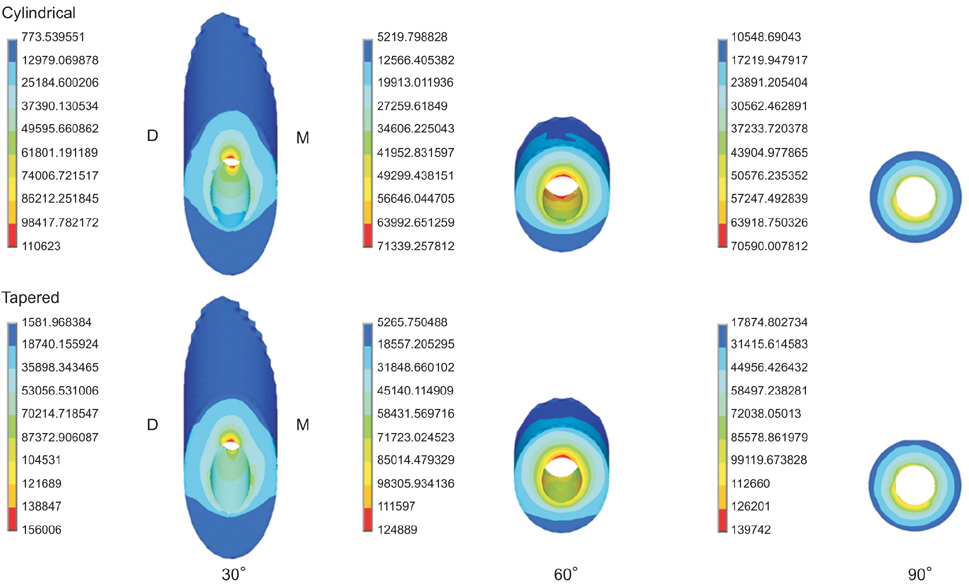
Figure 5 The von Mises stress distributions during miniscrew placement in cortical bone (buccal view). M, Mesial side; D, distal side.
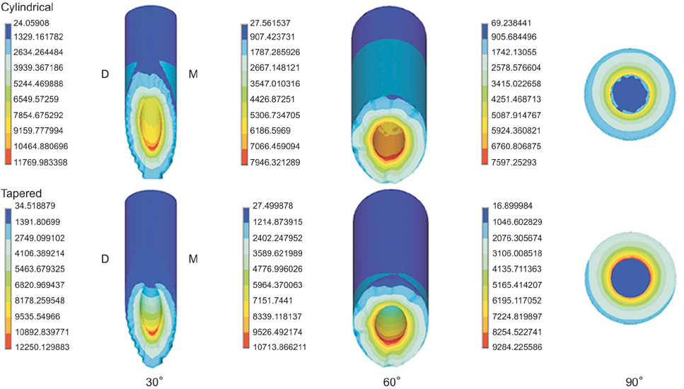
Figure 6 The von Mises stress distributions during miniscrew placement in cancellous bone (buccal view). M, Mesial side; D, distal side.
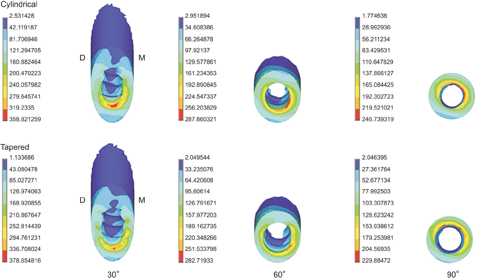
Figure 7 The von Mises stress distributions in cortical bone during force application (buccal view). M, Mesial side; D, distal side.
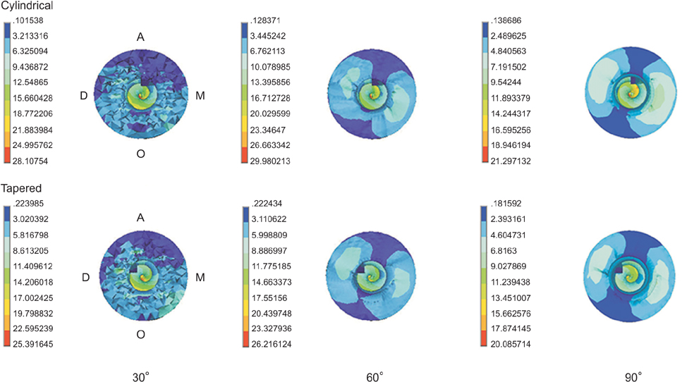
Figure 8 The von Mises stress distributions in cancellous bone during force application (buccal view). M, Mesial side; D, distal side; A, apical side; O, occlusal side.
Cited by 2 articles
-
Bone cutting capacity and osseointegration of surface-treated orthodontic mini-implants
Ho-Young Kim, Sang-Cheol Kim
Korean J Orthod. 2016;46(6):386-394. doi: 10.4041/kjod.2016.46.6.386.The effect of fluoride-containing oral rinses on the corrosion resistance of titanium alloy (Ti-6Al-4V)
Gui-Yue Huang, Heng Bo Jiang, Jung-Yul Cha, Kwang-Mahn Kim, Chung-Ju Hwang
Korean J Orthod. 2017;47(5):306-312. doi: 10.4041/kjod.2017.47.5.306.
Reference
-
1. Reynders R, Ronchi L, Bipat S. Mini-implants in orthodontics: a systematic review of the literature. Am J Orthod Dentofacial Orthop. 2009; 135:564.e1–564.e19. discussion 564-5.
Article2. Cha JY, Hwang CJ, Kwon SH, Jung HS, Kim KM, Yu HS. Strain of bone-implant interface and insertion torque regarding different miniscrew thread designs using an artificial bone model. Eur J Orthod. 2015; 37:268–274.
Article3. Miyawaki S, Koyama I, Inoue M, Mishima K, Sugahara T, Takano-Yamamoto T. Factors associated with the stability of titanium screws placed in the posterior region for orthodontic anchorage. Am J Orthod Dentofacial Orthop. 2003; 124:373–378.
Article4. Cheng SJ, Tseng IY, Lee JJ, Kok SH. A prospective study of the risk factors associated with failure of mini-implants used for orthodontic anchorage. Int J Oral Maxillofac Implants. 2004; 19:100–106.5. Park HS, Jeong SH, Kwon OW. Factors affecting the clinical success of screw implants used as orthodontic anchorage. Am J Orthod Dentofacial Orthop. 2006; 130:18–25.
Article6. Papadopoulos MA, Papageorgiou SN, Zogakis IP. Clinical effectiveness of orthodontic miniscrew implants: a meta-analysis. J Dent Res. 2011; 90:969–976.
Article7. Cha JY, Takano-Yamamoto T, Hwang CJ. The effect of miniscrew taper morphology on insertion and removal torque in dogs. Int J Oral Maxillofac Implants. 2010; 25:777–783.8. Katić V, Kamenar E, Blažević D, Špalj S. Geometrical design characteristics of orthodontic mini-implants predicting maximum insertion torque. Korean J Orthod. 2014; 44:177–183.
Article9. Lim SA, Cha JY, Hwang CJ. Insertion torque of orthodontic miniscrews according to changes in shape, diameter and length. Angle Orthod. 2008; 78:234–240.
Article10. Choi SH, Cha JY, Joo UH, Hwang CJ. Surface changes of anodic oxidized orthodontic titanium miniscrew. Angle Orthod. 2012; 82:522–528.
Article11. Cho YC, Cha JY, Hwang CJ, Park YC, Jung HS, Yu HS. Biologic stability of plasma ion-implanted miniscrews. Korean J Orthod. 2013; 43:120–126.
Article12. Motoyoshi M, Inaba M, Ono A, Ueno S, Shimizu N. The effect of cortical bone thickness on the stability of orthodontic mini-implants and on the stress distribution in surrounding bone. Int J Oral Maxillofac Surg. 2009; 38:13–18.
Article13. Kim JH, Park YC. Evaluation of mandibular cortical bone thickness for placement of temporary anchorage devices (TADs). Korean J Orthod. 2012; 42:110–117.
Article14. Kim JS, Choi SH, Cha SK, Kim JH, Lee HJ, Yeom SS, et al. Comparison of success rates of orthodontic mini-screws by the insertion method. Korean J Orthod. 2012; 42:242–248.
Article15. Jasmine MI, Yezdani AA, Tajir F, Venu RM. Analysis of stress in bone and microimplants during enmasse retraction of maxillary and mandibular anterior teeth with different insertion angulations: a 3-dimensional finite element analysis study. Am J Orthod Dentofacial Orthop. 2012; 141:71–80.
Article16. Perillo L, Jamilian A, Shafieyoon A, Karimi H, Cozzani M. Finite element analysis of miniscrew placement in mandibular alveolar bone with varied angulations. Eur J Orthod. 2015; 37:56–59.
Article17. Kyung HM, Park HS, Bae SM, Sung JH, Kim IB. Development of orthodontic micro-implants for intraoral anchorage. J Clin Orthod. 2003; 37:321–328. quiz 314.18. Zhao L, Xu Z, Wei X, Zhao Z, Yang Z, Zhang L, et al. Effect of placement angle on the stability of loaded titanium microscrews: a microcomputed tomographic and biomechanical analysis. Am J Orthod Dentofacial Orthop. 2011; 139:628–635.
Article19. Wilmes B, Su YY, Drescher D. Insertion angle impact on primary stability of orthodontic mini-implants. Angle Orthod. 2008; 78:1065–1070.
Article20. Woodall N, Tadepalli SC, Qian F, Grosland NM, Marshall SD, Southard TE. Effect of miniscrew angulation on anchorage resistance. Am J Orthod Dentofacial Orthop. 2011; 139:e147–e152.
Article21. Andrews LF. The six keys to normal occlusion. Am J Orthod. 1972; 62:296–309.
Article22. Kronfeld R. Histologic study of the influence of function on the human periodontal membrane. J Am Dent Assoc. 1931; 18:1242–1274.
Article23. Coolidge E. The thickness of the human periodontal membrane. J Am Dent Assoc. 1937; 24:1260–1270.
Article24. Tominaga JY, Ozaki H, Chiang PC, Sumi M, Tanaka M, Koga Y, et al. Effect of bracket slot and archwire dimensions on anterior tooth movement during space closure in sliding mechanics: a 3-dimensional finite element study. Am J Orthod Dentofacial Orthop. 2014; 146:166–174.
Article25. Reimann S, Keilig L, Jäger A, Bourauel C. Biomechanical finite-element investigation of the position of the centre of resistance of the upper incisors. Eur J Orthod. 2007; 29:219–224.
Article26. Ammar HH, Ngan P, Crout RJ, Mucino VH, Mukdadi OM. Three-dimensional modeling and finite element analysis in treatment planning for orthodontic tooth movement. Am J Orthod Dentofacial Orthop. 2011; 139:e59–e71.
Article27. Liu TC, Chang CH, Wong TY, Liu JK. Finite element analysis of miniscrew implants used for orthodontic anchorage. Am J Orthod Dentofacial Orthop. 2012; 141:468–476.
Article28. Yoo SH, Park YC, Hwang CJ, Kim JY, Choi EH, Cha JY. A comparison of tapered and cylindrical miniscrew stability. Eur J Orthod. 2014; 36:557–562.
Article29. Ueda M, Matsuki M, Jacobsson M, Tjellström A. Relationship between insertion torque and removal torque analyzed in fresh temporal bone. Int J Oral Maxillofac Implants. 1991; 6:442–447.30. Meredith N. Assessment of implant stability as a prognostic determinant. Int J Prosthodont. 1998; 11:491–501.31. Deguchi T, Nasu M, Murakami K, Yabuuchi T, Kamioka H, Takano-Yamamoto T. Quantitative evaluation of cortical bone thickness with computed tomographic scanning for orthodontic implants. Am J Orthod Dentofacial Orthop. 2006; 129:721.e7–721.e12.
Article32. Lin TS, Tsai FD, Chen CY, Lin LW. Factorial analysis of variables affecting bone stress adjacent to the orthodontic anchorage mini-implant with finite element analysis. Am J Orthod Dentofacial Orthop. 2013; 143:182–189.
Article
- Full Text Links
-
- Actions
-
Cited
- CITED
-
- Close
- Share
-
- Similar articles
-
- Comparison of insertion torque regarding changes in shape, diameter, and length of orthodontic miniscrews
- The comparison of torque values in two types of miniscrews placed in rabbits: tapered and cylindrical shapes: Preliminary study
- Removal torque and bone formation of orthodontic miniscrew implant
- A study on titanium miniscrew as orthodontic anchorage: An experimental investigation in dogs
- Identification of tumor necrosis factor-alpha levels around miniscrews during canine distalization
