Korean J Orthod.
2011 Feb;41(1):36-41. 10.4041/kjod.2011.41.1.36.
Identification of tumor necrosis factor-alpha levels around miniscrews during canine distalization
- Affiliations
-
- 1Department of Periodontology, Dicle University, Dlyarbakir, Turkey.
- 2Department of Orthodontics, Dicle University, Dlyarbakir, Turkey. nhamamci@dicle.edu.tr
- 3Department of Computarised Programming, Dicle University, Dlyarbakir, Turkey.
- 4Department of Biochemistry, Dicle University, Dlyarbakir, Turkey.
- KMID: 1762663
- DOI: http://doi.org/10.4041/kjod.2011.41.1.36
Abstract
OBJECTIVE
The aim of this study was to measure tumor necrosis factor-alpha (TNF-alpha) levels around miniscrews used for anchorage during a 3-month period of canine distalization.
METHODS
Sixteen patients (8 boys, 8 girls; mean age, 16.6 +/- 2.4 years) whose upper first premolars were extracted for orthodontic treatment were included in this study. Miniscrews were used as an anchorage unit in canine distalization. Thirty-two (32) miniscrew implants were placed bilaterally in the alveolar bone between the maxillary second premolars and first molars. The treatment, miniscrew, and control groups comprised upper canines, miniscrew implants, and upper first premolars, respectively. Peri-miniscrew implant crevicular fluid and gingival crevicular fluid were obtained before applying force and at 1, 24, and 48 hours, and at 7 and 21 days, and 3 months after applying force.
RESULTS
During the 3-month period, the TNF-alpha levels increased significantly at 24 hours only in the treatment group (p < 0.01). In the miniscrew and control groups, there were no statistically significant changes. No significant differences were observed between groups.
CONCLUSIONS
Miniscrews can be conveniently used for anchorage in orthodontics.
MeSH Terms
Figure
-
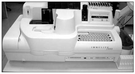
Fig. 1 Immunoassay analyzer used in the study.
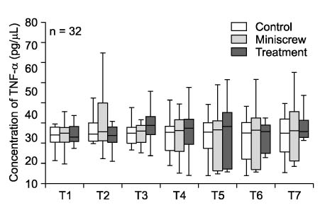
Fig. 2 Changes in the concentration of TNF-α during the observation period. T1, Before distalization (baseline); T2, T3, T4, T5, T6 and T7, 1 hour, 1 day, 2 days, 1 week, 3 weeks and 3 monts after activation, respectively.
Reference
-
1. Proffit WR, Fields HW, Sarver DM. Contemporary orthodontics. 2007. 4th ed. St Louis, Mo: Mosby;348–349.2. Sari E, Olmez H, Gürton AU. Comparison of some effects of acetylsalicylic acid and rofecoxib during orthodontic tooth movement. Am J Orthod Dentofacial Orthop. 2004. 125:310–315.
Article3. Teitelbaum SL. Bone resorption by osteoclasts. Science. 2000. 289:1504–1508.
Article4. Başaran G, Ozer T, Kaya FA, Kaplan A, Hamamci O. Interleukine-1β and tumor necrosis factor-α levels in the human gingival sulcus during orthodontic treatment. Angle Orthod. 2006. 76:830–836.5. Aggarwal BB, Samanta A, Feldman M. Oppenheim JJ, Feldman M, editors. TNF receptors. Cytokine. 2003. Vol 2. San Diego, Calif: Academic Press;1619–1632.6. Bertolini DR, Nedwin GE, Bringman TS, Smith DD, Mundy GR. Stimulation of bone resorption and inhibition of bone formation in vitro by human tumor necrosis factors. Nature. 1986. 319:516–518.
Article7. Lowney JJ, Norton LA, Shafer DM, Rossomando EF. Orthodontic forces increase tumor necrosis factor α in the human gingival sulcus. Am J Orthod Dentofacial Orthop. 1995. 108:519–524.
Article8. Delima AJ, Oates T, Assuma R, Schwartz Z, Cochran D, Amar S, et al. Soluble antagonists to interleukin-1 (IL-1) and tumor necrosis factor (TNF) inhibits loss of tissue attachment in experimental periodontitis. J Clin Periodontol. 2001. 28:233–240.
Article9. Graves DT, Cochran D. The contribution of interleukin-1 and tumor necrosis factor to periodontal tissue destruction. J Periodontol. 2003. 74:391–401.
Article10. Schierano G, Pejrone G, Brusco P, Trombetta A, Martinasso G, Preti G, et al. TNF-α TGF-β2 and IL-1β levels in gingival and peri-implant crevicular fluid before and after de novo plaque accumulation. J Clin Periodontol. 2008. 35:532–538.
Article11. Roberts WE, Engen DW, Schneider PM, Hohlt WF. Implant-anchored orthodontics for partially edentulous malocclusions in children and adults. Am J Orthod Dentofacial Orthop. 2004. 126:302–304.
Article12. Fukunaga T, Kuroda S, Kurosaka H, Takano-Yamamoto T. Skeletal anchorage for orthodontic correction of maxillary protrusion with adult periodontitis. Angle Orthod. 2006. 76:148–155.13. Kuroda S, Sugawara Y, Deguchi T, Kyung HM, Takano-Yamamoto T. Clinical use of miniscrew implants as orthodontic anchorage: success rates and postoperative discomfort. Am J Orthod Dentofacial Orthop. 2007. 131:9–15.
Article14. Park HS, Kwon TG, Sung JH. Nonextraction treatment with microscrew implants. Angle Orthod. 2004. 74:539–549.15. Kuroda S, Sugawara Y, Tamamura N, Takano-Yamamoto T. Anterior open bite with temporomandibular disorder treated with titanium screw anchorage: evaluation of morphological and functional improvement. Am J Orthod Dentofacial Orthop. 2007. 131:550–560.
Article16. Meffert RM, Langer B, Fritz ME. Dental implants: a review. J Periodontol. 1992. 63:859–870.
Article17. Lekholm U, Adell R, Lindhe J, Brånemark PI, Eriksson B, Rockler B, et al. Marginal tissue reactions at osseointegrated titanium fixtures. (II) A cross-sectional retrospective study. Int J Oral Maxillofac Surg. 1986. 15:53–61.18. Loe H, Silness J. Periodontal disease in pregnancy. I. Prevalence and severity. Acta Odontol Scand. 1963. 21:533–551.
Article19. Park HS, Bae SM, Kyung HM, Sung JH. Micro-implant anchorage for treatment of skeletal Class I bialveolar protrusion. J Clin Orthod. 2001. 35:417–422.20. Park HS, Kyung HM, Sung JH. A simple method of molar uprighting with micro-implant anchorage. J Clin Orthod. 2002. 36:592–596.21. Rüdin HJ, Overdiek HF, Rateitschak KH. Correlation between sulcus fluid rate and clinical and histological inflammation of the marginal gingiva. Helv Odontol Acta. 1970. 14:21–26.22. Sari E, Uçar C. Interleukin 1β levels around microscrew implants during orthodontic tooth movement. Angle Orthod. 2007. 77:881–884.23. Tzannetou S, Efstratiadis S, Nicolay O, Grbic J, Lamster I. Interleukin 1β and β-glucuronidase in gingival crevicular fluid from molars during rapid palatal expansion. Am J Orthod Dentofacial Orthop. 1999. 115:686–696.
Article24. Deguchi T, Takano-Yamamoto T, Kanomi R, Hartsfield JK Jr, Roberts WE, Garetto LP. The use of small titanium screws for orthodontic anchorage. J Dent Res. 2003. 82:377–381.
Article25. Tuncer BB, Ozmeriç N, Tuncer C, Teoman I, Cakilci B, Yücel A, et al. Levels of interleukin-8 during tooth movement. Angle Orthod. 2005. 75:631–636.26. Serra E, Perinetti G, D'Attilio M, Cordella C, Paolantonio M, Festa F, et al. Lactate dehydrogenase activity in gingival crevicular fluid during orthodontic treatment. Am J Orthod Dentofacial Orthop. 2003. 124:206–211.
Article27. Beutler B, Grau GE. Tumor necrosis factor in the pathogenesis of infectious diseases. Crit Care Med. 1993. 21:10 Suppl. S423–S435.
Article28. Holmes CL, Russell JA, Walley KR. Genetic polymorphisms in sepsis and septic shock: role in prognosis and potential for therapy. Chest. 2003. 124:1103–1115.29. Machtei EE, Oved-Peleg E, Peled M. Comparison of clinical, radiographic and immunological parameters of teeth and different dental implant platforms. Clin Oral Implants Res. 2006. 17:658–665.
Article30. Nowzari H, Botero JE, DeGiacomo M, Villacres MC, Rich SK. Microbiology and cytokine levels around healthy dental implants and teeth. Clin Implant Dent Relat Res. 2008. 10:166–173.
Article31. Jäger A, Zhang D, Kawarizadeh A, Tolba R, Braumann B, Lossdörfer S, et al. Soluble cytokine receptor treatment in experimental orthodontic tooth movement in the rat. Eur J Orthod. 2005. 27:1–11.
Article32. Listgarten MA. Soft and hard tissue response to endosseous dental implants. Anat Rec. 1996. 245:410–425.
Article
- Full Text Links
-
- Actions
-
Cited
- CITED
-
- Close
- Share
-
- Similar articles
-
- Kinetics of HMGB1 level changes in a canine endotoxemia model
- Elevated Tumor Necrosis Factor-alpha in Stable Angina Pectoris
- Changes of Serum Tumor Necrosis Factor a and Interleukin 1B in the Sepsis of Neonates
- Regulation of mouse macrophage Fc receptor-mediated phagocytosis by interferon-r, lipid a and tumor necrosis factor
- Regulation of tumor necrosis factor-alpha gene expression by gluco-corticoid, dehydroepiandrosterone, and 1,25-dihydroxyvitamin D3
