J Korean Soc Radiol.
2011 May;64(5):429-434. 10.3348/jksr.2011.64.5.429.
Angiomatous Meningioma: CT and MR Imaging Features
- Affiliations
-
- 1Department of Radiology, Eulji University Hospital, Daejeon, Korea. midosyu@eulji.ac.kr
- 2Department of Neurosurgery, Eulji University Hospital, Daejeon, Korea.
- KMID: 2040809
- DOI: http://doi.org/10.3348/jksr.2011.64.5.429
Abstract
- PURPOSE
To describe the computed tomography and magnetic resonance imaging features of angiomatous meningiomas.
MATERIALS AND METHODS
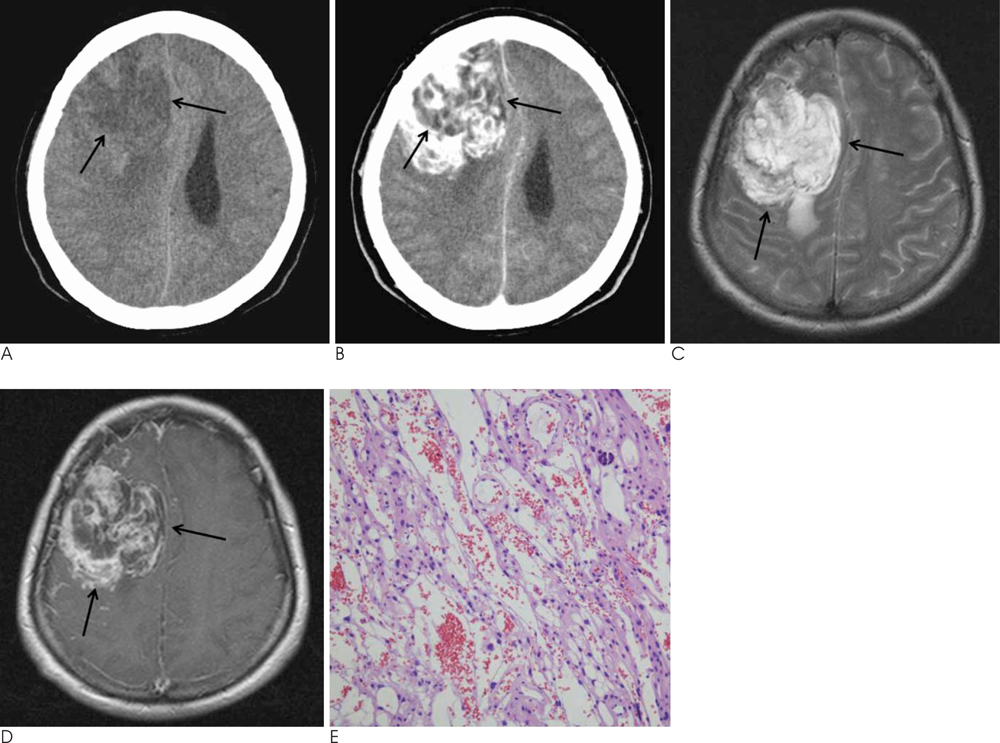
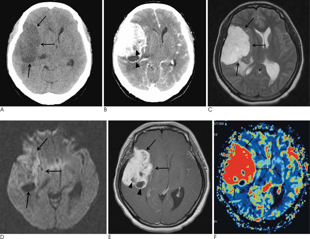
We reviewed the imaging findings of six patients with pathologically proven angiomatous meningiomas and characterized the location, margin, dura base, CT attenuation, MR signal intensity, intratumoral signal void, contrast enhancement, intratumoral cystic change, and peritumoral edema.
RESULTS
Most tumors showed high signal intensity on T2-weighted images, and low signal intensity on diffusion-weighted images. After intravenous contrast administration, the tumor showed heterogeneous strong enhancement. Most tumors had a lobulated margin with prominent intratumoral signal voids. Four patients showed marked or small intratumoral cystic changes.
CONCLUSION
Typically, angiomatous meningiomas were dura-based masses characterized by lobulated margins with high signal intensity on T2-weighted imaging (T2WI), low signal intensity on diffusion-weighted imaging (DWI), prominent intratumoral signal voids, intratumoral cystic changes, and marked enhancement after intravenous contrast administration.
Figure
Reference
-
1. Kim SH, Kim DG, Kim CY, Choe G, Chang KH, Jung HW. Microcystic meningioma: the characteristic neuroradiologic findings. J Korean Neurosurg Soc. 2003; 34:401–406.2. Hasselblatt M, Nolte KW, Paulus W. Angiomatous meningioma: a clinicopathology study of 38 cases. Am J Surg Pathol. 2004; 28:390–393.3. Swami R, Ghosh A, Verma-Pradhan S. Angiomatous meningioma. Nepal J Neurosci. 2007; 4:102.4. Hakyemez B, Yildirim N, Gokalp G, Erdogan C, Parlak M. The contribution of diffusion-weighted MR imaging to distinguishing typical from atypical meningiomas. Neuroradiology. 2006; 48:513–520.5. Elster AD, Challa VR, Gilbert TH, Richardson DN, Contento JC. Meningiomas: MR and histopathologic features. Radiology. 1989; 170:857–862.6. Kaplan RD, Coons S, Drayer BP, Bird CR, Johnson PC. MR characteristics of meningioma subtypes at 1.5 tesla. J Comput Assist Tomogr. 1992; 16:366–371.7. Taraszewska A, Bogucki J. A case of cystic form of angiomatous meningioma with prominent microvascular pattern mimicking haemangioblastoma. Folia Neuropathol. 2001; 39:119–123.8. Cushing H, Eisenhardt L. Meningiomas. Am J Med Sci. 1938; 196:741–742.9. Rao S, Rajkumar A, Kuruvilla S. Angiomatous meningioma: a diagnostic dilemma. Indian J Pathol Microbiol. 2008; 51:53–55.10. Alen JF, Lobato RD, Gomez PA, Boto GR, Lagares A, Ramos A, et al. Intracranial hemangiopericytoma: study of 12 cases. Acta Neurochir. 2001; 143:575–586.11. Chen TY, Lai PH, Ho JT, Wang JS, Chen WL, Pan HB, et al. Magnetic resonance imaging and diffusion-weighted images of cystic meningioma: correlating with histopathology. Clin Imaging. 2004; 28:10–19.12. Bydder GM, Kingsley DP, Brown J, Niendorf HP, Young IR. MR imaging of meningiomas including studies with and without gadolinium-DTPA. J Comput Assist Tomogr. 1985; 9:690–697.13. Schorner W, Schubeus P, Henkes H, Rottacker C, Hamm B, Felix R. Intracranial meningiomas. Comparison of plain and contrastenhanced examinations in CT and MRI. Neuroradiology. 1990; 32:12–18.14. Schubeus P, Schorner W, Rottacker C, Sander B. Intracranial meningiomas: how frequent are indicative findings in CT and MRI? Neuroradiology. 1990; 32:467–473.15. Buetow MP, Buetow PC, Smirniotopoulos JG. Typical, atypical, and misleading features in meningioma. Radiographics. 1991; 11:1087–1106.16. Sridhar K, Ravi R, Ramamurthi B, Vasudevan MC. Cystic meningiomas. Surg Neurol. 1995; 43:235–239.17. Zee CS, Chen T, Hinton DR, Tan M, Segall HD, Apuzzo ML. Magnetic resonance imaging of cystic meningiomas and its surgical implications. Neurosurgery. 1995; 36:482–488.18. Rengachary S, Batnitzky S, Kepes JJ, Morantz RA, O'Boynick P, Watanabe I. Cystic lesions associated with intracranial meningiomas. Neurosurgery. 1979; 4:107–114.19. Worthington C, Caron JL, Melanson D, Leblanc R. Meningioma cysts. Neurology. 1985; 35:1720–1724.20. Dell S, Ganti SR, Steinberger A, McMurtry J 3rd. Cystic meningiomas: a clinicoradiological study. J Neurosurg. 1982; 57:8–13.21. Fortuna A, Ferrante L, Acqui M, Guglielmi G, Mastronardi L. Cystic meningiomas. Acta Neurochir. 1988; 90:23–30.22. Parisi G, Tropea R, Giuffrida S, Lombardo M, Giuffre F. Cystic meningiomas. Report of seven cases. J Neurosurg. 1986; 64:35–38.23. Filippi CG, Edgar MA, Ulug AM, Prowda JC, Heier LA, Zimmerman RD. Appearance of meningiomas on diffusionweighted images: correlating diffusion constants with histopathologic findings. AJNR Am J Neuroradiol. 2001; 22:65–72.24. Kono K, Inoue Y, Nakayama K, Shakudo M, Morino M, Ohata K, et al. The role of diffusion-weighted imaging in patients with brain tumors. AJNR Am J Neuroradiol. 2001; 22:1081–1088.25. Whittle IR, Smith C, Navoo P, Collie D. Meningiomas. Lancet. 2004; 363:1535–1543.26. Pistolesi S, Fontanini G, Camacci T, De Ieso K, Boldrini L, Lupi G, et al. Meningioma-associated brain oedema: the role of angiogenic factors and pial blood supply. J Neurooncol. 2002; 60:159–164.27. Domingo Z, Rowe G, Blamire AM, Cadoux-Hudson TA. Role of ischaemia in the genesis of oedema surrounding meningiomas assessed using magnetic resonance imaging and spectroscopy. Br J Neurosurg. 1998; 12:414–418.28. Yoshioka H, Hama S, Taniguchi E, Sugiyama K, Arita K, Kurisu K. Peritumoral brain edema associated with meningioma: influence of vascular endothelial growth factor expression and vascular blood supply. Cancer. 1999; 85:936–944.29. Zhang H, Rodiger LA, Shen T, Miao J, Oudkerk M. Preoperative subtyping of meningiomas by perfusion MR imaging. Neuroradiology. 2008; 50:835–840.30. Kimura H, Takeuchi H, Koshimoto Y, Arishima H, Uematsu H, Kawamura Y, et al. Perfusion imaging of meningioma by using continuous arterial spin-labeling: comparison with dynamic susceptibility-weighted contrast-enhanced MR images and histopathologic features. AJNR Am J Neuroradiol. 2006; 27:85–93.31. Lupo JM, Cha S, Chang SM, Nelson SJ. Dynamic susceptibilityweighted perfusion imaging of high-grade gliomas: Characterization of spatial heterogeneity. AJNR Am J Neuroradiol. 2005; 26:1446–1454.
- Full Text Links
- Actions
-
Cited
- CITED
-
- Close
- Share
- Similar articles
-
- Imaging Features and Pathological Correlation in Mixed Microcystic and Angiomatous Meningioma: A Case Report
- Clinical and Radiological Characteristics of Angiomatous Meningiomas
- A Meningioma With Extensive Peritumoral Edema Mimicking Metastatic Brain Tumor: A Case Report
- Clear-Cell Meningioma: CT and MR Imaging Findings in Two Cases Involving the Spinal Canal and Cerebellopontine Angle
- Atypical CT findings of intracranial meningioma


