Clinical and Radiological Characteristics of Angiomatous Meningiomas
- Affiliations
-
- 1Department of Neurosurgery, Samsung Medical Center, Sungkyunkwan University College of Medicine, Seoul, Korea. jwchoins@gmail.com
- KMID: 2356976
- DOI: http://doi.org/10.14791/btrt.2016.4.2.94
Abstract
- BACKGROUND
Angiomatous meningioma is a rare histological subtype of meningioma. Therefore, this specific medical condition is rarely reviewed in the literature. In the present work, we report the clinical and radiological features with postoperative outcomes of angiomatous meningioma.
METHODS
This retrospective study included the patients who were pathologically diagnosed with angiomatous meningioma after surgical resection between February 2010 and September 2015 in our institute. We analyzed the clinical data, radiological manifestation, treatment and prognosis of all patients.
RESULTS
The 15 patients (5 males and 10 females) were diagnosed with angiomatous meningioma during the study period. The median age of patients at the time of surgery was 63 years (range: 40 to 80 years). According to Simpson classification, 7, 5, and 3 patients achieved Simpson grade I, II, and IV resection, respectively. In the follow-up period, recurrence was noted in one patient. Ten out of the 15 patients showed homogeneous enhancement. Two patients demonstrated cystic changes. There was no occurrence of calcification or hemorrhage in our patients. Characteristically, 14 out of 15 patients showed signal voids of vessels. Significant peritumoral edema was observed in the majority of tumors (67%).
CONCLUSION
Angiomatous meningiomas are rare benign meningioma. Brain images of angiomatous meningioma usually demonstrate signal void signs and peritumoral edema. In the present study, angiomatous meningiomas showed good prognosis after surgical resection.
MeSH Terms
Figure
-
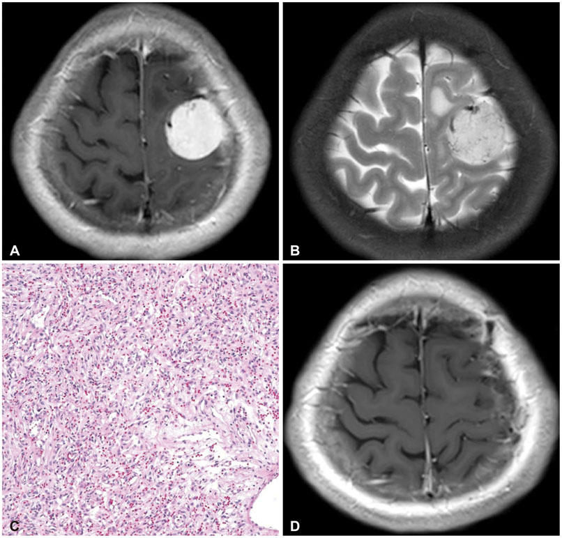
Fig. 1 MRI and histologic findings of case 4. A: The preoperative T1-weighted brain MRI with gadolinium enhancement demonstrates about 3 cm sized homogeneous enhancing mass. B: The preoperative T2-weighted brain MRI shows the signal voids of blood vessels. C: Histologic findings of angiomatous meningioma show predominence of vascular proliferations over the tumor cells. The vascular channels contain a mixture of delicate and thick walls with variable sizes (Hematoxylin and eosin stain, ×200). D: No recurrence is verified in the last follow-up images after 14 months from operation.

Fig. 2 MRI and histologic findings of case 15. A: The preoperative T1-weighted brain MRI with gadolinium enhancement shows a large cystic mass with mild midline shift. B: The preoperative T2-weighted brain MRI shows no definite signal voids of blood vessels and demonstrated significant brain edema. C: Histologic findings of angiomatous meningioma. In this case, most are small with hyalinized walls. Surrounding meningothelial cells have relatively uniform round to oval shape nuclei and nuclear pseudoinclusions with eosinophilic cytoplasm and indistinct cell borders (Hematoxylin and eosin stain, ×200). D: The postoperative T1-weighted MRI with gadolinium enhancement demonstrates that there is no residual mass at the tumorectomy site. E: The follow-up MRI after 13 months from surgery shows a small nodular enhancing lesion (T1-weighted with gadolinium enhancement). F: The latest follow-up MRI displays growth of recurrent tumor in inferior frontal gyrus (T1-weighted with gadolinium enhancement).
Reference
-
1. Riemenschneider MJ, Perry A, Reifenberger G. Histological classification and molecular genetics of meningiomas. Lancet Neurol. 2006; 5:1045–1054.
Article2. Louis DN, Ohgaki H, Wiestler OD, et al. The 2007 WHO classification of tumours of the central nervous system. Acta Neuropathol. 2007; 114:97–109.
Article3. Robson DK. Pathology and genetics of tumours of the nervous system. In : Kleihues P, Cavenee WK, editors. The Journal of pathology. Lyon: IARC Press;2000. p. 314.4. Tang H, Sun H, Chen H, et al. Clinicopathological analysis of metaplastic meningioma: report of 15 cases in Huashan Hospital. Chin J Cancer Res. 2013; 25:112–118.5. Simpson D. The recurrence of intracranial meningiomas after surgical treatment. J Neurol Neurosurg Psychiatry. 1957; 20:22–39.
Article6. Hasselblatt M, Nolte KW, Paulus W. Angiomatous meningioma: a clinicopathologic study of 38 cases. Am J Surg Pathol. 2004; 28:390–393.7. Liu Z, Wang C, Wang H, Wang Y, Li JY, Liu Y. Clinical characteristics and treatment of angiomatous meningiomas: a report of 27 cases. Int J Clin Exp Pathol. 2013; 6:695–702.8. Roser F, Nakamura M, Ritz R, et al. Proliferation and progesterone receptor status in benign meningiomas are not age dependent. Cancer. 2005; 104:598–601.
Article9. Radner H, Blümcke I, Reifenberger G, Wiestler OD. [The new WHO classification of tumors of the nervous system 2000. Pathology and genetics]. Pathologe. 2002; 23:260–283.10. Tamiya T, Ono Y, Matsumoto K, Ohmoto T. Peritumoral brain edema in intracranial meningiomas: effects of radiological and histological factors. Neurosurgery. 2001; 49:1046–1051. discussion 1051-2.
Article11. Whittle IR, Smith C, Navoo P, Collie D. Meningiomas. Lancet. 2004; 363:1535–1543.
Article12. Pistolesi S, Fontanini G, Camacci T, et al. Meningioma-associated brain oedema: the role of angiogenic factors and pial blood supply. J Neurooncol. 2002; 60:159–164.13. Domingo Z, Rowe G, Blamire AM, Cadoux-Hudson TA. Role of ischaemia in the genesis of oedema surrounding meningiomas assessed using magnetic resonance imaging and spectroscopy. Br J Neurosurg. 1998; 12:414–418.
Article14. Yoshioka H, Hama S, Taniguchi E, Sugiyama K, Arita K, Kurisu K. Peritumoral brain edema associated with meningioma: influence of vascular endothelial growth factor expression and vascular blood supply. Cancer. 1999; 85:936–944.15. Nanda A, Bir SC, Maiti TK, Konar SK, Missios S, Guthikonda B. Relevance of Simpson grading system and recurrence-free survival after surgery for World Health Organization Grade I meningioma. J Neurosurg. 2016; 04. 08. [Epub]. DOI: 10.3171/2016.1.JNS151842.
Article
- Full Text Links
-
- Actions
-
Cited
- CITED
-
- Close
- Share
-
- Similar articles
-
- Imaging Features and Pathological Correlation in Mixed Microcystic and Angiomatous Meningioma: A Case Report
- Multiple Intracranial Meningiomas: Case Report
- Angiomatous Meningioma: CT and MR Imaging Features
- Correlation of histopathologic classification with proliferative activity and DNA ploidy in 120 intracranial meningiomas, with special reference to atypical meningioma
- Antrochoanal Polyp's Variant, the Angiomatous Nasal Polyp: A Case Report
