Clear-Cell Meningioma: CT and MR Imaging Findings in Two Cases Involving the Spinal Canal and Cerebellopontine Angle
- Affiliations
-
- 1Department of Radiology Inha University Hospital College of Medicine, Korea. kanlim@chollian.net
- 2Department of Neurosurgery Inha University Hospital College of Medicine, Korea.
- 3Department of Pathology Inha University Hospital College of Medicine, Korea.
- KMID: 754071
- DOI: http://doi.org/10.3348/kjr.2002.3.2.125
Abstract
- Clear-cell meningioma is a rare subtype of meningioma which occurs at a younger age and has a higher recurrence rate than other subtypes. We report two cases of clear-cell meningioma, one in the thoracolumbar spinal canal and the other in the cerebellopontine angle. Though the CT and MR imaging findings were not different from those of ordinary meningioma, after surgical removal the condition recurred repeatedly in the patient with spinal canal involvement.
Keyword
MeSH Terms
Figure
-
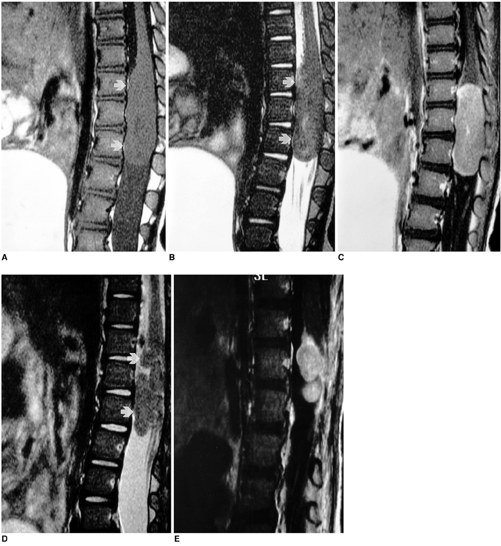
Fig. 1 Clear-cell meningioma in a 14-month-old girl. A, B. Initial T1- and T2-weighted sagittal MR images of the thoracolumbar spine reveal the presence of a well-demarcated, ovoid mass (arrows) in the intradural extramedullary spinal canal at the level of T12-L2. The mass is isointense to the spinal cord on both T1- and T2-weighted images. C. Initial contrast-enhanced T1-weighted sagittal MR image shows strong, homogeneous enhancement. D. T2-weighted sagittal MR image obtained eight months later shows a recurrent isointense signal of the intradural mass (arrows) at the site of initial surgery. E. Contrast-enhanced T1-weighted sagittal MR image obtained after a further interval of seven months depicts a recurrent well-enhanced intradural mass at the site of previous surgery.
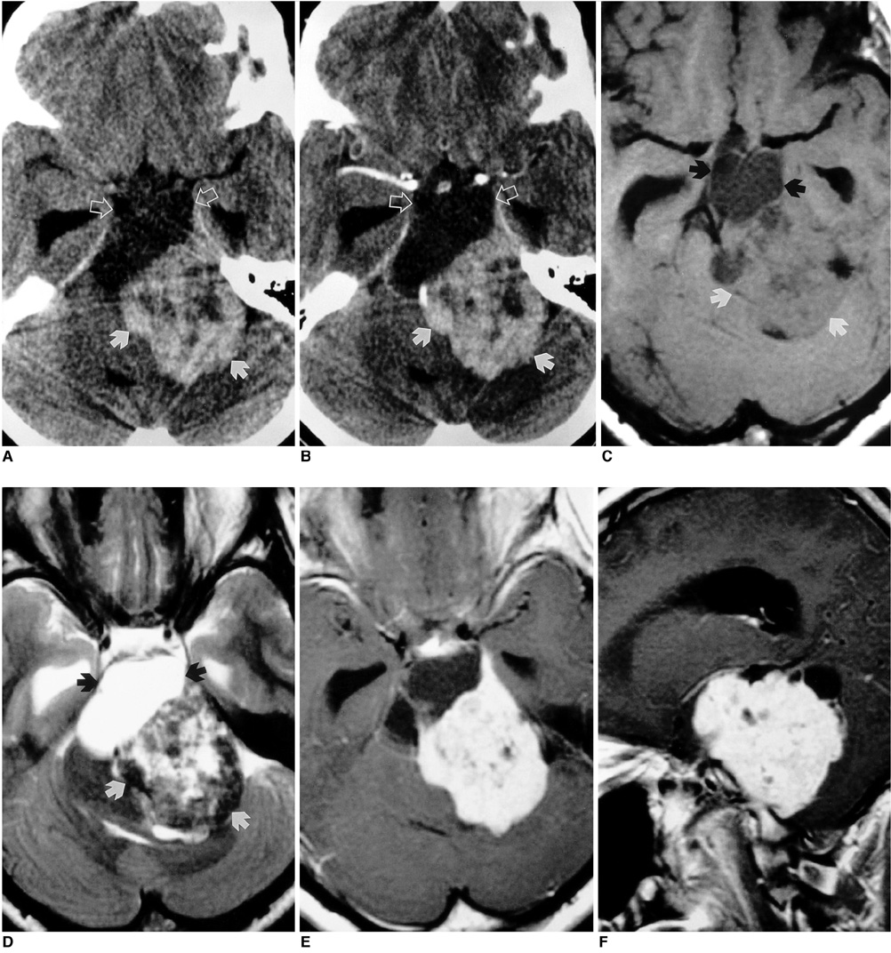
Fig. 2 Clear-cell meningioma in a 17-year-old girl. A. Precontrast axial CT scan shows a huge, irregular, mixed solid and cystic mass in the left cerebellopontine angle. The cystic portion of the mass (open arrows) is located anteriorly and is expansile. The clivus is grossly eroded by the mass, whose solid portion (white arrows) is slightly hyperdense on precontrast CT scans and contains multiple, small, irregular areas of low attenuation that suggest cystic change or necrosis. B. Contrast-enhanced axial CT scan. The solid portion of the mass (white arrows) is mildly and homogeneously enhanced. C. T1-weighted axial MR image depicts a large, multilobulated, mixed solid and cystic mass with a dorsal exophytic component in the upper medulla. It extends superiorly to the pons, and the brainstem is displaced to the right and posterosuperiorly. In the solid portion of the mass (white arrows), an isointense signal with a central area of high intensity is observed. D. T2-weighted axial MR image shows a heterogeneous isointense signal with a central area of high intensity in the solid portion of the mass (white arrows). There is associated mild hydrocephalus. E, F. Contrast-enhanced T1-weighted axial and sagittal MR images. After the use of contrast agent, the solid portion of the mass showed strong, homogeneous enhancement.
Reference
-
1. Alameda F, Lloreta J, Ferrer MD, Corominas JM, Galito E, Serrano S. Clear-cell meningioma of the lumbosacral spine with choroid features. Ultrastruct Pathol. 1999. 23:51–58.2. Zorludemir S, Scheithauer BW, Hirose T, Van Houten C, Miller G, Meyer FB. Clear-cell meningioma: a clinicopathologic study of a potentially aggressive variant of meningioma. Am J Surg Pathol. 1995. 19:493–505.3. Kleihues P, Burger PC, Scheithauer BW. The new WHO classification of brain tumors. Brain Pathol. 1993. 3:255–268.4. Shih DF, Wang JS, Pan RG, Tseng HH. Clear-cell meningioma: a case report. Chung Hua I Hsueh Tsa Chih (Taipei). 1996. 57:452–456.5. Matsui H, Kanamori M, Abe Y, Sakai T, Wakaki K. Multifocal clear-cell meningioma in the spine: a case report. Neurosurg Rev. 1998. 21:171–173.6. Lee W, Chang KH, Choe G, et al. MR imaging features of clear-cell meningioma with diffuse leptomeningeal seeding. AJNR. 2000. 21:130–132.7. Jellinger K, Slowik F. Histological subtypes and prognostic problems in meningiomas. J Neurol. 1975. 208:279–298.8. Prinz M, Patt S, Mitrovics T, Cervos-Navarro J. Clear-cell meningioma: report of a spinal case. Gen Diagn Pathol. 1996. 141:261–267.9. Li MH, Holtas S, Larsson EM. MR imaging of intradural extramedullary tumors. Acta Radiol. 1992. 33:207–212.
