Development of animal experimental periodontitis models
- Affiliations
-
- 1Department of Chemistry, Graduate School of Nanoscience and Technology (WCU), Korea Advanced Institute of Science and Technology, Seoul, Korea.
- 2Department of Periodontology, Dental Research Institute, Seoul National University School of Dentistry, Seoul, Korea. periopf@snu.ac.kr
- 3Department of Life Science, Dongguk University, Seoul, Korea.
- KMID: 2027809
- DOI: http://doi.org/10.5051/jpis.2013.43.4.147
Abstract
- PURPOSE
An animal periodontitis model is essential for research on the pathogenesis and treatment of periodontal disease. In this study, we have introduced a lipopolysaccharide (LPS) of a periodontal pathogen to the alveolar bone defect of experimental animals and investigated its suitability as a periodontitis model.
METHODS
Alveolar bone defects were made in both sides of the mandibular third premolar region of nine beagle dogs. Then, the animals were divided into the following groups: silk ligature tied on the cervical region of tooth group, Porphyromonas gingivalis LPS (P.g. LPS)-saturated collagen with silk ligature group, and no ligature or P.g. LPS application group as the control. The plaque index and gingival index were measured at 0 and 4 weeks postoperatively. The animals were then euthanized and prepared for histologic evaluation.
RESULTS
The silk ligature group and P.g. LPS with silk ligature group showed a significantly higher plaque index at 4 weeks compared to the control (P<0.05). No significant difference was found in the plaque index between the silk ligature group and P.g. LPS with silk ligature group. The P.g. LPS with silk ligature group showed a significantly higher gingival index compared to the silk ligature group or the control at 4 weeks (P<0.05). Histologic examination presented increased inflammatory cell infiltration in the gingival tissue and alveolar bone of the P.g. LPS with silk ligature group.
CONCLUSIONS
An additional P.g. LPS-saturated collagen with silk ligature ensured periodontal inflammation at 4 weeks. Therefore, P.g. LPS with silk ligature application to surgically created alveolar bone defects may be a candidate model for experimental periodontitis.
MeSH Terms
Figure
-
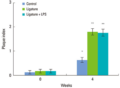
Figure 1 Plaque index for each group. There were no significant differences in any of the groups at baseline (week 0). After 4 weeks, the silk ligature group (Ligature) and silk ligature with Porphyromonas gingivalis lipopolysaccharide (P.g. LPS) group (Ligature+LPS) showed marked differences compared to the control (P<0.05). There were no significant differences between the silk ligature with P.g. LPS group and silk ligature group. Different asterisks indicate statistically significant differences. Data are presented as mean±standard error.
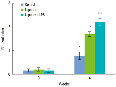
Figure 2 Gingival index for each group. There were no significant differences among the groups at week 0. After 4 weeks, the silk ligature with Porphyromonas gingivalis lipopolysaccharide (LPS) group (Ligature+LPS) showed significant differences compared to the other groups (P<0.05). There were also significant differences between the silk ligature group (Ligature) and the control. Different asterisks indicate statistically significant differences. Data are presented as mean±standard error.
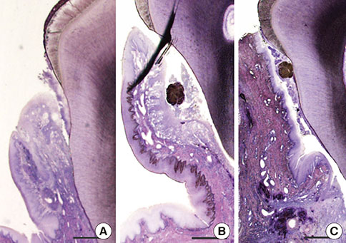
Figure 3 Histologic views of each group (H&E, bar=1 mm). (A) The control group showed minimal attachment loss and very little inflammation infiltration. (B) The silk ligature group showed mild gingival inflammation. (C) The Porphyromonas gingivalis lipopolysaccharide with silk ligature group showed severe inflammatory cell infiltration.

Figure 4 Alveolar bone images of each group (H&E, bar=100 µm). (A) There was no sign of inflammation in the control. (B) The silk ligature group showed reduced inflammation. (C) The Porphyromonas gingivalis lipopolysaccharide with silk ligature group showed a severe inflammatory cell presence in the alveolar bone tissue.
Cited by 2 articles
-
The influence of root surface distance to alveolar bone and periodontal ligament on periodontal wound healing
Marco Montevecchi, Annapaola Parrilli, Milena Fini, Maria Rosaria Gatto, Aurelio Muttini, Luigi Checchi
J Periodontal Implant Sci. 2016;46(5):303-319. doi: 10.5051/jpis.2016.46.5.303.Periodontal and endodontic pathology delays extraction socket healing in a canine model
Jung-Hoon Kim, Ki-Tae Koo, Joseph Capetillo, Jung-Ju Kim, Jung-Min Yoo, Heithem Ben Amara, Jung-Chul Park, Frank Schwarz, Ulf M.E. Wikesjö
J Periodontal Implant Sci. 2017;47(3):143-153. doi: 10.5051/jpis.2017.47.3.143.
Reference
-
1. Pihlstrom BL, Michalowicz BS, Johnson NW. Periodontal diseases. Lancet. 2005; 366:1809–1820.
Article2. Schou S, Holmstrup P, Kornman KS. Non-human primates used in studies of periodontal disease pathogenesis: a review of the literature. J Periodontol. 1993; 64:497–508.
Article3. Yamasaki A, Nikai H, Niitani K, Ijuhin N. Ultrastructure of the junctional epithelium of germfree rat gingiva. J Periodontol. 1979; 50:641–648.
Article4. Eggert FM, Germain JP, Cohen B. The gingival epithelium of rodent molars with limited eruption. Acta Anat (Basel). 1980; 107:297–306.
Article5. Attström R, Graf-de Beer M, Schroeder HE. Clinical and histologic characteristics of normal gingiva in dogs. J Periodontal Res. 1975; 10:115–127.
Article6. Haney JM, Zimmerman GJ, Wikesjo UM. Periodontal repair in dogs: evaluation of the natural disease model. J Clin Periodontol. 1995; 22:208–213.
Article7. Wikesjö UM, Kean CJ, Zimmerman GJ. Periodontal repair in dogs: supraalveolar defect models for evaluation of safety and efficacy of periodontal reconstructive therapy. J Periodontol. 1994; 65:1151–1157.
Article8. Holland M, Boring JG, Boyle CR, Pickrum HM, Jeffcoat MK. Radiographic bone loss correlations and technetium-99m-MDP bone uptake in ligature-induced periodontal disease in the beagle. Vet Radiol Ultrasound. 1998; 39:366–374.
Article9. Clergeau LP, Danan M, Clergeau-Guerithault S, Brion M. Healing response to anorganic bone implantation in periodontal intrabony defects in dogs. Part I. Bone regeneration. A microradiographic study. J Periodontol. 1996; 67:140–149.
Article10. Hayashi C, Kinoshita A, Oda S, Mizutani K, Shirakata Y, Ishikawa I. Injectable calcium phosphate bone cement provides favorable space and a scaffold for periodontal regeneration in dogs. J Periodontol. 2006; 77:940–946.
Article11. Andrian E, Grenier D, Rouabhia M. Porphyromonas gingivalis-epithelial cell interactions in periodontitis. J Dent Res. 2006; 85:392–403.
Article12. Dumitrescu AL, Abd-El-Aleem S, Morales-Aza B, Donaldson LF. A model of periodontitis in the rat: effect of lipopolysaccharide on bone resorption, osteoclast activity, and local peptidergic innervation. J Clin Periodontol. 2004; 31:596–603.
Article13. Silness J, Loe H. Periodontal disease in pregnancy: II. Correlation between oral hygiene and periodontal condtion. Acta Odontol Scand. 1964; 22:121–135.
Article14. Loe H, Silness J. Periodontal disease in pregnancy. I. Prevalence and severity. Acta Odontol Scand. 1963; 21:533–551.
Article15. Page RC, Schroeder HE. Pathogenesis of inflammatory periodontal disease: a summary of current work. Lab Invest. 1976; 34:235–249.16. Lallam-Laroye C, Escartin Q, Zlowodzki AS, Barritault D, Caruelle JP, Baroukh B, et al. Periodontitis destructions are restored by synthetic glycosaminoglycan mimetic. J Biomed Mater Res A. 2006; 79:675–683.
Article17. Peruzzo DC, Benatti BB, Antunes IB, Andersen ML, Sallum EA, Casati MZ, et al. Chronic stress may modulate periodontal disease: a study in rats. J Periodontol. 2008; 79:697–704.
Article18. Lindhe J, Hamp S, Loe H. Experimental periodontitis in the beagle dog. J Periodontal Res. 1973; 8:1–10.
Article19. Allaker RP, de Rosayro R, Young KA, Hardie JM. Prevalence of Porphyromonas and Prevotella species in the dental plaque of dogs. Vet Rec. 1997; 140:147–148.
Article20. Garrison SW, Nichols FC. LPS-elicited secretory responses in monocytes: altered release of PGE2 but not IL-1 beta in patients with adult periodontitis. J Periodontal Res. 1989; 24:88–95.
Article21. Page RC. The role of inflammatory mediators in the pathogenesis of periodontal disease. J Periodontal Res. 1991; 26(3 Pt 2):230–242.
Article22. Rogers JE, Li F, Coatney DD, Rossa C, Bronson P, Krieder JM, et al. Actinobacillus actinomycetemcomitans lipopolysaccharide-mediated experimental bone loss model for aggressive periodontitis. J Periodontol. 2007; 78:550–558.
Article23. Egelberg J. Local effect of diet on plaque formation and development of gingivitis in dogs: 3. Effect of frequency of meals and tube feeding. Odontol Revy. 1965; 16:50–60.24. Hamp SE, Lindhe J, Loe H. Experimental periodontitis in the beagle dog. J Periodontal Res. 1972; (10):13–14.
Article25. Lindhe J, Hamp SE, Loe H. Experimental periodontitis in the beagle dog. Int Dent J. 1973; 23:432–437.
Article26. Lindhe J, Ericsson I. Effect of ligature placement and dental plaque on periodontal tissue breakdown in the dog. J Periodontol. 1978; 49:343–350.
Article27. Lindemann RA, Economou JS, Rothermel H. Production of interleukin-1 and tumor necrosis factor by human peripheral monocytes activated by periodontal bacteria and extracted lipopolysaccharides. J Dent Res. 1988; 67:1131–1135.
Article28. Garrison SW, Holt SC, Nichols FC. Lipopolysaccharide-stimulated PGE2 release from human monocytes. Comparison of lipopolysaccharides prepared from suspected periodontal pathogens. J Periodontol. 1988; 59:684–687.29. Jiang Y, Mehta CK, Hsu TY, Alsulaimani FF. Bacteria induce osteoclastogenesis via an osteoblast-independent pathway. Infect Immun. 2002; 70:3143–3148.
Article30. Page RC, Schroeder HE. Spontaneous chronic periodontitis in adult dogs: a clinical and histopathological survey. J Periodontol. 1981; 52:60–73.
Article
