Effect of dentinal tubules orientation on penetration pattern of dentin adhesives using confocal laser scanning microscopy
- Affiliations
-
- 1Department of Conservative Dentistry, College of Dentistry, DSRI, Chonnam National University, Korea. hinso@jnu.ac.kr
- KMID: 1987265
- DOI: http://doi.org/10.5395/JKACD.2003.28.5.392
Abstract
- The purpose of this study was to evaluate the penetration pattern of dentin adhesives according to the orientation of dentinal tubules with confocal laser scanning microscopy. Specimens having perpendicular, parallel and oblique surface to dentinal tubules were fabricated. The primer of dentin adhesives (ALL BOND(R) 2, CLEARFIL(TM) SE BOND and PQ1) was mixed with fluorescent material, rhodamine B isothiocyanate (Aldrich Chem. CO., Milw., USA). It was applied to the specimens according to the instructions of manufactures. The specimens were covered with composite resin (Estelite, shade A2) and then cut to a thickness of 500 microm with low speed saw (Isomet(TM), Buehler, USA). The adhesive pattern of dentin adhesives were observed by fluorescence image using confocal laser scanning microscopy. The results were as follows. 1. For the groups with tubules perpendicular to bonded surface, funnel shape of resin tag was observed in all specimen. However, resin tags were more prominent in phosphoric acid etching system (ALL BOND(R) 2 and PQ1) than self etching system (CLEARFIL(TM) SE BOND). 2. For the groups with tubules parallel to bonded surface, rhodamine-labeled primer penetrated into peritubular dentin parallel to the orientation of dentinal tubules. But rhodamine-labeled primer of PQ1 diffused more radially into surrounding intertubular dentin than other dentin adhesive systems. 3. For the groups with tubules oblique to bonded surface, resin tags appeared irregular and discontinuous. But they penetrated deeper into dentinal tubules than other groups.
MeSH Terms
Figure
-

Fig. 1 Preparation of specimens with the bonding surface perpendicular, parallel or oblique to the dentinal tubules (B-L = bucco-lingual view, M-D = mesio-distal view).

Fig. 2 Confocal laser scanning microscopy (Olympus fluoview 300, Olympus, Japan).
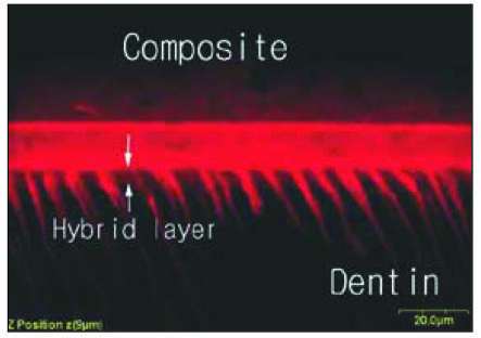
Fig. 3 Overlapped image of fluorescent mode of the interface between ALL-BOND® 2 and dentin. Resin tags with characteristic funnel cone shape were observed. ×1200.

Fig. 4 CLSM image scanned parallel to interface at a depth below 3 µm interval. The Red area indicate the penetration of rhodamine-labeled primer into dentinal tubules and White arrows also indicate their side branchs. ×2400.
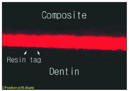
Fig. 5 Overlapped image of fluorescent mode of the interface between CLEARFIL™ SE BOND and dentin. Resin tags is less prominent than other dental adhesive systems. ×2100.
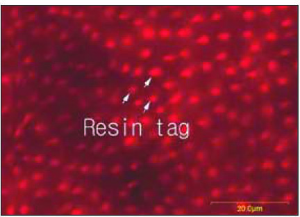
Fig. 6 CLSM image scanned parallel to interface at a depth below 3 µm interval. The Red area indicate the penetration of rhodamine-labeled primer into dnetinal tubules. ×2400.
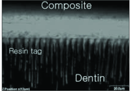
Fig. 7 Overlapped image of fluorescent mode of the interface between PQ1 and dentin. Resin tags with characteristic funnel cone shape were observed. ×1200.
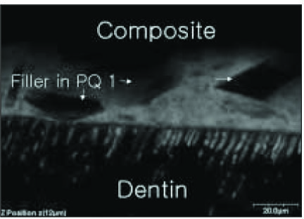
Fig. 8 Overlapped image of fluorescent mode of the interface between PQ1 and dentin. The white arrow indicate the filler of PQ1. ×1200.
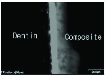
Fig. 9 Overlapped image of fluorescent mode of the interface betweenALL-BOND® 2 and dentin. The penetration pattern of rhodamine-labeled primer is different from perpendicular group. ×1200.

Fig. 10 CLSM image scanned parallel to interface at a depth below 3 µm interval. There were few resin tags like perpendicular group. ×2100.

Fig. 11 Overlapped image of fluorescent mode of the interface between CLEARFIL™ SE BOND and dentin. The direction of dentinal tubule and rhodamine-labeled primer is parallel. ×2100.

Fig. 12 CLSM image scanned parallel to interface at a depth below 3 µm interval. There were few resin tags like prependicular group. ×2100.
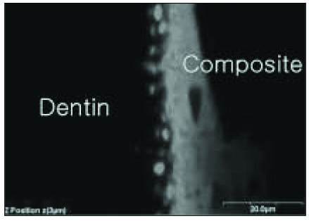
Fig. 13 Overlapped image of fluorescent mode of the interface between PQ1 and dentin. Rhodamine-labeled primer diffuses more radially into surrounding intertubular dentin. ×2100.
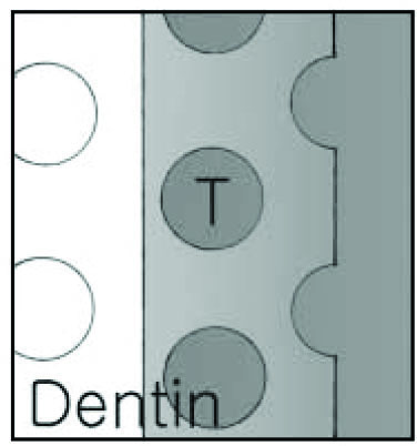
Fig. 14 Schematic illustration of Fig. 13.
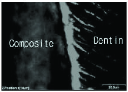
Fig. 15 Overlapped image of fluorescent mode of the interface betweenALL-BOND® 2 and dentin. Irregular shape resin tags were observed. ×1200.

Fig. 16 CLSM image scanned parallel to interface at a depth below 16 µm interval. The white arrow indicate resin tags. ×2100.
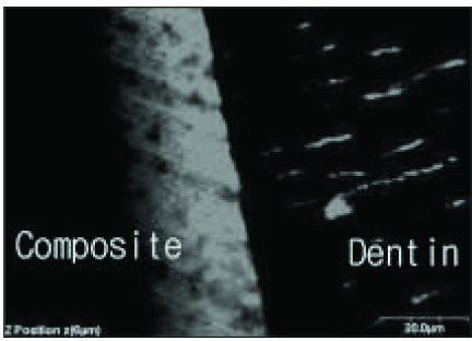
Fig. 17 Overlapped image of fluorescent mode of the interface between CLEARFIL™ SE BOND and dentin. Resin tags were not continuous. ×1200.
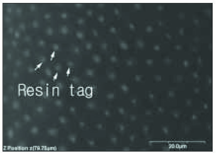
Fig. 18 CLSM image scanned parallel to interface at a depth below 16 µm interval. The white arrow indicate resin tags. ×2100.
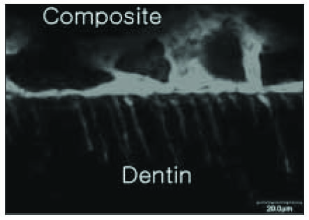
Fig. 19 Overlapped image of fluorescent mode of the interface between PQ1 and dentin. Irregular shape resin tags were observed. ×1200.
Cited by 1 articles
-
Bonding efficacy of cured or uncured dentin adhesives in indirect resin
Ji-Hyun Jang, Bin-Na Lee, Hoon-Sang Chang, Yun-Chan Hwang, Won-Mann Oh, In-Nam Hwang
J Korean Acad Conserv Dent. 2011;36(6):490-497. doi: 10.5395/JKACD.2011.36.6.490.
Reference
-
1. Heymann HO, Bayne SC. Current concepts in dentin bonding:Focusing on dental adhesion factors. J Am Dent Assoc. 1993. 124:26–36.2. Pashley DH, Horner JA, Brewer PD. Interactions of conditioners on the dentin surface. Oper Dent. 1992. Suppl 5. 137–150.3. Burrow MF, Takakura H, Nakajima M, Inai N, Tagami J, Takatsu T. The influence of age and depth of dentin on bonding. Dent Mater. 1994. 10:241–246.
Article4. Perdigao J, Swift EJ, Denehy GE, Wefel JS, Donly KJ. In vitro bond strengths and SEM evaluation of dentin bonding systems to different dentin substrates. J Dent Res. 1994. 73:44–55.
Article5. Nakajima M, Sano H, Burrow MF, Tagami J, Yoshiyama M, Ebisu S, Ciucchi B, Russell CM, Pashley DH. Tensile bond strength and SEM evaluation of caries-affected dentin using dentin adhesives. J Dent Res. 1995. 74:1679–1688.
Article6. Harnirattisai C, Inokoshi S, Shimada Y, Hosoda H. Interfacial morphology of an adhesive composite resin and etched caries-affected dentin. Oper Dent. 1992. 17:222–228.7. Nakabayashi N, Takarada K. Effect of HEMA on bonding to dentin. Dent Mater. 1992. 8:125–130.
Article8. Watson TF, Wilmot DM. A confocal microscopic evaluation of the interface between syntac adhesive and tooth tissue. J Dent. 1992. 20:302–310.
Article9. Meerbeek B, Braem M, Lambrechts P, Vanherle G. Morphological characterization of the interface between resin and sclerotic dentin. J Dent. 1994. 22:141–146.10. Spencer P, Byerley TJ, Eick JD, Witt JD. Chemical characterization of the dentin/adhesive interface by fourier transform infrared photoacoustic spectroscopy. Dent Mater. 1992. 8:10–15.
Article11. Uno S, Finger WJ. Functional of the hybrid zone as stress absorbing layer in resin-dentin bonding. Quintessence Int. 1995. 26:733–738.12. Gwinnett AJ, Tay FR, Pang KM. Quantitative contribution of the collagen network in dentin hybridization. Am J Dent. 1996. 9:140–144.13. Ferrari M, Davison CL. In vivo resin-dentin interdiffusion and tag formation with lateral branches of two adhesive systems. J Prosthet Dent. 1996. 76:250–253.
Article14. Meerbeek B, Inokishi S, Braem M, Lambrechts P, Vanherle G. Morphological aspects of the resin-dentin interdiffusion zone with different dentin adhesive systems. J Dent Res. 1992. 71:1530–1540.
Article15. Watson TF, Boyde A. The use of fluorescent makers for studying the distribution of a dentin bonding agent between a composite restoration and tooth. Clin Mater. 1987. 2:45–53.
Article16. Phrukkanon S, Burrow MF, Tyas MJ. The effect of dentin location and tubule orientation on the bond strengths between resin and dentin. J Dent. 1999. 27:265–274.
Article17. Schupbach P, Krejci I, Felix L. Dentin bonding: effect of tubule orientation on hybrid layer formation. Eur J Oral Sci. 1997. 105:344–352.18. Gwinnett AJ, Kanca JA III. Micromorphology of the bonded dentin interface and its relationship to bond strength. Am J Dent. 1992. 5:73–77.19. Kim MS, Ohn YS, Lee KW, Son HH. Morphologic Change of Dentin Surface According to the Difference in Concentration and Application Time of Phosphoric Acid. J Korean Acad Conserv Dent. 1998. 23:141–155.20. Nakabayashi N, Saimi Y. Bonding to intact dentin. J Dent Res. 1996. 75:1706–1715.
Article21. Finger WJ, Inoue M, Asmussen E. Effect of wettability of adhesive resins on bonding to dentin. Am J Dent. 1994. 7:35–38.22. Toledano M, Osorio R, de Leonardi G, Rosales-Leal JI, Ceballos L, Cabrerizo-Vilchez MA. Influence of self-etching primer on the resin adhesion to enamel and dentin. Am J Dent. 2001. 14:205–210.23. Ogata M, Okuda M, Nakajima M, PNR Pereira, sano H, Tagami J. Influence of the direction of tubules on bond strength to dentin. Oper Dent. 2001. 26:27–35.24. Pioch T, Stotz S, Staehle HJ, Duschner H. Application of confocal laser scanning microscopy to dental bonding. Adv Dent Res. 1997. 11:453–461.25. Griffiths BM, Watson TF. Resin-dentin interface of Scotchbond multipurpose dentin adhesive. Am J Dent. 1995. 8:212–216.26. Chappell RP, Cobb CM, Spencer P, Eick JD. Dentinal tubule anastomosis-a potential factor in adhesive bonding. J Prosthet Dent. 1994. 72:183–188.
- Full Text Links
-
- Actions
-
Cited
- CITED
-
- Close
- Share
-
- Similar articles
-
- Evaluation of penetration depth of 2% chlorhexidine digluconate into root dentinal tubules using confocal laser scanning microscope
- Effect of three different irrigation solutions applied by passive ultrasonic irrigation
- Microorganism penetration in dentinal tubules of instrumented and retreated root canal walls. In vitro SEM study
- A confocal microscopic study on dentinal infiltration of one-bottle adhesive systems and self-etching priming system bonded to class V cavities
- A Scanning electron microscopic study of the dentinal tubule obliteration effect by the different irradiations of a pulsed Nd:YAG laser
