Microorganism penetration in dentinal tubules of instrumented and retreated root canal walls. In vitro SEM study
- Affiliations
-
- 1Division of Endodontics, Department of Restorative Dental Science, King Saud University College of Dentistry, Riyadh, kingdom of Saudi Arabia. snazhan@ksu.edu.sa
- 2Riyadh Colleges of Dentistry and Pharmacy, College of Dentistry, Riyadh, Kingdom of Saudi Arabia.
- 3Royal Clinics for the Custodian of The Two Holy Mosques, Riyadh, Kingdom of Saudi Arabia.
- 4Microbiology Lab, King Saud University College of Dentistry, Riyadh, kingdom of Saudi Arabia.
- KMID: 2316905
- DOI: http://doi.org/10.5395/rde.2014.39.4.258
Abstract
OBJECTIVES
This in vitro study aimed to investigate the ability of Candida albicans (C. albicans) and Enterococcus faecalis (E. faecalis) to penetrate dentinal tubules of instrumented and retreated root canal surface of split human teeth.
MATERIALS AND METHODS
Sixty intact extracted human single-rooted teeth were divided into 4 groups, negative control, positive control without canal instrumentation, instrumented, and retreated. Root canals in the instrumented group were enlarged with endodontic instruments, while root canals in the retreated group were enlarged, filled, and then removed the canal filling materials. The teeth were split longitudinally after canal preparation in 3 groups except the negative control group. The teeth were inoculated with both microorganisms separately and in combination. Teeth specimens were examined by scanning electron microscopy (SEM), and the depth of penetration into the dentinal tubules was assessed using the SMILE view software (JEOL Ltd).
RESULTS
Penetration of C. albicans and E. faecalis into the dentinal tubules was observed in all 3 groups, although penetration was partially restricted by dentin debris of tubules in the instrumented group and remnants of canal filling materials in the retreated group. In all 3 groups, E. faecalis penetrated deeper into the dentinal tubules by way of cell division than C. albicans which built colonies and penetrated by means of hyphae.
CONCLUSIONS
Microorganisms can easily penetrate dentinal tubules of root canals with different appearance based on the microorganism size and status of dentinal tubules.
Keyword
MeSH Terms
Figure
-
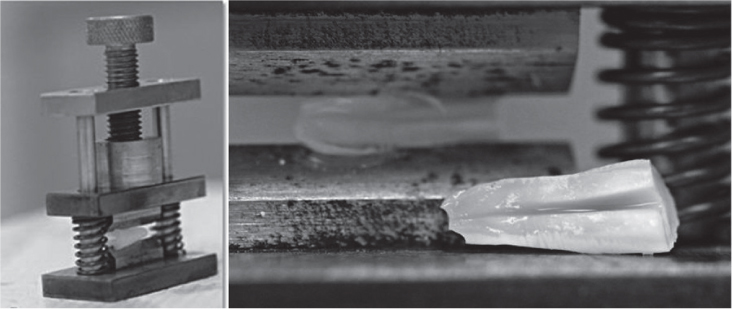
Figure 1 The tool used to split the root into two halves.
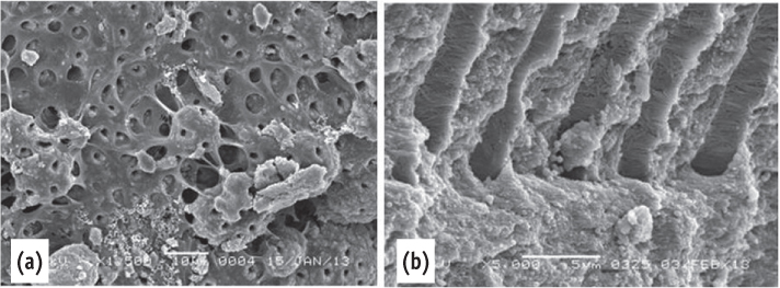
Figure 2 Scanning electron microscope images of the negative control. (a) The dentin surface and the dentinal tubules were covered by pulp tissue; (b) Collagen fibers were seen inside the dentinal tubules.
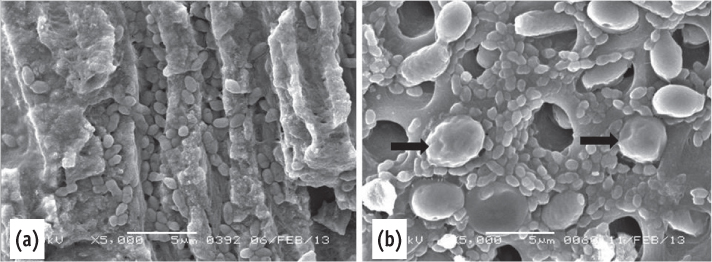
Figure 3 Scanning electron microscope images of the positive control. (a) Deep penetration of E. faecalis into dentinal tubules. Note the size of the E. faecalis and the C. albicans; (b) The C. albicans blocked the opening of the dentinal tubules (arrow) mixed microorganisms.
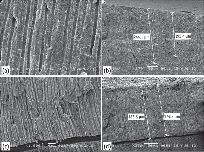
Figure 4 (a) Scanning electron microscope images of the instrumented group showing E. faecalis inside the dentinal tubules; (b) It can be traced up to 244 µm - low magnification; (c) C. albicans inside the dentinal tubules; (d) Penetrated up to 184 µm.

Figure 5 Scanning electron microscope images of the re-treated group. (a) Note the depth of penetration of the dentinal tubules by C. albicans; (b) It can be traced up to 74 µm; (c) E. faecalis in dentinal tubules; (d) Penetrated up to 123 µm.
Cited by 1 articles
-
Evaluation of penetration depth of 2% chlorhexidine digluconate into root dentinal tubules using confocal laser scanning microscope
Sekar Vadhana, Jothi Latha, Natanasabapathy Velmurugan
Restor Dent Endod. 2015;40(2):149-154. doi: 10.5395/rde.2015.40.2.149.
Reference
-
1. Kakehashi S, Stanley HR, Fitzgerald RJ. The effects of surgical exposures of dental pulps in germ-free and conventional laboratory rats. Oral Surg Oral Med Oral Pathol. 1965; 20:340–349.
Article2. Akpata ES, Blechman H. Bacterial invasion of pulpal dentin wall in vitro. J Dent Res. 1982; 61:435–438.3. Haapasalo M, Orstavik D. In vitro infection and disinfection of dentinal tubules. J Dent Res. 1987; 66:1375–1379.4. Ricucci D, Siqueira JF Jr. Fate of the tissue in lateral canals and apical ramifications in response to pathologic conditions and treatment procedures. J Endod. 2010; 36:1–15.
Article5. Engström B, Lundberg M. The correlation between positive culture and the prognosis of root canal therapy after pulpectomy. Odontol Revy. 1965; 16:193–203.6. Nair PN, Sjögren U, Krey G, Kahnberg KE, Sundqvist G. Intraradicular bacteria and fungi in root-filled, asymptomatic human teeth with therapy-resistant periapical lesions: a long-term light and electron microscope follow-up study. J Endod. 1990; 16:580–588.
Article7. Sen BH, Piskin B, Demirci T. Observation of bacteria and fungi in infected root canals and dentinal tubules by SEM. Endod Dent Traumatol. 1995; 11:6–9.
Article8. Armitage GC, Ryder MI, Wilcox SE. Cemental changes in teeth with heavily infected root canals. J Endod. 1983; 9:127–130.
Article9. Orstavik D, Haapasalo M. Disinfection by endodontic irrigants and dressing of experimentally infected dentinal tubules. Endod Dent Traumatol. 1990; 6:142–149.
Article10. Valera MC, de Moraes Rego J, Jorge AO. Effect of sodium hypochlorite and five intracanal medications on Candida albicans in root canals. J Endod. 2001; 27:401–403.
Article11. Saleh IM, Ruyter IE, Haapasalo M, Ørstavik D. Survival of Enterococcus faecalis in infected dentinal tubules after root canal filling with different root canal sealers in vitro. Int Endod J. 2004; 37:193–198.
Article12. Gurgel-Filho ED, Vivacqua-Gomes N, Gomes BP, Ferraz CC, Zaia AA, Souza-Filho FJ. In vitro evaluation of the effectiveness of the chemomechanical preparation against Enterococcus faecalis after single- or multiple-visit root canal treatment. Braz Oral Res. 2007; 21:308–313.
Article13. Michelich VJ, Schuster GS, Pashley DH. Bacterial Penetration of human dentin in vitro. J Dent Res. 1980; 59:1398–1403.14. Siqueira JF Jr. Aetiology of root canal treatment failure: why well-treated teeth can fail. Int Endod J. 2001; 34:1–10.
Article15. Lee YJ, Kim MK, Hwang HK, Kook JK. Isolation and identification of bacteria from the root canal of the teeth diagnosed as the acute pulpitis and acute periapical abscess. J Korean Acad Conserv Dent. 2005; 30:409–422.
Article16. Siqueira JF Jr, Rôças IN. Clinical implications and microbiology of bacterial persistence after treatment procedures. J Endod. 2008; 34:1291–1301.
Article17. Poptani B, Sharaff M, Archana G, Parekh V. Detection of Enterococcus faecalis and Candida albicans in previously root-filled teeth in a population of Gujarat with polymerase chain reaction. Contemp Clin Dent. 2013; 4:62–66.
Article18. Kim HS, Jang SW, Shon WJ, Lee ST, Kim CH, Lee WC, Lim SS. Effects of Enterococcus faecalis sonicated extracts on IL-2, IL-4 and TGF-β1 production from human lymphocytes. J Korean Acad Conserv Dent. 2005; 30:1–6.
Article19. Peciuliene V, Reynaud AH, Balciuniene I, Haapasalo M. Isolation of yeasts and enteric bacteria in root-filled teeth with chronic apical periodontitis. Int Endod J. 2001; 34:429–434.
Article20. Molander A, Reit C, Dahlén G, Kvist T. Microbiological status of root-filled teeth with apical periodontitis. Int Endod J. 1998; 31:1–7.
Article21. Kim HJ, Park SH, Cho KM, Kim JW. Evaluation of time-dependent antimicrobial effect of sodium dichloroisocyanurate (NaDCC) on Enterococcus faecalis in the root canal. J Korean Acad Conserv Dent. 2007; 32:121–129.
Article22. Love RM. Enterococcus faecalis-a mechanism for its role in endodontic failure. Int Endod J. 2001; 34:399–405.23. Zapata RO, Bramante CM, de Moraes IG, Bernardineli N, Gasparoto TH, Graeff MS, Campanelli AP, Garcia RB. Confocal laser scanning microscopy is appropriate to detect viability of Enterococcus faecalis in infected dentin. J Endod. 2008; 34:1198–1201.
Article24. Sen BH, Safavi KE, Spångberg LS. Growth patterns of Candida albicans in relation to radicular dentin. Oral Surg Oral Med Oral Pathol Oral Radiol Endod. 1997; 84:68–73.25. Hagihara Y, Kaminishi H, Cho T, Tanaka M, Kaita H. Degradation of human dentine collagen by an enzyme produced by the yeast Candida albicans. Arch Oral Biol. 1988; 33:617–619.
Article26. Sen BH, Safavi KE, Spångberg LS. Colonisation of Candida albicans on cleaned human dental hard tissues. Arch Oral Biol. 1997; 42:513–520.27. Waltimo TM, Ørstavik D, Sirén EK, Haapasalo MP. In vitro yeast infection of human dentin. J Endod. 2000; 26:207–209.28. Siqueira JF Jr, Rôças IN, Lopes HP, Elias CN, de Uzeda M. Fungal infection of the radicular dentin. J Endod. 2002; 28:770–773.
Article29. Jeon IS, Kum KY, Park SH, Yoon TC. Scanning electron microscopic study on the efficacy of root canal wall debridement of rotary Ni-Ti instruments with different cutting angle. J Korean Acad Conserv Dent. 2002; 27:577–586.
Article30. Hülsmann M, Schade M, Schäfers F. A comparative study of root canal preparation with HERO 642 and Quantec SC rotary Ni-Ti instruments. Int Endod J. 2001; 34:538–546.
Article31. Biesterfeld RC, Taintor JF. A comparison of periapical seals of root canals with RC-Prep or Salvizol. Oral Surg Oral Med Oral Path. 1980; 49:532–537.
Article32. Meryon SD, Brook AM. Penetration of dentine by three oral bacteria in vitro and their associated cytotoxicity. Int Endod J. 1990; 23:196–202.
Article33. Ando N, Hoshino E. Predominant obligate anaerobes invading the deep layers of root canal dentine. Int Endod J. 1990; 23:20–27.
Article34. Peters LB, Wesselink PR, Buijs JF, van Winkelhoff AJ. Viable bacteria in root dentinal tubules of teeth with apical periodontitis. J Endod. 2001; 27:76–81.
Article
- Full Text Links
-
- Actions
-
Cited
- CITED
-
- Close
- Share
-
- Similar articles
-
- Scanning electron microscopic study on the efficacy of root canal wall debridement of rotary Ni-Ti instruments with different cutting angle
- Evaluation of penetration depth of 2% chlorhexidine digluconate into root dentinal tubules using confocal laser scanning microscope
- Effect of Tetracycline-HCL in Root Conditioning : A SEM Study
- Outcomes of the GentleWave system on root canal treatment: a narrative review
- Effect of different application modalities of EDTA on dentinal tubule opening: a SEM study
