Effects of various toothpastes on remineralization of white spot lesions
- Affiliations
-
- 1Department of Orthodontics, College of Dentistry, Wonkwang University, Iksan, Korea.
- 2Private Practice, Iksan, Korea.
- 3Department of Orthodontics, College of Dentistry, Wonkwang University Dental Hospital, Sanbon, Korea.
- 4Department of Orthodontics, College of Dentistry, Wonkwang University Dental Hospital, Daejeon, Korea.
- 5Department of Orthodontics, College of Dentistry, Wonkwang Dental Research Institute, Wonkwang University, Iksan, Korea. pigtail@wku.ac.kr
- KMID: 1974996
- DOI: http://doi.org/10.4041/kjod.2014.44.3.113
Abstract
OBJECTIVE
The purpose of this in vitro study was to examine the effects of fluoridated, casein phosphopeptide.amorphous calcium phosphate complex (CPP-ACP)-containing, and functionalized beta-tricalcium phosphate (fTCP)-containing toothpastes on remineralization of white spot lesions (WSLs) by using Quantitative light-induced fluorescence (QLF-D) Biluminator(TM) 2.
METHODS
Forty-eight premolars, extracted for orthodontic reasons from 12 patients, with artificially induced WSLs were randomly and equally assigned to four treatment groups: fluoride (1,000 ppm), CPP-ACP, fTCP (with sodium fluoride), and control (deionized water) groups. Specimens were treated twice daily for 2 weeks and stored in saliva solution (1:1 mixture of artificial and human stimulated saliva) otherwise. QLF-D Biluminator(TM) 2 was used to measure changes in fluorescence, indicating alterations in the mineral contents of the WSLs, immediately before and after the 2 weeks of treatment.
RESULTS
Fluorescence greatly increased in the fTCP and CPP-ACP groups compared with the fluoride and control groups, which did not show significant differences.
CONCLUSIONS
fTCP- and CPP-ACP-containing toothpastes seem to be more effective in reducing WSLs than 1,000-ppm fluoride-containing toothpastes.
MeSH Terms
Figure
-
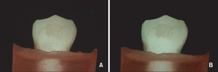
Figure 1 Representative light-induced fluorescent images of the control group. Pretreatment (A) and post-treatment (B).
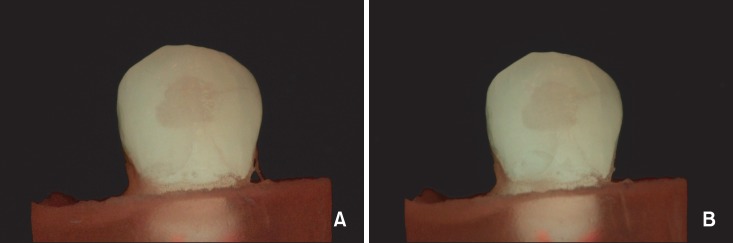
Figure 2 Representative light-induced fluorescent images of the fluoride group. Pretreatment (A) and post-treatment (B).
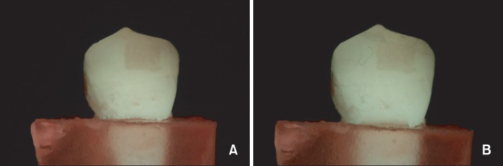
Figure 3 Representative light-induced fluorescent images of the casein phosphopeptide.amorphous calcium phosphate complex group. Pretreatment (A) and post-treatment (B).
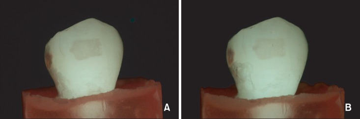
Figure 4 Representative light-induced fluorescent images of the functionalized β-tricalcium phosphate group. Pretreatment (A) and post-treatment (B).
Cited by 2 articles
-
Colorimetric evaluation of white spot lesions following external bleaching with fluoridation: An in-vitro study
Yoon-Young Choi, Dong-Yul Lee, Yae-Jin Kim
Korean J Orthod. 2018;48(6):377-383. doi: 10.4041/kjod.2018.48.6.377.Micro-computed tomographic evaluation of the effect of fluoride agents on white spot lesions: An
in vitro study
Sook-Chan Hong, Dong-Yul Lee, Yae-Jin Kim
Korean J Orthod. 2022;52(1):75-79. doi: 10.4041/kjod.2022.52.1.75.
Reference
-
1. Øgaard B. White spot lesions during orthodontic treatment: mechanisms and fluoride preventive aspects. Semin Orthod. 2008; 14:183–193.
Article2. Lundström F, Krasse B. Streptococcus mutans and lactobacilli frequency in orthodontic patients; the effect of chlorhexidine treatments. Eur J Orthod. 1987; 9:109–116. PMID: 3472888.3. Chatterjee R, Kleinberg I. Effect of orthodontic band placement on the chemical composition of human incisor tooth plaque. Arch Oral Biol. 1979; 24:97–100. PMID: 45361.
Article4. Gorelick L, Geiger AM, Gwinnett AJ. Incidence of white spot formation after bonding and banding. Am J Orthod. 1982; 81:93–98. PMID: 6758594.
Article5. Artun J, Brobakken BO. Prevalence of carious white spots after orthodontic treatment with multibonded appliances. Eur J Orthod. 1986; 8:229–234. PMID: 3466795.
Article6. O'Reilly MM, Featherstone JD. Demineralization and remineralization around orthodontic appliances: an in vivo study. Am J Orthod Dentofacial Orthop. 1987; 92:33–40. PMID: 3300270.7. Geiger AM, Gorelick L, Gwinnett AJ, Griswold PG. The effect of a fluoride program on white spot formation during orthodontic treatment. Am J Orthod Dentofacial Orthop. 1988; 93:29–37. PMID: 3276146.
Article8. Ogaard B. Prevalence of white spot lesions in 19-year-olds: a study on untreated and orthodontically treated persons 5 years after treatment. Am J Orthod Dentofacial Orthop. 1989; 96:423–427. PMID: 2816842.9. Ogaard B, Rølla G, Arends J. Orthodontic appliances and enamel demineralization. Part 1. Lesion development. Am J Orthod Dentofacial Orthop. 1988; 94:68–73. PMID: 3164585.10. Mitchell L. Decalcification during orthodontic treatment with fixed appliances-an overview. Br J Orthod. 1992; 19:199–205. PMID: 1390575.
Article11. Baysan A, Lynch E, Ellwood R, Davies R, Petersson L, Borsboom P. Reversal of primary root caries using dentifrices containing 5,000 and 1,100 ppm fluoride. Caries Res. 2001; 35:41–46. PMID: 11125195.
Article12. Schirrmeister JF, Gebrande JP, Altenburger MJ, Mönting JS, Hellwig E. Effect of dentifrice containing 5000 ppm fluoride on non-cavitated fissure carious lesions in vivo after 2 weeks. Am J Dent. 2007; 20:212–216. PMID: 17907481.13. Artun J, Thylstrup A. Clinical and scanning electron microscopic study of surface changes of incipient caries lesions after debonding. Scand J Dent Res. 1986; 94:193–201. PMID: 3526528.14. Reynolds EC. Remineralization of enamel subsurface lesions by casein phosphopeptide-stabilized calcium phosphate solutions. J Dent Res. 1997; 76:1587–1595. PMID: 9294493.
Article15. Shi XQ, Tranaeus S, Angmar-Månsson B. Comparison of QLF and DIAGNOdent for quantification of smooth surface caries. Caries Res. 2001; 35:21–26. PMID: 11125192.
Article16. Murray JJ, Shaw L. Classification and prevalence of enamel opacities in the human deciduous and permanent dentitions. Arch Oral Biol. 1979; 24:7–13. PMID: 292365.
Article17. Mizrahi E. Enamel demineralization following orthodontic treatment. Am J Orthod. 1982; 82:62–67. PMID: 6984291.
Article18. Dirks OB. Posteruptive changes in dental enamel. J Dent Res. 1966; 45:503–511.
Article19. Zachrisson BU, Zachrisson S. Caries incidence and oral hygiene during orthodontic treatment. Scand J Dent Res. 1971; 79:394–401. PMID: 5288673.20. Geiger AM, Gorelick L, Gwinnett AJ, Benson BJ. Reducing white spot lesions in orthodontic populations with fluoride rinsing. Am J Orthod Dentofacial Orthop. 1992; 101:403–407. PMID: 1590288.
Article21. Bröchner A, Christensen C, Kristensen B, Tranæus S, Karlsson L, Sonnesen L, et al. Treatment of post-orthodontic white spot lesions with casein phosphopeptide-stabilised amorphous calcium phosphate. Clin Oral Investig. 2011; 15:369–373.
Article22. Bhat SS, Hegde SK, Habibullah MA, Bernhardt V. Incipient enamel lesions remineralization using casein phosphopeptide amorphous calcium phosphate cream with and without fluoride: a laser fluorescence study. J Clin Pediatr Dent. 2012; 36:353–355. PMID: 23019831.
Article23. Ogata K, Warita S, Shimazu K, Kawakami T, Aoyagi K, Karibe H. Combined effect of paste containing casein phosphopeptide-amorphous calcium phosphate and fluoride on enamel lesions: an in vitro pH-cycling study. Pediatr Dent. 2010; 32:433–438. PMID: 21070712.24. Karlinsey RL, Mackey AC. Solid-state preparation and dental application of an organically modified calcium phosphate. J Mater Sci. 2009; 44:346–349.
Article25. Derks A, Katsaros C, Frencken JE, van't Hof MA, Kuijpers-Jagtman AM. Caries-inhibiting effect of preventive measures during orthodontic treatment with fixed appliances. A systematic review. Caries Res. 2004; 38:413–420. PMID: 15316184.26. Silverstone LM. Remineralization and enamel caries: significance of fluoride and effect on crystal diameters. In : Leach SA, Edgar WM, editors. Demineralisation and remineralisation of the teeth. Oxford: IRL Press;1983. p. 185–205.
- Full Text Links
-
- Actions
-
Cited
- CITED
-
- Close
- Share
-
- Similar articles
-
- Non-destructive management of white spot lesions by using tooth jewelry
- Management of white spots: resin infiltration technique and microabrasion
- Remineralisation effect of 1,500 ppm fluoride-containing toothpaste in enamel early caries lesion
- Evaluation of Total and Soluble Fluoride Concentrations in Ten Toothpastes for Children
- Micro-computed tomographic evaluation of the effect of fluoride agents on white spot lesions: An in vitro study
