Restor Dent Endod.
2012 Nov;37(4):236-239.
Non-destructive management of white spot lesions by using tooth jewelry
- Affiliations
-
- 1Department of Conservative Dentistry, Wonkwang University School of Dentistry, Iksan, Korea. conspsj@wonkwang.ac.kr
Abstract
- Although several methods including composite resin restoration and microabrasion have been used for management of white spot lesion, tooth jewelry can be considered as another noninvasive option. This case report describes the management of white spot lesions by using tooth jewelry. This report also highlights the patients' preference for tooth jewelry as an esthetic concern.
Keyword
MeSH Terms
Figure
-
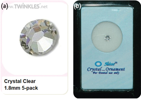
Figure 1 Materials used. (a) Twinkles: Clear, ø1.8 mm; (b) Shine: A style, ø1.8 mm.

Figure 2 Clinical appearance before (a and c) and after (b and d) placement of tooth jewelry.
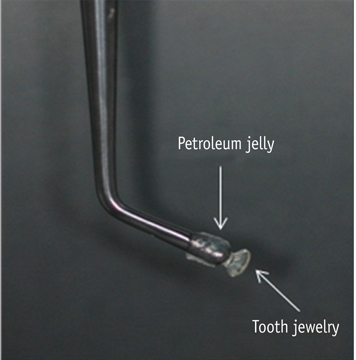
Figure 3 Carrying method for tooth jewelry.
Reference
-
1. Malterud MI. Minimally invasive restorative dentistry: a biomimetic approach. Pract Proced Aesthet Dent. 2006. 18:409–414.2. Son JH, Hur B, Kim HC, Park JK. Management of white spots: resin infiltration technique and microabrasion. J Korean Acad Conserv Dent. 2011. 36:66–71.
Article3. American Academy of Pediatric Dentistry Council on Clinical Affairs. Policy on intraoral and perioral piercing. Pediatr Dent. 2008-2009. 30:56–57.4. Jeger F, Lussi A, Zimmerli B. Oral jewelry: a review. Schweiz Monatsschr Zahnmed. 2009. 119:615–631.5. Murphy TC, Willmot DR, Rodd HD. Management of postorthodontic demineralized white lesions with microabrasion: a quantitative assessment. Am J Orthod Dentofacial Orthop. 2007. 131:27–33.
Article6. Paris S, Meyer-Lueckel H. Masking of labial enamel white spot lesions by resin infiltration-a clinical report. Quintessence Int. 2009. 40:713–718.7. Shivanna V, Shivakumar B. Novel treatment of white spot lesions: a report of two cases. J Conserv Dent. 2011. 14:423–426.
Article8. Torlakovic L, Olsen I, Petzold C, Tiainen H, Øgaard B. Clinical color intensity of white spot lesions might be a better predictor of enamel demineralization depth than traditional clinical grading. Am J Orthod Dentofacial Orthop. 2012. 142:191–198.
Article9. Reston EG, Corba DV, Ruschel K, Tovo MF, Barbosa AN. Conservative approach for esthetic treatment of enamel hypoplasia. Oper Dent. 2011. 36:340–343.
Article10. Greene JC, Vermillion JR. The oral hygiene index: a method for classifying oral hygiene status. J Am Dent Assoc. 1960. 61:172–179.
Article11. Chan YL, Ngan AH, King NM. Degraded prism sheaths in the transition region of hypomineralized teeth. J Dent. 2010. 38:237–244.
Article12. Willmot DR. White lesions after orthodontic treatment: does low fluoride make a difference? J Orthod. 2004. 31:235–242.
Article13. Wright JT. The etch-bleach-seal technique for managing stained enamel defects in young permanent incisors. Pediatr Dent. 2002. 24:249–252.14. Haywood VB, Leonard RH, Nelson CF, Brunson WD. Effectiveness, side effects and long-term status of nightguard vital bleaching. J Am Dent Assoc. 1994. 125:1219–1226.
Article
- Full Text Links
-
- Actions
-
Cited
- CITED
-
- Close
- Share
-
- Similar articles
-
- Management of white spots: resin infiltration technique and microabrasion
- Minimally invasive treatment for esthetic enhancement of white spot lesion in adjacent tooth
- Colorimetric evaluation of white spot lesions following external bleaching with fluoridation: An in-vitro study
- A global overview of enamel microabrasion for white spot lesions: a bibliometric review
- Modification of surface pretreatment of white spot lesions to improve the safety and efficacy of resin infiltration
