Restor Dent Endod.
2024 Aug;49(3):e29. 10.5395/rde.2024.49.e29.
A global overview of enamel microabrasion for white spot lesions: a bibliometric review
- Affiliations
-
- 1Department of Dentistry, Federal University of Santa Catarina, Florianopolis, SC, Brazil
- KMID: 2559742
- DOI: http://doi.org/10.5395/rde.2024.49.e29
Abstract
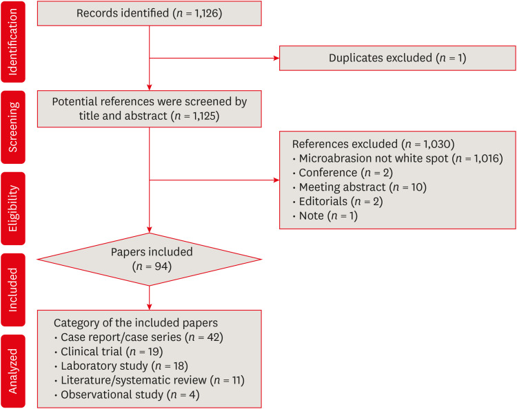
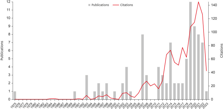
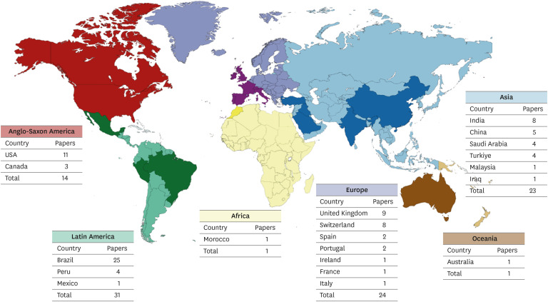
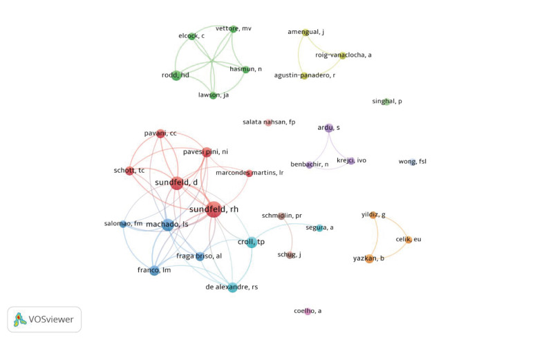
- This study aimed to identify and analyze articles on enamel microabrasion for the treatment of white spot lesions. A search was conducted on the Web of Science. The following parameters were recorded and analyzed: number of citations, year, journal, impact factor, study design, theme, country and continent, institution, authors, and keywords. Data was analyzed using VOSviewer software. The initial search resulted in 1,126 documents, of which 94 articles were included. The highest number of citations an article received was 65. The oldest article was published in 1975, and the most recent in 2023. The most frequent study design was case report (n = 42). Regarding the themes, it was observed that the main objective of the studies was to evaluate the clinical performance of enamel microabrasion (n = 75), primarily using Opalustre (Ultradent Products Inc., South Jordan, UT, USA) (n = 37) for treating white stains caused by dental fluorosis (n = 41). Most articles originated from Latin America (n = 31), mainly from Brazil (n = 26). The most frequent author was Sundfeld RH (n = 10). This study reveals research trends in the field of enamel microabrasion. The publications were mainly case reports/series using Opalustre for the removal of fluorosis stains.
Figure
Reference
-
1. Perdigão J. Resin infiltration of enamel white spot lesions: an ultramorphological analysis. J Esthet Restor Dent. 2020; 32:317–324. PMID: 31742888.2. Gupta A, Dhingra R, Chaudhuri P, Gupta A. A comparison of various minimally invasive techniques for the removal of dental fluorosis stains in children. J Indian Soc Pedod Prev Dent. 2017; 35:260–268. PMID: 28762354.3. Bernardi LG, Favoreto MW, de Souza Carneiro T, Borges CP, Pulido C, Loguercio AD. Effects of microabrasion association to at-home bleaching on hydrogen peroxide penetration and color change. J Esthet Restor Dent. 2022; 34:335–341. PMID: 34723443.4. Croll TP, Cavanaugh RR. Enamel color modification by controlled hydrochloric acid-pumice abrasion. II. Further examples. Quintessence Int. 1986; 17:157–164. PMID: 3458266.5. Pini NI, Sundfeld-Neto D, Aguiar FH, Sundfeld RH, Martins LR, Lovadino JR, et al. Enamel microabrasion: an overview of clinical and scientific considerations. World J Clin Cases. 2015; 3:34–41. PMID: 25610848.6. Bahadir HS, Haberal M, Çelik Ç. Effect of microabrasion on the staining susceptibility of enamel: an in vitro study. J Dent Res Dent Clin Dent Prospect. 2022; 16:95–100.7. Sundfeld D, Pavani CC, Pini N, Machado LS, Schott TC, Sundfeld RH. Enamel microabrasion and dental bleaching on teeth presenting severe-pitted enamel fluorosis: a case report. Oper Dent. 2019; 44:566–573. PMID: 30702410.8. Elkhazindar MM, Welbury RR. Enamel microabrasion. Dent Update. 2000; 27:194–196. PMID: 11218456.9. Croll TP. Enamel microabrasion: 10 years experience. Asian J Aesthet Dent. 1995; 3:9–15. PMID: 9063104.10. Sundfeld RH, Croll TP, Briso AL, de Alexandre RS, Sundfeld Neto D. Considerations about enamel microabrasion after 18 years. Am J Dent. 2007; 20:67–72. PMID: 17542197.11. Karobari MI, Maqbool M, Ahmad P, Abdul MS, Marya A, Venugopal A, et al. Endodontic microbiology: a bibliometric analysis of the top 50 classics. BioMed Res Int. 2021; 2021:6657167. PMID: 34746305.12. Al-Moraissi EA, Christidis N, Ho YS. Publication performance and trends in temporomandibular disorders research: a bibliometric analysis. J Stomatol Oral Maxillofac Surg. 2023; 124:101273. PMID: 36057419.13. Donthu N, Kumar S, Mukherjee D, Pandey N, Lim WM. How to conduct a bibliometric analysis: an overview and guidelines. J Bus Res. 2021; 133:285–296.14. Ahmad P, Slots J. A bibliometric analysis of periodontology. Periodontol 2000. 2021; 85:237–240. PMID: 33226679.15. Van Noorden R, Maher B, Nuzzo R. The top 100 papers. Nature. 2014; 514:550–553. PMID: 25355343.16. Tong LS, Pang MK, Mok NY, King NM, Wei SH. The effects of etching, micro-abrasion, and bleaching on surface enamel. J Dent Res. 1993; 72:67–71. PMID: 8418110.17. Schmucker CM, Blümle A, Schell LK, Schwarzer G, Oeller P, Cabrera L, et al. Systematic review finds that study data not published in full text articles have unclear impact on meta-analyses results in medical research. PLoS One. 2017; 12:e0176210. PMID: 28441452.18. Scherer RW, Saldanha IJ. How should systematic reviewers handle conference abstracts? A view from the trenches. Syst Rev. 2019; 8:264. PMID: 31699124.19. Wong FS, Winter GB. Effectiveness of microabrasion technique for improvement of dental aesthetics. Br Dent J. 2002; 193:155–158. PMID: 12213009.20. Chandra S, Chawla TN. Clinical evaluation of the sandpaper disk method for removing fluorosis stains from teeth. J Am Dent Assoc. 1975; 90:1273–1276. PMID: 1056394.21. Goebel MC, Rocha AO, Santos PS, Bolan M, Martins-Júnior PA, Santana CM, et al. A bibliometric analysis of the top 100 most-cited papers concerning dental fluorosis. Caries Res. 2023; 57:509–515. PMID: 37100040.22. Page MJ, McKenzie JE, Bossuyt PM, Boutron I, Hoffmann TC, Mulrow CD, et al. The PRISMA 2020 statement: an updated guideline for reporting systematic reviews. BMJ. 2021; 372:n71. PMID: 33782057.23. Murrin JR, Barkmeier WW. Chemical treatment of endemic dental fluorosis. Quintessence Int Dent Dig. 1982; 13:363–369. PMID: 6955830.24. Croll TP. Hastening the enamel microabrasion procedure eliminating defects, cutting treatment time. J Am Dent Assoc. 1993; 124:87–90.25. Kamp AA. Removal of white spot lesions by controlled acid-pumice abrasion. J Clin Orthod. 1989; 23:690–693. PMID: 2639881.26. Donly KJ, O’Neill M, Croll TP. Enamel microabrasion: a microscopic evaluation of the “abrosion effect”. Quintessence Int. 1992; 23:175–179. PMID: 1641458.27. Neurath C, Limeback H, Osmunson B, Connett M, Kanter V, Wells CR. Dental fluorosis trends in US oral health surveys: 1986 to 2012. JDR Clin Trans Res. 2019; 4:298–308. PMID: 30931722.28. Rocha AO, Anjos LM, Vitali FC, Santos PS, Bolan M, Santana CM, et al. Tooth bleaching: a bibliometric analysis of the top 100 most-cited papers. Braz Dent J. 2023; 34:41–55. PMID: 37194856.29. Baldiotti AL, Amaral-Freitas G, Barcelos JF, Freire-Maia J, Perazzo MF, Freire-Maia FB, et al. The top 100 most-cited papers in cariology: a bibliometric analysis. Caries Res. 2021; 55:32–40. PMID: 33341798.30. Dos Anjos LM, Rocha AO, Magrin GL, Kammer PV, Benfatti CA, Matias de Souza JC, et al. Bibliometric analysis of the 100 most cited articles on bone grafting in dentistry. Clin Oral Implants Res. 2023; 34:1198–1216. PMID: 37577958.
- Full Text Links
- Actions
-
Cited
- CITED
-
- Close
- Share
- Similar articles
-
- Management of white spots: resin infiltration technique and microabrasion
- Non-destructive management of white spot lesions by using tooth jewelry
- Application of quantitative light-induced fluorescence to determine the depth of demineralization of dental fluorosis in enamel microabrasion: a case report
- Color and hardness changes in artificial white spot lesions after resin infiltration
- Colorimetric evaluation of white spot lesions following external bleaching with fluoridation: An in-vitro study

