Application of quantitative light-induced fluorescence to determine the depth of demineralization of dental fluorosis in enamel microabrasion: a case report
- Affiliations
-
- 1Department of Conservative Dentistry, Chosun University School of Dentistry, Gwangju, Korea. minjb@chosun.ac.kr
- KMID: 2346772
- DOI: http://doi.org/10.5395/rde.2016.41.3.225
Abstract
- Enamel microabrasion has become accepted as a conservative, nonrestorative method of removing intrinsic and superficial dysmineralization defects from dental fluorosis, restoring esthetics with minimal loss of enamel. However, it can be difficult to determine if restoration is necessary in dental fluorosis, because the lesion depth is often not easily recognized. This case report presents a method for analysis of enamel hypoplasia that uses quantitative light-induced fluorescence (QLF) followed by a combination of enamel microabrasion with carbamide peroxide home bleaching. We describe the utility of QLF when selecting a conservative treatment plan and confirming treatment efficacy. In this case, the treatment plan was based on QLF analysis, and the selected combination treatment of microabrasion and bleaching had good results.
MeSH Terms
Figure
-

Figure 1 The QLF device is based on a DSLR camera equipped with an illumination tube with white and blue light-emitting diodes positioned in a ring around the lens opening. QLF, quantitative light-induced fluorescence; DSLR, digital single lens reflex.
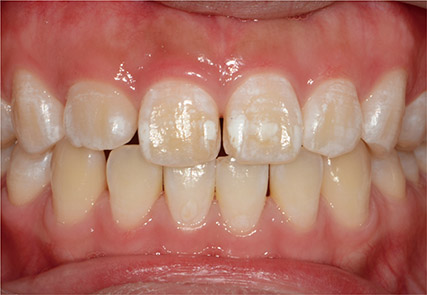
Figure 2 Initial photograph from the first examination showing white spots on the upper dentition.
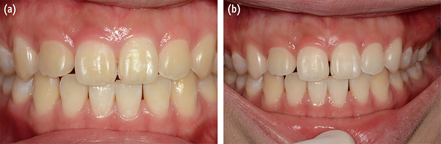
Figure 3 Results of QLF analysis before treatment. (a) Right maxillary central incisor; (b) Left maxillary central incisor. QLF, quantitative light-induced fluorescence.
Figure 4 Photograph taken after treatment showing improved esthetics of the upper dentition. (a) Intraoral photograph after microabrasion; (b) Intraoral photograph after home bleaching.
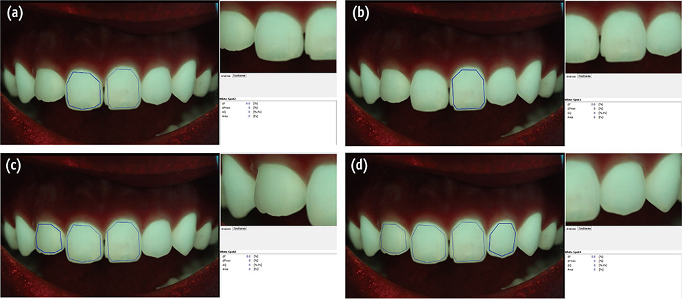
Figure 5 Results of QLF analysis after treatment. (a) Right maxillary central incisor; (b) Left maxillary central incisor; (c) Right maxillary lateral incisor; (d) Left maxillary lateral incisor. QLF, quantitative light-induced fluorescence.
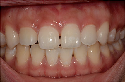
Figure 6 Photograph of maxillary lateral incisors after completion of the treatment with composite restoration of left central incisor.
Reference
-
1. Castro KS, Ferreira AC, Duarte RM, Sampaio FC, Meireles SS. Acceptability, efficacy and safety of two treatment protocols for dental fluorosis: a randomized clinical trial. J Dent. 2014; 42:938–944.
Article2. Son JH, Hur B, Kim HC, Park JK. Management of white spots: resin infiltration technique and microabrasion. J Korean Acad Conserv Dent. 2011; 36:66–71.
Article3. Kim JH, Son HH, Chang J. Color and hardness changes in artificial white spot lesions after resin infiltration. Restor Dent Endod. 2012; 37:90–95.
Article4. Kim HJ, Karanxha L, Park SJ. Non-destructive management of white spot lesions by using tooth jewelry. Restor Dent Endod. 2012; 37:236–239.
Article5. Pretty IA, Edgar WM, Higham SM. The effect of ambient light on QLF analyses. J Oral Rehabil. 2002; 29:369–373.
Article6. Pretty IA, Hall AF, Smith PW, Edgar WM, Higham SM. The intra- and inter-examiner reliability of quantitative light-induced fluorescence (QLF) analyses. Br Dent J. 2002; 193:105–109.
Article7. Kim HE, Kwon HK, Kim BI. Recovery percentage of remineralization according to severity of early caries. Am J Dent. 2013; 26:132–136.8. Nakata K, Nikaido T, Ikeda M, Foxton RM, Tagami J. Relationship between fluorescence loss of QLF and depth of demineralization in an enamel erosion model. Dent Mater J. 2009; 28:523–529.
Article9. Wu J, Donly ZR, Donly KJ, Hackmyer S. Demineralization depth using QLF and a novel image processing software. Int J Dent. 2010; 2010:958264.
Article10. Murphy TC, Willmot DR, Rodd HD. Management of postorthodontic demineralized white lesions with microabrasion: a quantitative assessment. Am J Orthod Dentofacial Orthop. 2007; 131:27–33.
Article11. Paris S, Meyer-Lueckel H. Masking of labial enamel white spot lesions by resin infiltration-a clinical report. Quintessence Int. 2009; 40:713–718.12. Kendell RL. Hydrochloric acid removal of brown fluorosis stains: clinical and scanning electron micrographic observations. Quintessence Int. 1989; 20:837–839.13. Jacobsson-Hunt U, Harrison MR. The effect of hydrochloric acid pumice abrasion on fluorosed teeth. J Dent Res. 1989; 68:Supplement 2. 982. Abstract #924.14. Welbury RR, Shaw L. A simple technique for removal of mottling, opacities and pigmentation from enamel. Dent Update. 1990; 17:161–163.15. Bussadori SK, do Rego MA, da Silva PE, Pinto MM, Pinto AC. Esthetic alternative for fluorosis blemishes with the usage of a dual bleaching system based on hydrogen peroxide at 35%. J Clin Pediatr Dent. 2004; 28:143–146.
Article16. Knösel M, Attin R, Becker K, Attin T. External bleaching effect on the color and luminosity of inactive whitespot lesions after fixed orthodontic appliances. Angle Orthod. 2007; 77:646–652.
Article
- Full Text Links
-
- Actions
-
Cited
- CITED
-
- Close
- Share
-
- Similar articles
-
- A global overview of enamel microabrasion for white spot lesions: a bibliometric review
- The effects of a sealant resin on enamel demineralization in orthodontic bracket bonding
- A study of enamel demineralization related to bonded orthodontic bracket and improved method of enamel demineralization: in vivo study
- Effect of fluoride releasing orthodontic sealants on enamel demineralization in vitro
- Comparison of Prevention Methods against Enamel Demineralization adjacent to Orthodontic Bracket Using Fluoride
