Modification of surface pretreatment of white spot lesions to improve the safety and efficacy of resin infiltration
- Affiliations
-
- 1Department of Preventive Dentistry and Public Oral Health, Brain Korea 21 PLUS Project, College of Dentistry, Yonsei University, Seoul, Korea. drkbi@yuhs.ac
- KMID: 2273276
- DOI: http://doi.org/10.4041/kjod.2014.44.4.195
Abstract
OBJECTIVE
A low-viscosity resin (infiltrant) was used to inhibit the progression of white spot lesions (WSLs) and resolve associated esthetic issues. An alternative pretreatment was explored to increase the pore volume of the surface layer of the WSLs. Also, the penetration effects of the infiltrant were evaluated for various pretreatments.
METHODS
Sixty two artificial lesions were fabricated on bovine teeth. As a positive control, 15% HCl gel was applied for 120 seconds. Further, 37% H3PO4 gel was applied for 30 seconds using three methods. The samples were divided as follows: H3PO4 only group, H3PO4 sponge group, and H3PO4 brush group. The acid was gently rubbed with the applicators (i.e., a sponge or brush) throughout the application time. To compare the effects of resin infiltration, twenty paired halves of specimens were treated with an infiltrant (ICON(R)).
RESULTS
Thicknesses of the removed surface layers and infiltrated areas were evaluated by confocal laser scanning microscope. The positive control and the 37% H3PO4 brush group failed to show significant differences in the removed thickness (p > 0.05); however, the mean percentage of the infiltrated area was higher in the 37% H3PO4 brush group (84.13 +/- 7.58%) than the positive control (63.51 +/- 7.62%, p < 0.001). Scanning electron microscope observations indicate higher pore volumes for the 37% H3PO4 brush group than for the positive control.
CONCLUSIONS
Application of 37% H3PO4 with a brush for 30 seconds increased the pore volume of WSL surface layers and the percentage of infiltrated areas in comparison to the use of 15% HCl for 120 seconds.
Keyword
MeSH Terms
Figure
-
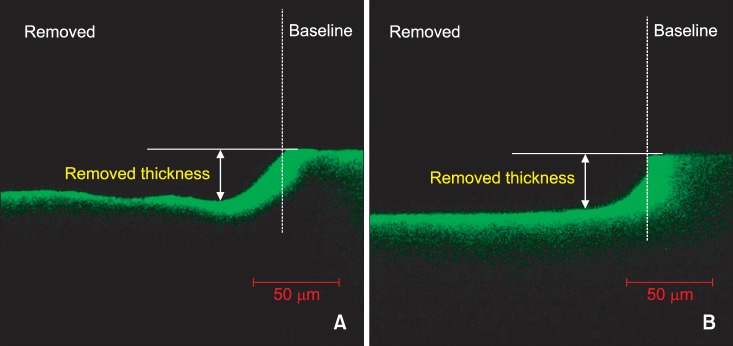
Figure 1 Confocal laser scanning microscope images of the removed surfaces (magnification ×400). A, Positive control showing lesion treated with 15% HCl gel for 120 seconds; B, lesion treated with 37% H3PO4 gel using the brush applicator for 30 seconds. Baseline, unetched lesion surface.
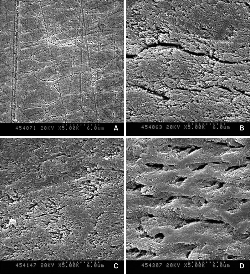
Figure 2 Scanning electron microscope images of the lesion surfaces after acid treatment (magnification ×5,000). A, Baseline showing unetched lesion surface; B, lesion surfaces treated with 15% HCl gel for 120 seconds; C, 37% H3PO4 gel using the sponge applicator for 30 seconds; and D, 37% H3PO4 gel using the brush applicator for 30 seconds.
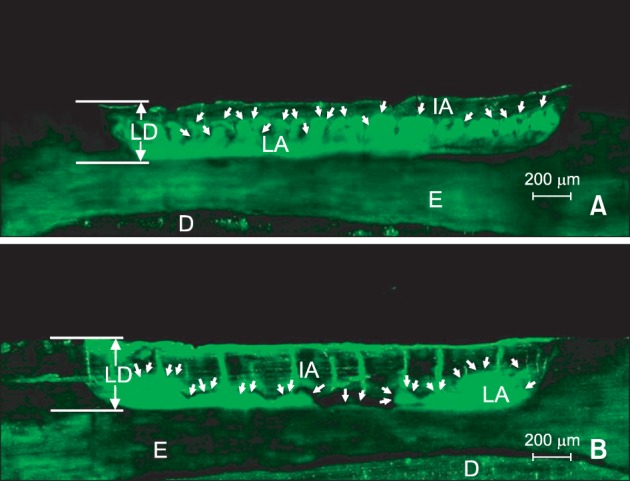
Figure 3 Confocal laser scanning microscope images of the lesions treated with infiltrant (magnification ×100). The lesion surfaces were pretreated with either A, 15% HCl for 120 seconds; or B, 37% H3PO4 using the brush applicator for 30 seconds. In the lesions, the infiltrated area (IA) as well as the sound enamel is displayed as dark areas and the lesion area (LA) is displayed in green. White arrows indicate the boundaries of LAs and IAs. LD, Lesion depth; E, enamel; D, dentin.
Reference
-
1. Ekizer A, Zorba YO, Uysal T, Ayrikcila S. Effects of demineralizaton-inhibition procedures on the bond strength of brackets bonded to demineralized enamel surface. Korean J Orthod. 2012; 42:17–22. PMID: 23112927.
Article2. Wagner L, Szepietowska M. Fluoride penetration from three orthodontic adhesives: an experimental study. Korean J Orthod. 2013; 43:29–34. PMID: 23502591.
Article3. O'Reilly MT, De Jesús Viñas J, Hatch JP. Effectiveness of a sealant compared with no sealant in preventing enamel demineralization in patients with fixed orthodontic appliances: a prospective clinical trial. Am J Orthod Dentofacial Orthop. 2013; 143:837–844. PMID: 23726334.4. Tufekci E, Dixon JS, Gunsolley JC, Lindauer SJ. Prevalence of white spot lesions during orthodontic treatment with fixed appliances. Angle Orthod. 2011; 81:206–210. PMID: 21208070.
Article5. Neuhaus KW, Graf M, Lussi A, Katsaros C. Late infiltration of post-orthodontic white spot lesions. J Orofac Orthop. 2010; 71:442–447. PMID: 21082307.
Article6. Paris S, Meyer-Lueckel H. Masking of labial enamel white spot lesions by resin infiltration--a clinical report. Quintessence Int. 2009; 40:713–718. PMID: 19862396.7. Kim S, Kim EY, Jeong TS, Kim JW. The evaluation of resin infiltration for masking labial enamel white spot lesions. Int J Paediatr Dent. 2011; 21:241–248. PMID: 21401750.
Article8. Paris S, Hopfenmuller W, Meyer-Lueckel H. Resin infiltration of caries lesions: an efficacy randomized trial. J Dent Res. 2010; 89:823–826. PMID: 20505049.9. Meyer-Lueckel H, Paris S. Improved resin infiltration of natural caries lesions. J Dent Res. 2008; 87:1112–1116. PMID: 19029077.
Article10. Paris S, Meyer-Lueckel H. Infiltrants inhibit progression of natural caries lesions in vitro. J Dent Res. 2010; 89:1276–1280. PMID: 20739697.
Article11. Meyer-Lueckel H, Paris S. Infiltration of natural caries lesions with experimental resins differing in penetration coefficients and ethanol addition. Caries Res. 2010; 44:408–414. PMID: 20714153.
Article12. Meyer-Lueckel H, Paris S, Kielbassa AM. Surface layer erosion of natural caries lesions with phosphoric and hydrochloric acid gels in preparation for resin infiltration. Caries Res. 2007; 41:223–230. PMID: 17426404.
Article13. Meireles SS, Andre Dde A, Leida FL, Bocangel JS, Demarco FF. Surface roughness and enamel loss with two microabrasion techniques. J Contemp Dent Pract. 2009; 10:58–65. PMID: 19142257.14. Croll TP, Killian CM, Miller AS. Effect of enamel microabrasion compound on human gingiva: report of a case. Quintessence Int. 1990; 21:959–963. PMID: 2082423.15. Nyvad B, Machiulskiene V, Baelum V. Reliability of a new caries diagnostic system differentiating between active and inactive caries lesions. Caries Res. 1999; 33:252–260. PMID: 10343087.16. Rocha Gomes Torres C, Borges AB, Torres LM, Gomes IS, de Oliveira RS. Effect of caries infiltration technique and fluoride therapy on the colour masking of white spot lesions. J Dent. 2011; 39:202–207. PMID: 21172402.17. Kugel G, Ferrari M. The science of bonding: from first to sixth generation. J Am Dent Assoc. 2000; 131(Suppl):20S–25S. PMID: 10860341.
Article18. Attin T, Knöfel S, Buchalla W, Tütüncü R. In situ evaluation of different remineralization periods to decrease brushing abrasion of demineralized enamel. Caries Res. 2001; 35:216–222. PMID: 11385203.19. White DJ. Use of synthetic polymer gels for artificial carious lesion preparation. Caries Res. 1987; 21:228–242. PMID: 3471346.
Article20. Paris S, Meyer-Lueckel H, Cölfen H, Kielbassa AM. Resin infiltration of artificial enamel caries lesions with experimental light curing resins. Dent Mater J. 2007; 26:582–588. PMID: 17886464.
Article21. Hermsen RJ, Vrijhoef MM. Loss of enamel due to etching with phosphoric or maleic acid. Dent Mater. 1993; 9:332–336. PMID: 7995486.
Article22. Croll TP. Bonded resin sealant for smooth surface enamel defects: new concepts in "microrestorative" dentistry. Quintessence Int. 1987; 18:5–10. PMID: 3469683.23. Poitevin A, De Munck J, Van Ende A, Suyama Y, Mine A, Peumans M, et al. Bonding effectiveness of self-adhesive composites to dentin and enamel. Dent Mater. 2013; 29:221–230. PMID: 23107191.
Article24. Carstensen W. The effects of different phosphoric acid concentrations on surface enamel. Angle Orthod. 1992; 62:51–58. PMID: 1554164.25. Neuhaus KW, Schlafer S, Lussi A, Nyvad B. Infiltration of natural caries lesions in relation to their activity status and acid pretreatment in vitro. Caries Res. 2013; 47:203–210. PMID: 23235388.
Article26. Meyer-Lueckel H, Paris S. Progression of artificial enamel caries lesions after infiltration with experimental light curing resins. Caries Res. 2008; 42:117–124. PMID: 18305389.
Article
- Full Text Links
-
- Actions
-
Cited
- CITED
-
- Close
- Share
-
- Similar articles
-
- Management of white spots: resin infiltration technique and microabrasion
- Alternative Pretreatment Methods for Resin Infiltration in Primary Anterior Teeth
- Color and hardness changes in artificial white spot lesions after resin infiltration
- Non-destructive management of white spot lesions by using tooth jewelry
- Minimally invasive treatment for esthetic enhancement of white spot lesion in adjacent tooth
