Micro-computed tomographic evaluation of the effect of fluoride agents on white spot lesions: An in vitro study
- Affiliations
-
- 1Major in Dentistry, Department of Medicine, Graduate School of Korea University, Seoul, Korea
- 2Department of Dentistry, College of Medicine, Korea University, Seoul, Korea
- 3Department of Orthodontics, Korea University Guro Hospital, Seoul, Korea
- KMID: 2524904
- DOI: http://doi.org/10.4041/kjod.2022.52.1.75
Abstract
Objective
To investigate remineralizing effect of three fluoride regimens on artificially demineralized enamel around orthodontic bracket by analyzing mineral density (MD) acquired from micro-computed tomography (micro-CT).
Methods
Forty-eight bracket bonded bovine incisors were prepared to create demineralized enamel (DE) surface. The samples were divided into four groups according to the fluoride regimen: 1) no fluoridation, 2) 1.23% acidulated phosphate fluoride (APF) gel, 3) fluoridated toothpaste, and 4) 0.05% sodium fluoride mouthwash. Micro-CT was scanned after demineralization (T0), and 2 weeks (T1) and 4 weeks (T2) of fluoridation.
Results
APF gel showed highest remineralization of DE during T1–T0 interval among the groups (p < 0.05); followed by toothpaste, mouthwash and no fluoridation. APF gel and toothpaste demonstrated significant increase in MD after 4 weeks of application (p < 0.05).
Conclusions
Remineralization effects of three fluoride regimens were depicted through micro-CT analysis, of which APF gel was most effective.
Keyword
Figure
-
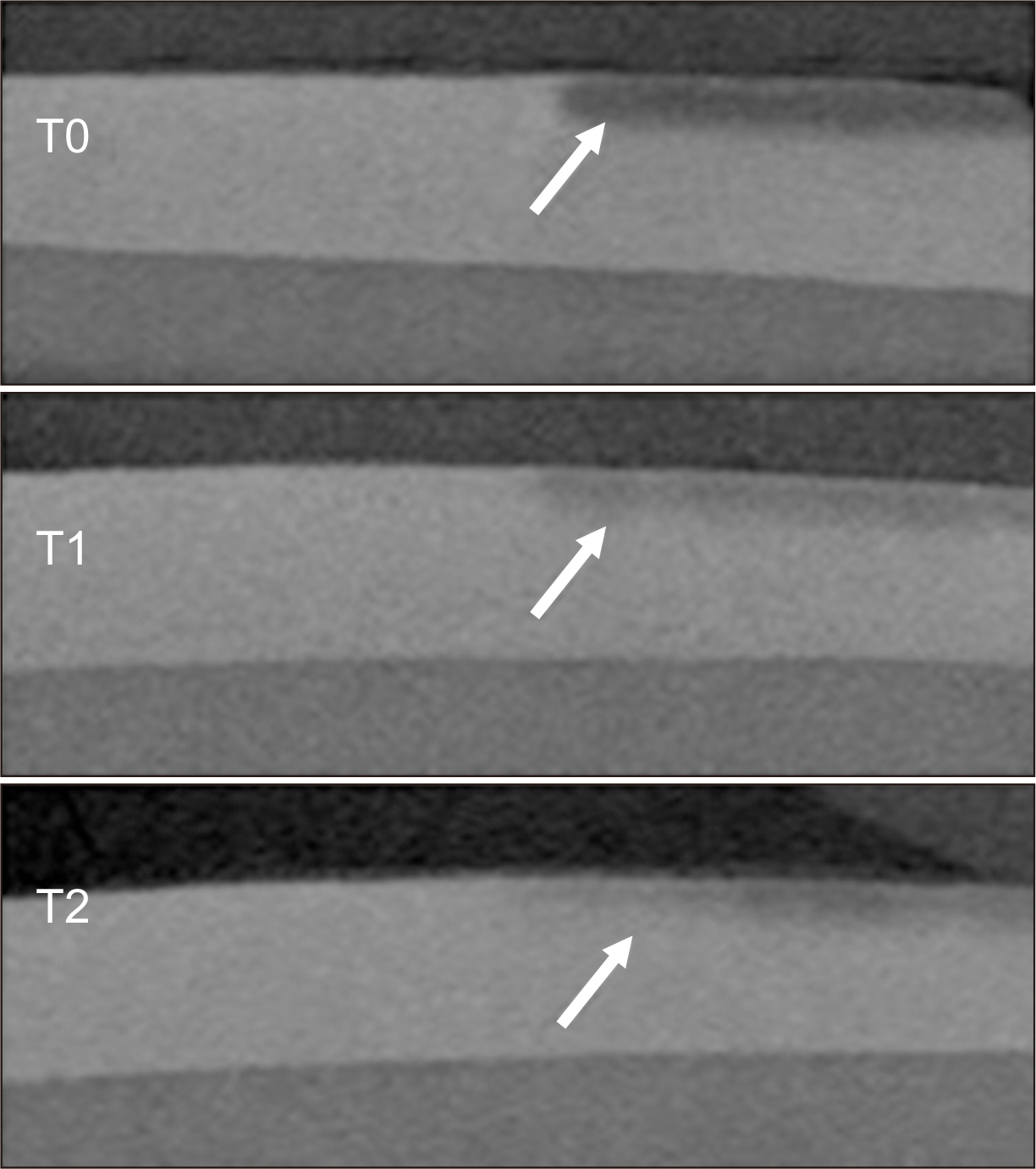
Figure 1 Two dimensional micro-computed tomography images. The arrows indicate mineral density changes as depicted by the gray scale at T0, after demineralization; and T1, 2 weeks and T2, 4 weeks of fluoridation.
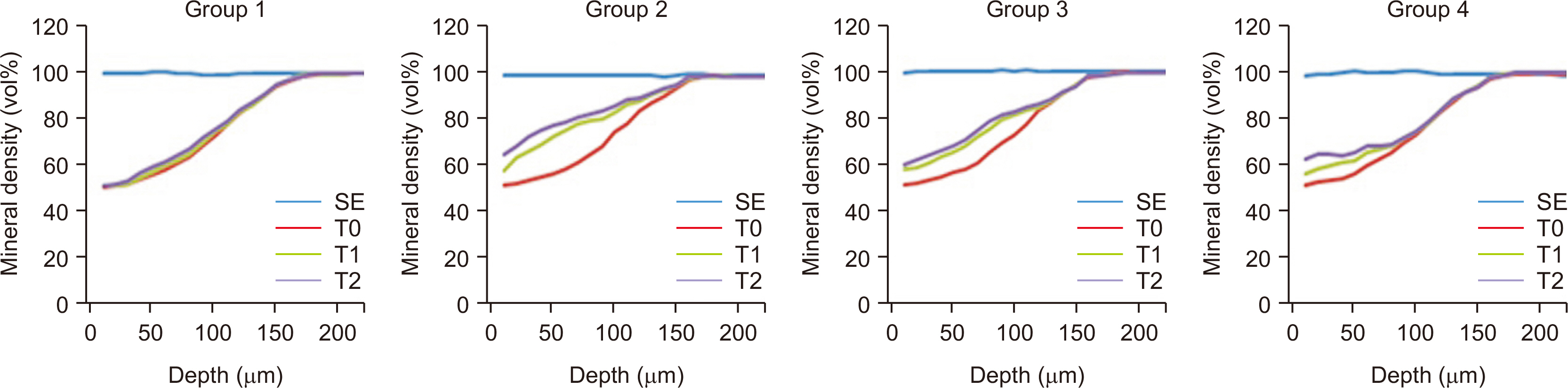
Figure 2 Mean mineral density (vol%) at various depths of enamel in each group. Group 1, no treatment; Group 2, 1.23% acidulated phosphate fluoride gel; Group 3, fluoridated toothpaste; Group 4, 0.05% sodium fluoride mouthwash solution; SE, sound enamel; T0, after demineralization; T1, 2 weeks of fluoridation; T2, 4 weeks of fluoridation.
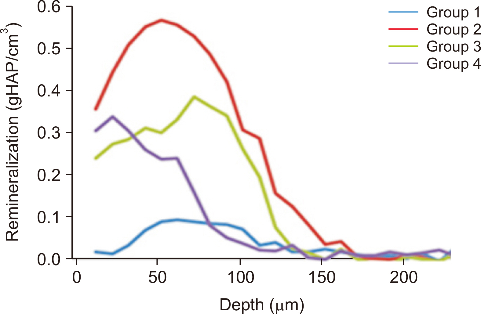
Figure 3 Total remineralization changes in each group over the course of the 4-week fluoridation regimen at various enamel depths. Group 1, no treatment; Group 2, 1.23% acidulated phosphate fluoride gel; Group 3, fluoridated toothpaste; Group 4, 0.05% sodium fluoride mouthwash solution.
Reference
-
1. Gorelick L, Geiger AM, Gwinnett AJ. 1982; Incidence of white spot formation after bonding and banding. Am J Orthod. 81:93–8. DOI: 10.1016/0002-9416(82)90032-X. PMID: 6758594.
Article2. Hadler-Olsen S, Sandvik K, El-Agroudi MA, Øgaard B. 2012; The incidence of caries and white spot lesions in orthodontically treated adolescents with a comprehensive caries prophylactic regimen--a prospective study. Eur J Orthod. 34:633–9. DOI: 10.1093/ejo/cjr068. PMID: 21750245.
Article3. Boersma JG, van der Veen MH, Lagerweij MD, Bokhout B, Prahl-Andersen B. 2005; Caries prevalence measured with QLF after treatment with fixed orthodontic appliances: influencing factors. Caries Res. 39:41–7. DOI: 10.1159/000081655. PMID: 15591733.
Article4. Jo SY, Chong HJ, Lee EH, Chang NY, Chae JM, Cho JH, et al. 2014; Effects of various toothpastes on remineralization of white spot lesions. Korean J Orthod. 44:113–8. DOI: 10.4041/kjod.2014.44.3.113. PMID: 24892024. PMCID: PMC4040358.
Article5. Sonesson M, Bergstrand F, Gizani S, Twetman S. 2017; Management of post-orthodontic white spot lesions: an updated systematic review. Eur J Orthod. 39:116–21. DOI: 10.1093/ejo/cjw023. PMID: 27030284.
Article6. Yetkiner E, Wegehaupt F, Wiegand A, Attin R, Attin T. 2014; Colour improvement and stability of white spot lesions following infiltration, micro-abrasion, or fluoride treatments in vitro. Eur J Orthod. 36:595–602. DOI: 10.1093/ejo/cjt095. PMID: 24385411.
Article7. Bishara SE, Ostby AW. 2008; White spot lesions: formation, prevention, and treatment. Semin Orthod. 14:174–82. DOI: 10.1053/j.sodo.2008.03.002.
Article8. Farhadian N, Miresmaeili A, Eslami B, Mehrabi S. 2008; Effect of fluoride varnish on enamel demineralization around brackets: an in-vivo study. Am J Orthod Dentofacial Orthop. 133(4 Suppl):S95–8. DOI: 10.1016/j.ajodo.2006.09.050. PMID: 18407027.
Article9. Lee YE, Baek HJ, Choi YH, Jeong SH, Park YD, Song KB. 2010; Comparison of remineralization effect of three topical fluoride regimens on enamel initial carious lesions. J Dent. 38:166–71. DOI: 10.1016/j.jdent.2009.10.002. PMID: 19819290.
Article10. Mielczarek A, Gedrange T, Michalik J. 2015; An in vitro evaluation of the effect of fluoride products on white spot lesion remineralization. Am J Dent. 28:51–6. PMID: 25864243.11. Pai N, McIntyre J, Tadic N, Laparidis C. 2007; Comparative uptake of fluoride ion into enamel from various topical fluorides in vitro. Aust Dent J. 52:41–6. DOI: 10.1111/j.1834-7819.2007.tb00464.x. PMID: 17500163.
Article12. Willmot D. 2008; White spot lesions after orthodontic treatment. Semin Orthod. 14:209–19. DOI: 10.1053/j.sodo.2008.03.006. PMID: 23452972.
Article13. Songsiripradubboon S, Hamba H, Trairatvorakul C, Tagami J. 2014; Sodium fluoride mouthrinse used twice daily increased incipient caries lesion remineralization in an in situ model. J Dent. 42:271–8. DOI: 10.1016/j.jdent.2013.12.012. PMID: 24394584.
Article14. al-Khateeb S, Oliveby A, de Josselin de Jong E, Angmar-Månsson B. 1997; Laser fluorescence quantification of remineralisation in situ of incipient enamel lesions: influence of fluoride supplements. Caries Res. 31:132–40. DOI: 10.1159/000262388. PMID: 9118185.
Article15. ten Cate JM, van de Plassche-Simons YM, van Strijp AJ. 1992; Importance of model parameters in the assessment of intra-oral remineralization. J Dent Res. 71 Spec No:879–83. DOI: 10.1177/002203459207100S18. PMID: 1592979.
Article16. Damen JJ, Exterkate RA, ten Cate JM. 1997; Reproducibility of TMR for the determination of longitudinal mineral changes in dental hard tissues. Adv Dent Res. 11:415–9. DOI: 10.1177/08959374970110040601. PMID: 9470498.
Article17. Nakata K, Nikaido T, Nakashima S, Nango N, Tagami J. 2012; An approach to normalizing micro-CT depth profiles of mineral density for monitoring enamel remineralization progress. Dent Mater J. 31:533–40. DOI: 10.4012/dmj.2011-228. PMID: 22864205.
Article18. Hamba H, Nikaido T, Sadr A, Nakashima S, Tagami J. 2012; Enamel lesion parameter correlations between polychromatic micro-CT and TMR. J Dent Res. 91:586–91. DOI: 10.1177/0022034512444127. PMID: 22476867.
Article19. White DJ. 1995; The application of in vitro models to research on demineralization and remineralization of the teeth. Adv Dent Res. 9:175–93. discussion 194–7. DOI: 10.1177/08959374950090030101. PMID: 8615942.
Article
- Full Text Links
-
- Actions
-
Cited
- CITED
-
- Close
- Share
-
- Similar articles
-
- Non-destructive management of white spot lesions by using tooth jewelry
- Management of white spots: resin infiltration technique and microabrasion
- Evaluation of the Remineralization Capacity of Water-based Silver Fluoride
- Effects of various toothpastes on remineralization of white spot lesions
- Colorimetric evaluation of white spot lesions following external bleaching with fluoridation: An in-vitro study
