Computed Tomography Guided Percutaneous Injection of a Mixture of Lipiodol and Methylene Blue in Rabbit Lungs: Evaluation of Localization Ability for Video-Assisted Thoracoscopic Surgery
- Affiliations
-
- 1Department of Radiology, Seoul Metropolitan Government-Seoul National University Boramae Medical Center, Seoul, Korea.
- 2Department of Radiology, Seoul National University Bundang Hospital, Seongnam, Korea. lkwrad@radiol.snu.ac.kr
- 3Department of Radiology, Seoul National University Hospital, Seoul, Korea.
- 4Department of Pathology, Green Cross Laboratories, Yongin, Korea.
- KMID: 1796926
- DOI: http://doi.org/10.3346/jkms.2014.29.1.129
Abstract
- Preoperative localization is necessary prior to video assisted thoracoscopic surgery for the detection of small or deeply located lung nodules. We compared the localization ability of a mixture of lipiodol and methylene blue (MLM) (0.6 mL, 1:5) to methylene blue (0.5 mL) in rabbit lungs. CT-guided percutaneous injections were performed in 21 subjects with MLM and methylene blue. We measured the extent of staining on freshly excised lung and evaluated the subjective localization ability with 4 point scales at 6 and 24 hr after injections. For MLM, radio-opacity was evaluated on the fluoroscopy. We considered score 2 (acceptable) or 3 (excellent) as appropriate for localization. The staining extent of MLM was significantly smaller than methylene blue (0.6 vs 1.0 cm, P<0.001). MLM showed superior staining ability over methylene blue (2.8 vs 2.2, P=0.010). Excellent staining was achieved in 17 subjects (81%) with MLM and 8 (38%) with methylene blue (P=0.011). An acceptable or excellent radio-opacity of MLM was found in 13 subjects (62%). An appropriate localization rate of MLM was 100% with the use of the directly visible ability and radio-opacity of MLM. MLM provides a superior pulmonary localization ability over methylene blue.
MeSH Terms
-
Animals
Ethiodized Oil/*administration & dosage
Fluoroscopy
Injections, Subcutaneous
Lung/*radiography/surgery
Methylene Blue/*administration & dosage
Preoperative Care
Rabbits
Solitary Pulmonary Nodule/*surgery
Staining and Labeling/methods
Thoracic Surgery, Video-Assisted/*methods
Thoracoscopy/methods
Tomography, X-Ray Computed
Ethiodized Oil
Methylene Blue
Figure
-
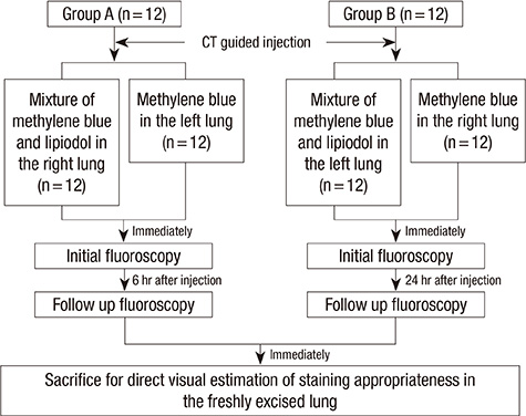
Fig. 1 Overview of the experimental design. Animals were randomly divided into two groups: Group A (n = 12) was sacrificed 6 hr after percutaneous injection and Group B (n = 12) was sacrificed 24 hr after a CT guided percutaneous injection of MLM and methylene blue.
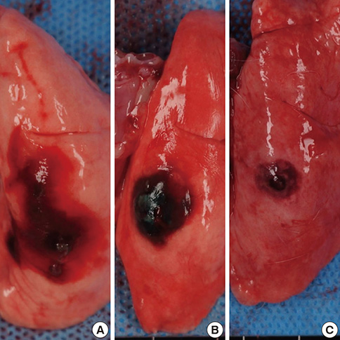
Fig. 2 Examples of evaluation of staining on the lung surface. Photographs show (A) the extensive staining (score 1), (B) localized dispersion of staining (score 2), and (C) minimal dispersion of staining (score 3). The white lines on the bottom of the figure are markings of the ruler. The distance between two lines is one centimeter.
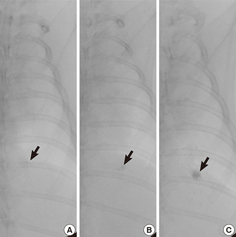
Fig. 3 Examples of assessment of radio-opacity on the fluoroscopic examinations. The fluoroscopic images show (A) a minimally increased opacity (arrow) (score 1), (B) a low density of increased opacity (arrow) (score 2), and (C) a compact nodular increased opacity (arrow) (score 3).
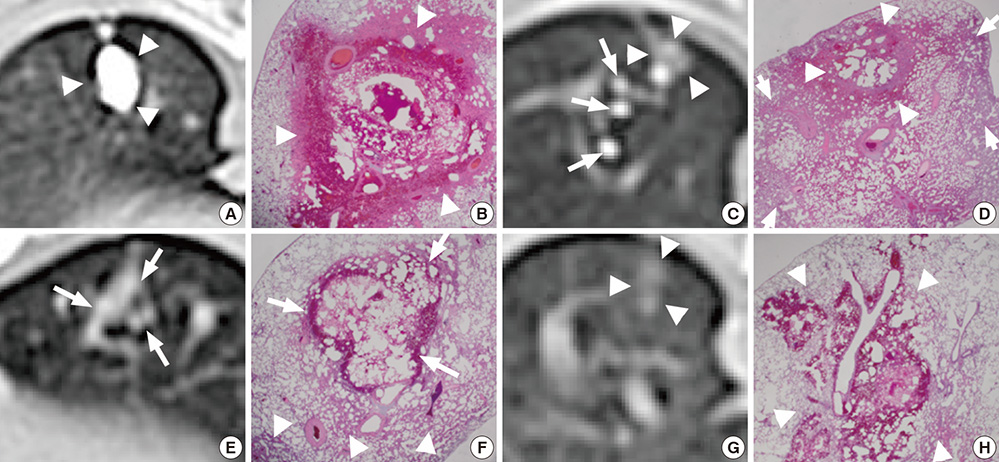
Fig. 4 CT and corresponding photomicrograph of lung specimen. MLM in Group B (A-D); (A) discrete and compact nodular opacity (arrowheads), (B) focal neutrophil infiltration, necrosis, and hemorrhage (arrowheads) (H&E, ×12.5), (C) scattered small nodular opacities of lipiodol (long arrows) and faint nodular opacity (arrowheads), (D) focal hemorrhage and necrosis (arrowheads) with diffuse neutrophil infiltration (short arrows) (H&E, ×12.5). MLM in Group A (E, F); (E) faint nodular lipiodol opacity (arrows), (F) focal hemorrhage (arrows) with diffuse neutrophil infiltration (arrowheads) (H&E, ×12.5). Methylene blue in Group A (G, H); (G) faint nodular opacity (arrowheads), and (H) focal extent of neutrophil infiltration, necrosis and hemorrhage (arrowheads) (H&E, ×12.5).
Reference
-
1. Nakashima S, Watanabe A, Obama T, Yamada G, Takahashi H, Higami T. Need for preoperative computed tomography-guided localization in video-assisted thoracoscopic surgery pulmonary resections of metastatic pulmonary nodules. Ann Thorac Surg. 2010; 89:212–218.2. Chen S, Zhou J, Zhang J, Hu H, Luo X, Zhang Y, Chen H. Video-assisted thoracoscopic solitary pulmonary nodule resection after CT-guided hookwire localization: 43 cases report and literature review. Surg Endosc. 2011; 25:1723–1729.3. Ciriaco P, Negri G, Puglisi A, Nicoletti R, Del Maschio A, Zannini P. Video-assisted thoracoscopic surgery for pulmonary nodules: rationale for preoperative computed tomography-guided hookwire localization. Eur J Cardiothorac Surg. 2004; 25:429–433.4. Suzuki K, Nagai K, Yoshida J, Ohmatsu H, Takahashi K, Nishimura M, Nishiwaki Y. Video-assisted thoracoscopic surgery for small indeterminate pulmonary nodules: indications for preoperative marking. Chest. 1999; 115:563–568.5. Seo JM, Lee HY, Kim HK, Choi YS, Kim J, Shim YM, Lee KS. Factors determining successful computed tomography-guided localization of lung nodules. J Thorac Cardiovasc Surg. 2012; 143:809–814.6. Gossot D, Miaux Y, Guermazi A, Celerier M, Friga J. The hook-wire technique for localization of pulmonary nodules during thoracoscopic resection. Chest. 1994; 105:1467–1469.7. Pittet O, Christodoulou M, Pezzetta E, Schmidt S, Schnyder P, Ris HB. Video-assisted thoracoscopic resection of a small pulmonary nodule after computed tomography-guided localization with a hook-wire system: experience in 45 consecutive patients. World J Surg. 2007; 31:575–578.8. Chen W, Chen L, Yang S, Chen Z, Qian G, Zhang S, Jing J. A novel technique for localization of small pulmonary nodules. Chest. 2007; 131:1526–1531.9. Bernard A. Resection of pulmonary nodules using video-assisted thoracic surgery: the Thorax Group. Ann Thorac Surg. 1996; 61:202–204.10. Martin AE, Chen JY, Muratore CS, Mayo-Smith WW, Luks FI. Dual localization technique for thoracoscopic resection of lung lesions in children. J Laparoendosc Adv Surg Tech A. 2009; 19:S161–S164.11. Kawanaka K, Nomori H, Mori T, Ikeda K, Ikeda O, Tomiguchi S, Yamashita Y. Marking of small pulmonary nodules before thoracoscopic resection: injection of lipiodol under CT-fluoroscopic guidance. Acad Radiol. 2009; 16:39–45.12. Yamagami T, Miura H, Yoshimatsu R, Tanaka O, Ono S, Iehara T, Hosoi H, Nishimura T. Experience of fluoroscopy-aided thoracoscopic resection of pulmonary nodule localised with Lipiodol in a child. J Med Imaging Radiat Oncol. 2011; 55:401–403.13. Iwasaki Y, Nagata K, Yuba T, Hosogi S, Kohno K, Ohsugi S, Kuwahara H, Takemura Y, Yokomura I. Fluoroscopy-guided barium marking for localizing small pulmonary lesions before video-assisted thoracic surgery. Respir Med. 2005; 99:285–289.14. Yoshida J, Nagai K, Nishimura M, Takahashi K. Computed tomography-fluoroscopy guided injection of cyanoacrylate to mark a pulmonary nodule for thoracoscopic resection. Jpn J Thorac Cardiovasc Surg. 1999; 47:210–213.15. Nomori H, Horio H. Colored collagen is a long-lasting point marker for small pulmonary nodules in thoracoscopic operations. Ann Thorac Surg. 1996; 61:1070–1073.16. McConnell PI, Feola GP, Meyers RL. Methylene blue-stained autologous blood for needle localization and thoracoscopic resection of deep pulmonary nodules. J Pediatr Surg. 2002; 37:1729–1731.17. Hu J, Zhang C, Sun L. Localization of small pulmonary nodules for videothoracoscopic surgery. ANZ J Surg. 2006; 76:649–651.18. Wicky S, Mayor B, Cuttat JF, Schnyder P. CT-guided localizations of pulmonary nodules with methylene blue injections for thoracoscopic resections. Chest. 1994; 106:1326–1328.19. Vandoni RE, Cuttat JF, Wicky S, Suter M. CT-guided methylene-blue labelling before thoracoscopic resection of pulmonary nodules. Eur J Cardiothorac Surg. 1998; 14:265–270.20. Lenglinger FX, Schwarz CD, Artmann W. Localization of pulmonary nodules before thoracoscopic surgery: value of percutaneous staining with methylene blue. AJR Am J Roentgenol. 1994; 163:297–300.21. Ikeda K, Nomori H, Mori T, Kobayashi H, Iwatani K, Yoshimoto K, Kawanaka K. Impalpable pulmonary nodules with ground-glass opacity: success for making pathologic sections with preoperative marking by lipiodol. Chest. 2007; 131:502–506.22. Nomori H, Horio H, Naruke T, Suemasu K. Fluoroscopy-assisted thoracoscopic resection of lung nodules marked with lipiodol. Ann Thorac Surg. 2002; 74:170–173.23. Watanabe K, Nomori H, Ohtsuka T, Kaji M, Naruke T, Suemasu K. Usefulness and complications of computed tomography-guided lipiodol marking for fluoroscopy-assisted thoracoscopic resection of small pulmonary nodules: experience with 174 nodules. J Thorac Cardiovasc Surg. 2006; 132:320–324.24. Kim YD, Jeong YJ, I H, Cho JS, Lee JW, Kim HJ, Lee SH, Kim DH. Localization of pulmonary nodules with lipiodol prior to thoracoscopic surgery. Acta Radiol. 2011; 52:64–69.25. Mayo JR, Clifton JC, Powell TI, English JC, Evans KG, Yee J, McWilliams AM, Lam SC, Finley RJ. Lung nodules: CT-guided placement of microcoils to direct video-assisted thoracoscopic surgical resection. Radiology. 2009; 250:576–585.26. Lee NK, Park CM, Kang CH, Jeon YK, Choo JY, Lee HJ, Goo JM. CT-guided percutaneous transthoracic localization of pulmonary nodules prior to video-assisted thoracoscopic surgery using barium suspension. Korean J Radiol. 2012; 13:694–701.27. Kamiyoshihara M, Ishikawa S, Morishita Y. Pulmonary cryptococcosis diagnosed by video-assisted thoracoscopic surgery with CT-guided localization: report of a case. Kyobu Geka. 2000; 53:795–797.28. Kwon WJ, Kim HJ, Jeong YJ, Lee CH, Kim KI, Kim YD, Lee JH. Direct lipiodol injection used for a radio-opaque lung marker: stability and histopathologic effects. Exp Lung Res. 2011; 37:310–317.29. Jang HS. Effect of drugs for preoperative localization of thoracoscopy to histopathologic change in rabbit lung. Seoul: the Catholic University of Korea;2000. 27. Dissertation.30. Okumura T, Kondo H, Suzuki K, Asamura H, Kobayashi T, Kaneko M, Tsuchiya R. Fluoroscopy-assisted thoracoscopic surgery after computed tomography-guided bronchoscopic barium marking. Ann Thorac Surg. 2001; 71:439–442.
- Full Text Links
-
- Actions
-
Cited
- CITED
-
- Close
- Share
-
- Similar articles
-
- Planting Seeds into the Lung: Image-Guided Percutaneous Localization to Guide Minimally Invasive Thoracic Surgery
- Localization of Nonpalpable Breast lesion with Ultrasonoguided Dye Injection
- Indocyanine Green-Guided Video-Assisted Thoracoscopic Surgery for Resection of an Ectopic Mediastinal Parathyroid Adenoma
- Methylene Blue for Localization of Sentinel Lymph Nodes in Breast Cancer: A Comparison with Isosulfan Blue
- The Usefulness of Methylene Blue Infusion in Parathyroidectomy for Secondary Hyperparathyroidism
