Planting Seeds into the Lung: Image-Guided Percutaneous Localization to Guide Minimally Invasive Thoracic Surgery
- Affiliations
-
- 1Department of Radiology, Pusan National University Hospital, Busan, Korea.
- 2Department of Radiology and Research Institute of Radiological Science, Gangnam Severance Hospital, Yonsei University College of Medicine, Seoul, Korea.
- 3Department of Radiology, Asan Medical Center, University of Ulsan College of Medicine, Seoul, Korea.
- 4Department of Radiology and Research Institute of Radiological Science, Severance Hospital, Yonsei University College of Medicine, Seoul, Korea. khuhz@yuhs.ac
- KMID: 2459418
- DOI: http://doi.org/10.3348/kjr.2019.0155
Abstract
- Image-guided localization materials are constantly evolving, providing options for the localization of small pulmonary nodules to guide minimally invasive thoracic surgery. Several preoperative methods have been developed to localize small pulmonary lesions prior to video-assisted thoracic surgery. These localization techniques can be categorized into 4 groups according to the materials used: localization with metallic materials (hook-wire, microcoil, or spiral coil), localization with dye (methylene blue or indigo carmine), localization with contrast agents (lipiodol, barium, or iodine contrast agents), and radiotracers (technetium-99m). However, the optimal localization method has not yet been established. In this review article, we discuss the various localization techniques and the advantages and disadvantages of localization techniques as well as the available safety and efficacy data on these techniques.
Keyword
MeSH Terms
Figure
-
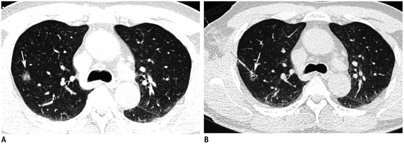
Fig. 1 Preoperative hook-wire localization for part-solid nodule in right upper lobe in 59-year-old man.A. Axial CT image with lung window shows 15-mm part-solid nodule (arrow) in right upper lobe. B. Post-procedure axial CT image with lung window shows that horn of hook-wire is positioned in target nodule (arrow).
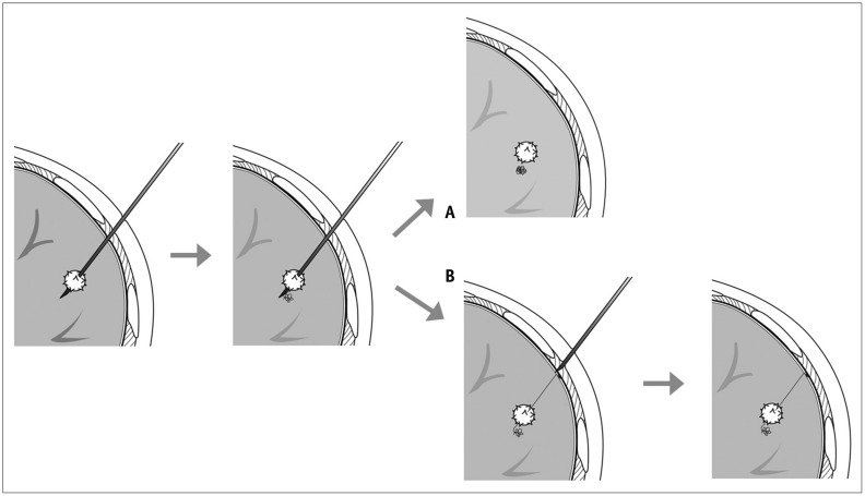
Fig. 2 Diagrams showing two localization methods using microcoil.A. Drawing illustrates entire microcoil was inserted into lung tissue. B. Drawing illustrates that microcoil was partially inserted, and end tail of microcoil remained above visceral pleura.
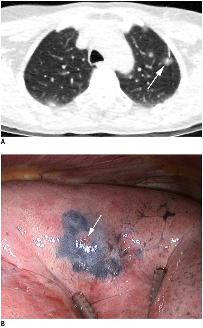
Fig. 3 Preoperative localization with indigo carmine for part-solid nodule in left upper lobe in 60-year-old woman.A. Axial CT image with lung window during localization shows 22-gauge Chiba needle introduced into nodule (arrow). B. Video-assisted thoracoscopic image shows deposition of indigo carmine as purple area around inserted area of needle (arrow).

Fig. 4 Preoperative lipiodol localization for part-solid nodule in right upper lobe in 48-year-old woman.A. Axial CT image with lung window during lipiodol localization in prone position shows 22-gauge Chiba needle introduced into nodule (arrow) in left upper lobe. B. Post-procedure axial CT image with lung window shows lipiodol accumulation (arrow) in target nodule after procedure.
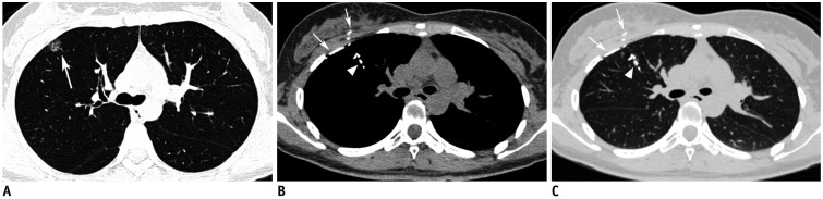
Fig. 5 Preoperative lipiodol localization for part-solid nodule in right upper lobe in 35-year-old woman.A. Axial CT image with lung window shows 15-mm part-solid nodule (arrow) in right upper lobe. Post-procedure axial CT images with mediastinal (B) and lung (C) windows demonstrate lipiodol spillage to right pleural, right chest wall (arrows) and bronchi of right upper lobe (arrowheads). Thoracoscopic wedge resection was successfully performed.
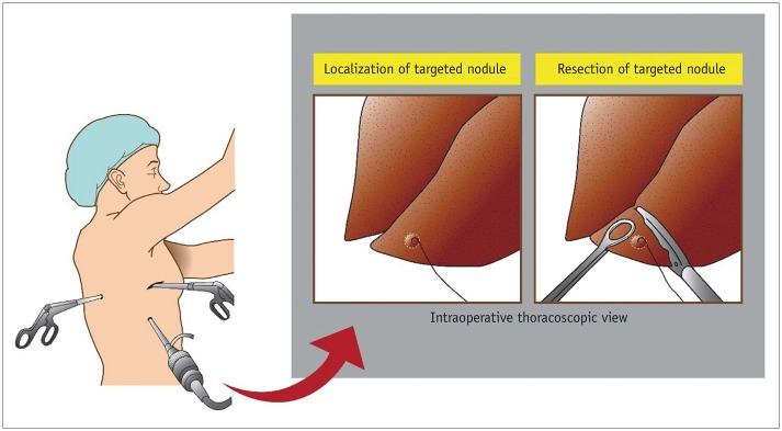
Fig. 6 Video-assisted thoracoscopic surgery after localization using hook-wire.Thoracoscopic surgery is performed with 3–5-cm utility incision at anterior axillary line at fourth or fifth intercostal space using endoscopic instruments without rib spreading and additional 2 or 3 ports for camera, stapler insertion, and assistant. After identification of marked nodule by thoracoscopic vision, thoracoscopic wedge resection is performed using endoscopic stapler with 1–2-cm margin from lesion.
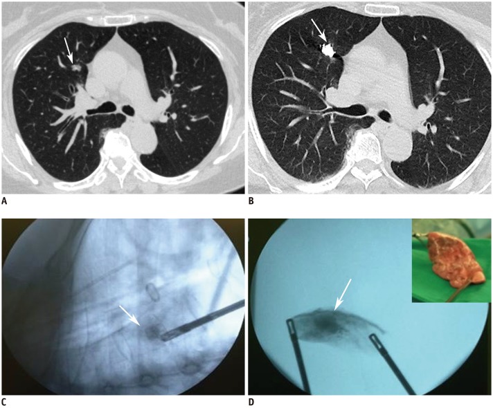
Fig. 7 Images in 65-year-old woman undergoing lipiodol localization for part-solid nodule in right upper lobe.A. Axial CT image with lung window shows 11-mm part-solid nodule (arrow) in right upper lobe. B. Post-procedure axial CT image with lung window shows lipiodol accumulation (arrow) in right upper lobe. C. Intraoperative fluoroscopic images obtained during resection demonstrate radiopaque spot (arrow) representing lipiodol accumulation. D. After wedge resection, successful resection can be confirmed by identifying radiopaque spot (arrow) by intraoperative fluoroscopy.
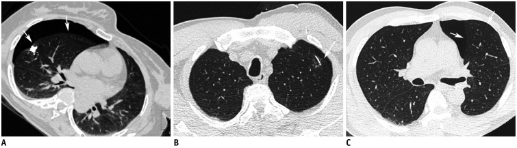
Fig. 8 Pneumothorax as complication after localization.A. Lipiodol localization for right middle lobe part-solid nodule in 56-year-old woman. Post-procedure CT image shows right pneumothorax (arrows) after lipiodol localization. B, C. Hook-wire localization for left upper lobe part-solid nodule in 57-year-old man. Post-procedure CT image shows right pneumothorax (arrows) after hook-wire insertion. Thoracoscopic wedge resections were successfully performed in both patients.
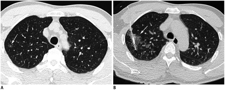
Fig. 9 Pulmonary hemorrhage as complication after hook-wire localization.A. Axial CT image shows 10-mm ground glass nodules (arrow) in right upper lobe. B. Post-procedure axial CT image after hook-wire localization shows ground glass opacity around hook-wire, suggesting pulmonary hemorrhage (arrow) in right upper lobe. Patient was asymptomatic and required no treatment.
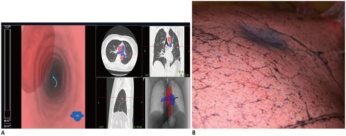
Fig. 10 ENB localization of lung nodule.A. ENB can convert preoperative CT data into virtual bronchial map. Steerable probe contains position sensor and allows navigation of turns in endobronchial tree. After guidance of steerable probe tip to targeted lung nodule, dye is injected to target nodule. B. Video-assisted thoracoscopic image shows deposition of dye as purple area around targeted nodule position after ENB localization. ENB = electromagnetic navigation bronchography
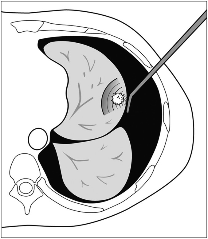
Fig. 11 Diagram showing intraoperative ultrasonography localization for left upper lobe nodule.Lung is collapsed and probe is placed firmly in area of target nodule.
Reference
-
1. National Lung Screening Trial Research Team. Aberle DR, Adams AM, Berg CD, Black WC, Clapp JD, Fagerstrom RM, et al. Reduced lung-cancer mortality with low-dose computed tomographic screening. N Engl J Med. 2011; 365:395–409. PMID: 21714641.
Article2. Godoy MC, Naidich DP. Subsolid pulmonary nodules and the spectrum of peripheral adenocarcinomas of the lung: recommended interim guidelines for assessment and management. Radiology. 2009; 253:606–622. PMID: 19952025.
Article3. Infante M, Lutman RF, Imparato S, Di Rocco M, Ceresoli GL, Torri V, et al. Differential diagnosis and management of focal ground-glass opacities. Eur Respir J. 2009; 33:821–827. PMID: 19047318.
Article4. Mack MJ, Aronoff RJ, Acuff TE, Douthit MB, Bowman RT, Ryan WH. Present role of thoracoscopy in the diagnosis and treatment of diseases of the chest. Ann Thorac Surg. 1992; 54:403–408. discussion 407–409. PMID: 1510505.
Article5. Suzuki K, Nagai K, Yoshida J, Ohmatsu H, Takahashi K, Nishimura M, et al. Video-assisted thoracoscopic surgery for small indeterminate pulmonary nodules: indications for preoperative marking. Chest. 1999; 115:563–568. PMID: 10027460.6. Plunkett MB, Peterson MS, Landreneau RJ, Ferson PF, Posner MC. Peripheral pulmonary nodules: preoperative percutaneous needle localization with CT guidance. Radiology. 1992; 185:274–276. PMID: 1523323.
Article7. Finley RJ, Mayo JR, Grant K, Clifton JC, English J, Leo J, et al. Preoperative computed tomography-guided microcoil localization of small peripheral pulmonary nodules: a prospective randomized controlled trial. J Thorac Cardiovasc Surg. 2015; 149:26–31. PMID: 25293355.
Article8. Park CH, Han K, Hur J, Lee SM, Lee JW, Hwang SH, et al. Comparative effectiveness and safety of preoperative lung localization for pulmonary nodules: a systematic review and meta-analysis. Chest. 2017; 151:316–328. PMID: 27717643.9. Lin MW, Chen JS. Image-guided techniques for localizing pulmonary nodules in thoracoscopic surgery. J Thorac Dis. 2016; 8(Suppl 9):S749–S755. PMID: 28066679.
Article10. Zaman M, Bilal H, Woo CY, Tang A. In patients undergoing video-assisted thoracoscopic surgery excision, what is the best way to locate a subcentimetre solitary pulmonary nodule in order to achieve successful excision? Interact Cardiovasc Thorac Surg. 2012; 15:266–272. PMID: 22572410.
Article11. Horan TA, Pinheiro PM, Araújo LM, Santiago FF, Rodrigues MR. Massive gas embolism during pulmonary nodule hook wire localization. Ann Thorac Surg. 2002; 73:1647–1649. PMID: 12022575.
Article12. Sakiyama S, Kondo K, Matsuoka H, Yoshida M, Miyoshi T, Yoshida S, et al. Fatal air embolism during computed tomography-guided pulmonary marking with a hook-type marker. J Thorac Cardiovasc Surg. 2003; 126:1207–1209. PMID: 14566279.
Article13. Asamura H, Kondo H, Naruke T, Tsuchiya R, Wakao F, Kaneko M, et al. Computed tomography-guided coil injection and thoracoscopic pulmonary resection under roentgenographic fluoroscopy. Ann Thorac Surg. 1994; 58:1542–1544. PMID: 7979696.
Article14. Su TH, Fan YF, Jin L, He W, Hu LB. CT-guided localization of small pulmonary nodules using adjacent microcoil implantation prior to video-assisted thoracoscopic surgical resection. Eur Radiol. 2015; 25:2627–2633. PMID: 25773939.
Article15. Mayo JR, Clifton JC, Powell TI, English JC, Evans KG, Yee J, et al. Lung nodules: CT-guided placement of microcoils to direct video-assisted thoracoscopic surgical resection. Radiology. 2009; 250:576–585. PMID: 19188326.
Article16. Sui X, Zhao H, Yang F, Li JL, Wang J. Computed tomography guided microcoil localization for pulmonary small nodules and ground-glass opacity prior to thoracoscopic resection. J Thorac Dis. 2015; 7:1580–1587. PMID: 26543605.17. Lenglinger FX, Schwarz CD, Artmann W. Localization of pulmonary nodules before thoracoscopic surgery: value of percutaneous staining with methylene blue. AJR Am J Roentgenol. 1994; 163:297–300. PMID: 7518642.
Article18. Keating J, Singhal S. Novel methods of intraoperative localization and margin assessment of pulmonary nodules. Semin Thorac Cardiovasc Surg. 2016; 28:127–136. PMID: 27568150.
Article19. Vandoni RE, Cuttat JF, Wicky S, Suter M. CT-guided methylene-blue labelling before thoracoscopic resection of pulmonary nodules. Eur J Cardiothorac Surg. 1998; 14:265–270. PMID: 9761435.
Article20. Lee NK, Park CM, Kang CH, Jeon YK, Choo JY, Lee HJ, et al. CT-guided percutaneous transthoracic localization of pulmonary nodules prior to video-assisted thoracoscopic surgery using barium suspension. Korean J Radiol. 2012; 13:694–701. PMID: 23118567.
Article21. Okumura T, Kondo H, Suzuki K, Asamura H, Kobayashi T, Kaneko M, et al. Fluoroscopy-assisted thoracoscopic surgery after computed tomography-guided bronchoscopic barium marking. Ann Thorac Surg. 2001; 71:439–442. PMID: 11235684.
Article22. Asano F, Shindoh J, Shigemitsu K, Miya K, Abe T, Horiba M, et al. Ultrathin bronchoscopic barium marking with virtual bronchoscopic navigation for fluoroscopy-assisted thoracoscopic surgery. Chest. 2004; 126:1687–1693. PMID: 15539745.
Article23. Kobayashi T, Kaneko M, Kondo H, Nakayama H, Asamura H, Tsuchiya R, et al. CT-guided bronchoscopic barium marking for resection of a fluoroscopically invisible peripheral pulmonary lesion. Jpn J Clin Oncol. 1997; 27:204–205. PMID: 9255280.
Article24. Nomori H, Horio H, Naruke T, Suemasu K. Fluoroscopy-assisted thoracoscopic resection of lung nodules marked with lipiodol. Ann Thorac Surg. 2002; 74:170–173. PMID: 12118752.
Article25. Moon SW, Wang YP, Jo KH, Kwack MS, Kim SW, Kwon OK, et al. Fluoroscopy-aided thoracoscopic resection of pulmonary nodule localized with contrast media. Ann Thorac Surg. 1999; 68:1815–1820. PMID: 10585064.
Article26. Kawanaka K, Nomori H, Mori T, Ikeda K, Ikeda O, Tomiguchi S, et al. Marking of small pulmonary nodules before thoracoscopic resection: injection of lipiodol under CT-fluoroscopic guidance. Acad Radiol. 2009; 16:39–45. PMID: 19064210.27. Kim YD, Jeong YJ, I H, Cho JS, Lee JW, Kim HJ, et al. Localization of pulmonary nodules with lipiodol prior to thoracoscopic surgery. Acta Radiol. 2011; 52:64–69. PMID: 21498328.
Article28. Ambrogi MC, Melfi F, Zirafa C, Lucchi M, De Liperi A, Mariani G, et al. Radio-guided thoracoscopic surgery (RGTS) of small pulmonary nodules. Surg Endosc. 2012; 26:914–919. PMID: 22011947.
Article29. Chella A, Lucchi M, Ambrogi MC, Menconi G, Melfi FM, Gonfiotti A, et al. A pilot study of the role of TC-99 radionuclide in localization of pulmonary nodular lesions for thoracoscopic resection. Eur J Cardiothorac Surg. 2000; 18:17–21. PMID: 10869935.
Article30. Starnes SL, Wolujewicz M, Guitron J, Williams V, Scheler J, Ristagno R. Radiotracer localization of nonpalpable pulmonary nodules: a single-center experience. J Thorac Cardiovasc Surg. 2018; 156:1986–1992. PMID: 29778333.
Article31. Bellomi M, Veronesi G, Trifirò G, Brambilla S, Bonello L, Preda L, et al. Computed tomography-guided preoperative radiotracer localization of nonpalpable lung nodules. Ann Thorac Surg. 2010; 90:1759–1764. PMID: 21095303.
Article32. Koike T, Koike T, Yoshiya K, Tsuchida M, Toyabe S. Risk factor analysis of locoregional recurrence after sublobar resection in patients with clinical stage IA non-small cell lung cancer. J Thorac Cardiovasc Surg. 2013; 146:372–378. PMID: 23870323.
Article33. El-Sherif A, Fernando HC, Santos R, Pettiford B, Luketich JD, Close JM, et al. Margin and local recurrence after sublobar resection of non-small cell lung cancer. Ann Surg Oncol. 2007; 14:2400–2405. PMID: 17505859.
Article34. Wolf AS, Swanson SJ, Yip R, Liu B, Tarras ES, Yankelevitz DF, et al. I-ELCAP Investigators. The impact of margins on outcomes after wedge resection for stage I non-small cell lung cancer. Ann Thorac Surg. 2017; 104:1171–1178. PMID: 28669499.
Article35. Dendo S, Kanazawa S, Ando A, Hyodo T, Kouno Y, Yasui K, et al. Preoperative localization of small pulmonary lesions with a short hook wire and suture system: experience with 168 procedures. Radiology. 2002; 225:511–518. PMID: 12409589.
Article36. Miyoshi K, Toyooka S, Gobara H, Oto T, Mimura H, Sano Y, et al. Clinical outcomes of short hook wire and suture marking system in thoracoscopic resection for pulmonary nodules. Eur J Cardiothorac Surg. 2009; 36:378–382. PMID: 19414272.
Article37. Li W, Wang Y, He X, Li G, Wang S, Xu L, et al. Combination of CT-guided hookwire localization and video-assisted thoracoscopic surgery for pulmonary nodular lesions: analysis of 103 patients. Oncol Lett. 2012; 4:824–828. PMID: 23205107.
Article38. Seo JM, Lee HY, Kim HK, Choi YS, Kim J, Shim YM, et al. Factors determining successful computed tomography-guided localization of lung nodules. J Thorac Cardiovasc Surg. 2012; 143:809–814. PMID: 22104686.
Article39. Ichinose J, Kohno T, Fujimori S, Harano T, Suzuki S. Efficacy and complications of computed tomography-guided hook wire localization. Ann Thorac Surg. 2013; 96:1203–1208. PMID: 23895891.
Article40. Hanauer M, Perentes JY, Krueger T, Ris HB, Bize P, Schmidt S, et al. Pre-operative localization of solitary pulmonary nodules with computed tomography-guided hook wire: report of 181 patients. J Cardiothorac Surg. 2016; 11:5. PMID: 26772183.
Article41. Huang HZ, Wang GZ, Xu LC, Li GD, Wang Y, Wang YH, et al. CT-guided Hookwire localization before video-assisted thoracoscopic surgery for solitary ground-glass opacity dominant pulmonary nodules: radiologic-pathologic analysis. Oncotarget. 2017; 8:108118–108129. PMID: 29296228.
Article42. Klinkenberg TJ, Dinjens L, Wolf RFE, van der Wauwer C, van der Wekken AJ, de Bock GH, et al. CT-guided percutaneous hookwire localization increases the efficacy and safety of VATS for pulmonary nodules. J Surg Oncol. 2017; 115:898–904. PMID: 28230245.
Article43. Yao F, Wang J, Yao J, Xu L, Wang J, Gao L. Reevaluation of the efficacy of preoperative computed tomography-guided hook wire localization: a retrospective analysis. Int J Surg. 2018; 51:24–30. PMID: 29367030.
Article44. Suzuki K, Shimohira M, Hashizume T, Ozawa Y, Sobue R, Mimura M, et al. Usefulness of CT-guided hookwire marking before video-assisted thoracoscopic surgery for small pulmonary lesions. J Med Imaging Radiat Oncol. 2014; 58:657–662. PMID: 25088355.
Article45. Iguchi T, Hiraki T, Gobara H, Fujiwara H, Matsui Y, Sugimoto S, et al. Simultaneous multiple preoperative localizations of small pulmonary lesions using a short hook wire and suture system. Cardiovasc Intervent Radiol. 2015; 38:971–976. PMID: 25465062.
Article46. Mullan BF, Stanford W, Barnhart W, Galvin JR. Lung nodules: improved wire for CT-guided localization. Radiology. 1999; 211:561–565. PMID: 10228543.
Article47. Hajjar W, Al-Nassar S, Almousa O, Rahal S, Al-Aqeed A, Ahmed I, et al. Thoracoscopic resection of suspected metastatic pulmonary nodules after microcoil localization technique: a prospective study. J Cardiovasc Surg (Torino). 2017; 58:606–612.
Article48. Kha LC, Hanneman K, Donahoe L, Chung T, Pierre AF, Yasufuku K, et al. Safety and efficacy of modified preoperative lung nodule microcoil localization without pleural marking: a pilot study. J Thorac Imaging. 2016; 31:15–22. PMID: 26502347.49. Wang ZX, Li L, Zhang Z, Wang GH, Kong DM, Wang XD, et al. High-resolution computed tomography features and CT-guided microcoil localization of subcentimeter pulmonary ground-glass opacities: radiological processing prior to video-assisted thoracoscopic surgery. J Thorac Dis. 2018; 10:2676–2684. PMID: 29997929.
Article50. Sui X, Zhao H, Yang F, Liu G, Hu L, Chen C, et al. Analysis of factors affecting successful microcoil localization for pulmonary nodules. J Surg Res. 2018; 224:193–199. PMID: 29506840.
Article51. Stephenson JA, Mahfouz A, Rathinam S, Nakas A, Bajaj A. A simple and safe technique for CT guided lung nodule marking prior to video assisted thoracoscopic surgical resection revisited. Lung Cancer Int. 2015; 2015:235720. PMID: 26579236.
Article52. Shimamura Y, Sasaki S, Shimohira M, Ogino H, Yuki D, Nakamae K, et al. New technique of percutaneous CT fluoroscopy-guided marking before video-assisted thoracoscopic surgery for small lung lesions: feasibility of using a 25-gauge needle without local anaesthesia. Br J Radiol. 2018; 91:20170692. PMID: 29172683.
Article53. Lin MW, Tseng YH, Lee YF, Hsieh MS, Ko WC, Chen JY, et al. Computed tomography-guided patent blue vital dye localization of pulmonary nodules in uniportal thoracoscopy. J Thorac Cardiovasc Surg. 2016; 152:535–544.e2. PMID: 27189890.
Article54. McConnell PI, Feola GP, Meyers RL. Methylene blue-stained autologous blood for needle localization and thoracoscopic resection of deep pulmonary nodules. J Pediatr Surg. 2002; 37:1729–1731. PMID: 12483642.
Article55. Kleedehn M, Kim DH, Lee FT, Lubner MG, Robbins JB, Ziemlewicz TJ, et al. Preoperative pulmonary nodule localization: a comparison of methylene blue and hookwire techniques. AJR Am J Roentgenol. 2016; 207:1334–1339. PMID: 27657546.
Article56. Watanabe K, Nomori H, Ohtsuka T, Kaji M, Naruke T, Suemasu K. Usefulness and complications of computed tomography-guided lipiodol marking for fluoroscopy-assisted thoracoscopic resection of small pulmonary nodules: experience with 174 nodules. J Thorac Cardiovasc Surg. 2006; 132:320–324. PMID: 16872957.
Article57. Mogi A, Yajima T, Tomizawa K, Onozato R, Tanaka S, Kuwano H. Video-assisted thoracoscopic surgery after preoperative CT-guided lipiodol marking of small or impalpable pulmonary nodules. Ann Thorac Cardiovasc Surg. 2015; 21:435–439. PMID: 26004116.
Article58. Miura H, Yamagami T, Tanaka O, Yoshimatsu R, Ichijo Y, Kato D, et al. CT findings after lipiodol marking performed before video-assisted thoracoscopic surgery for small pulmonary nodules. Acta Radiol. 2016; 57:303–310. PMID: 25795703.
Article59. Doo KW, Yong HS, Kim HK, Kim S, Kang EY, Choi YH. Needlescopic resection of small and superficial pulmonary nodule after computed tomographic fluoroscopy-guided dual localization with radiotracer and hookwire. Ann Surg Oncol. 2015; 22:331–337. PMID: 25008029.
Article60. Iguchi T, Hiraki T, Gobara H, Fujiwara H, Matsui Y, Miyoshi S, et al. CT fluoroscopy-guided preoperative short hook wire placement for small pulmonary lesions: evaluation of safety and identification of risk factors for pneumothorax. Eur Radiol. 2016; 26:114–121. PMID: 25991483.
Article61. Nour-Eldin NE, Alsubhi M, Naguib NN, Lehnert T, Emam A, Beeres M, et al. Risk factor analysis of pulmonary hemorrhage complicating CT-guided lung biopsy in coaxial and non-coaxial core biopsy techniques in 650 patients. Eur J Radiol. 2014; 83:1945–1952. PMID: 25063212.
Article62. Wu CC, Maher MM, Shepard JA. Complications of CT-guided percutaneous needle biopsy of the chest: prevention and management. AJR Am J Roentgenol. 2011; 196:W678–W682. PMID: 21606253.
Article63. Cham MD, Lane ME, Henschke CI, Yankelevitz DF. Lung biopsy: special techniques. Semin Respir Crit Care Med. 2008; 29:335–349. PMID: 18651353.
Article64. Yi JH, Choi PJ, Bang JH, Jeong SS, Cho JH. Systemic air embolism after computed tomography-guided hook wire localization: two case reports and literature review. J Thorac Dis. 2018; 10:E59–E64. PMID: 29600106.
Article65. Endo M, Kotani Y, Satouchi M, Takada Y, Sakamoto T, Tsubota N, et al. CT fluoroscopy-guided bronchoscopic dye marking for resection of small peripheral pulmonary nodules. Chest. 2004; 125:1747–1752. PMID: 15136386.
Article66. Krimsky WS, Minnich DJ, Cattaneo SM, Sarkar SA, Harley DP, Finley DJ, et al. Thoracoscopic detection of occult indeterminate pulmonary nodules using bronchoscopic pleural dye marking. J Community Hosp Intern Med Perspect. 2014; 2. 17. [Epub]. DOI: 10.3402/jchimp.v4.23084.
Article67. Tay JH, Wallbridge PD, Larobina M, Russell PA, Irving LB, Steinfort DP. Electromagnetic navigation bronchoscopydirected pleural tattoo to aid surgical resection of peripheral pulmonary lesions. J Bronchology Interv Pulmonol. 2016; 23:245–250. PMID: 26496089.
Article68. Kuo SW, Tseng YF, Dai KY, Chang YC, Chen KC, Lee JM. Electromagnetic navigation bronchoscopy localization versus percutaneous CT-guided localization for lung resection via video-assisted thoracoscopic surgery: a propensity-matched study. J Clin Med. 2019; 8:E379. PMID: 30889927.
Article69. Bolton WD, Howe H 3rd, Stephenson JE. The utility of electromagnetic navigational bronchoscopy as a localization tool for robotic resection of small pulmonary nodules. Ann Thorac Surg. 2014; 98:471–475. discussion 475–476. PMID: 24968769.
Article70. Marino KA, Sullivan JL, Weksler B. Electromagnetic navigation bronchoscopy for identifying lung nodules for thoracoscopic resection. Ann Thorac Surg. 2016; 102:454–457. PMID: 27173068.
Article71. Georgiou HD, Taverner J, Irving LB, Steinfort DP. Safety and efficacy of radial EBUS for the investigation of peripheral pulmonary lesions in patients with advanced COPD. J Bronchology Interv Pulmonol. 2016; 23:192–198. PMID: 27454473.
Article72. Wada H, Anayama T, Hirohashi K, Nakajima T, Kato T, Waddell TK, et al. Thoracoscopic ultrasonography for localization of subcentimetre lung nodules. Eur J Cardiothorac Surg. 2016; 49:690–697. PMID: 25855597.
Article73. Kondo R, Yoshida K, Hamanaka K, Hashizume M, Ushiyama T, Hyogotani A, et al. Intraoperative ultrasonographic localization of pulmonary ground-glass opacities. J Thorac Cardiovasc Surg. 2009; 138:837–842. PMID: 19660350.
Article74. Piolanti M, Coppola F, Papa S, Pilotti V, Mattioli S, Gavelli G. Ultrasonographic localization of occult pulmonary nodules during video-assisted thoracic surgery. Eur Radiol. 2003; 13:2358–2364. PMID: 12736756.
Article75. Mattioli S, D'Ovidio F, Daddi N, Ferruzzi L, Pilotti V, Ruffato A, et al. Transthoracic endosonography for the intraoperative localization of lung nodules. Ann Thorac Surg. 2005; 79:443–449. discussion 443–449. PMID: 15680811.
Article76. Khereba M, Ferraro P, Duranceau A, Martin J, Goudie E, Thiffault V, et al. Thoracoscopic localization of intraparenchymal pulmonary nodules using direct intracavitary thoracoscopic ultrasonography prevents conversion of VATS procedures to thoracotomy in selected patients. J Thorac Cardiovasc Surg. 2012; 144:1160–1165. PMID: 22980667.
Article
- Full Text Links
-
- Actions
-
Cited
- CITED
-
- Close
- Share
-
- Similar articles
-
- Establishment of Minimally Invasive Thoracic Surgery Program
- Percutaneous Ablation of Osteoid Osteoma under Image Intensifier Guidance: A Case Report
- Minimally Invasive Thoracic Surgery in Lung Cancer Operation
- Percutaneous image-guided surgery
- Experience with an Image-Guided Surgery Device-CANS Navigator-
