A Case of Concurrent Thymic Carcinoma with Systemic Lupus Erythematosus
- Affiliations
-
- 1Department of Internal Medicine, Yonsei University College of Medicine, Seoul, Korea. kjhang@yumc.yonsei.ac.kr
- KMID: 1518492
- DOI: http://doi.org/10.4046/trd.2007.62.1.67
Abstract
- A thymic carcinoma is a rare malignant neoplasm of the thymus epithelium, which can be distinguished from a benign or invasive thymoma. Contrary to a thymoma, the association of a thymic carcinoma and autoimmune disease is rare, with only a few cases having been reported. Herein, a case of thymic carcinoma diagnosed concurrently with systemic lupus erythematosus (SLE) is reported. A 49 year-old man presented at our clinic with myalgia. He was diagnosed with SLE, based on an oral ulcer, lymphopenia, and positive ANA and anti-Sm antibodies. Incidentally, a routine chest X-ray showed a large mediastinal mass. Pathological examination of the mediastinal mass revealed an undifferentiated thymic carcinoma, of WHO classification type C. Further work-up for staging showed multiple bone and lung metastases. With a palliative aim, he received systemic chemotherapy, but refused further chemotherapy after the 2nd course. Currently, the patient has not been followed up since the chemotherapy.
MeSH Terms
Figure
-
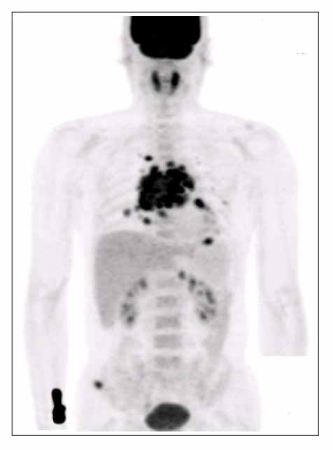
Figure 1 Positron emission tomography revealed the strong FDG uptake on a large irregular mass in anterior mediastinum, multiple lung nodules with bilateral lymphadenopathy, the 8th T-spine, right iliac crest, and the posterior pararenal area. It suggested malignant mediastinal tumor with multiple lung and bone metastases.

Figure 2 Chest computed tomography revealed a large irregular mass of 60 × 50 mm, in anterior mediastinum with the involvement of pericardiac recess, prericardium and subcarina. It also revealed the hematogenous lung metastases with hilar lymphadenopathy.

Figure 3 Photomicograph of the specimen. Sheets of highly atypical cells with pleomorphic nuclei and a high mitotic rate are seen. This finding is compatible with thymic carcinoma, undifferentiated type. (H&E stain, original magnification A: × 12, B: × 200)
Reference
-
1. Levine GD, Rosai J. Thymic hyperplasia and neoplasia: a review of current concepts. Hum Pathol. 1978. 9:495–515.2. Cameron RB. Devita , Hellman , Rosenberg , editors. Neoplasms of the mediastinum. Principles and practice of oncology. 2000. 6th ed. 1024.3. Sungur A, Ruacan S, Gungen Y, Dalkara T. Myasthenia gravis and primary squamous cell carcinoma of the thymus. Arch Pathol Lab Med. 1993. 117:937–938.4. Yonekura S, Nagao T, Arimori S, Kobayashi I, Fukuhara N, Mori T. Thymic carcinoma associated with pinealoma and terminating with peroxidase-negative acute myeloid leukemia. Intern Med. 1992. 31:825–827.5. Thomas CV, Manivel JC. Thymic carcinoma and aplastic anemia: report of a previously undocumented association. Am J Hematol. 1987. 25:333–335.6. Negron-Soto JM, Cascade PN. Squamous cell carcinoma of the thymus with paraneoplastic hypercalcemia. Clin Imaging. 1995. 19:122–124.7. Suster S, Rosai J. Thymic carcinoma: a clinicopathologic study of 60 cases. Cancer. 1991. 67:1025–1032.8. Eng TY, Fuller CD, Jagirdar J, Bains Y, Thomas CR Jr. Thymic carcinoma: state of the art review. Int J Radiat Oncol Biol Phys. 2004. 59:654–664.9. Fong PH, Wee A, Chan HL, Tan YO. Primary thymic carcinoma and its association with dermatomyositis and pure red cell aplasia. Int J Dermatol. 1992. 31:426–428.10. Di Cataldo A, Villari L, Milone P, Miano AE, Sambataro MP, Florio G, et al. Thymic carcinoma, systemic lupus erythematosus, and hypertrophic pulmonary osteoarthtopathy. Pediatr Hematol Oncol. 2000. 17:701–706.
- Full Text Links
-
- Actions
-
Cited
- CITED
-
- Close
- Share
-
- Similar articles
-
- Systemic Lupus Erythematosus And Thymic Hyperplasia: A Case Report
- A Case of Transverse Myelitis as a First Manifestation of Systemic Lupus Erythematosus
- A Case Of Systemic Lupus Erythematosus Associated With Hyperthyroidism And Severe Retinopathy
- A Case of Lupus Enteritis That Developed during the Treatment of Systemic Lupus Erythematosus
- Multiple Dermatofibromas in a woman with Systemic Lupus Erythematosus
