Korean J Radiol.
2013 Feb;14(1):126-131. 10.3348/kjr.2013.14.1.126.
Optimizing Imaging Quality and Radiation Dose by the Age-Dependent Setting of Tube Voltage in Pediatric Chest Digital Radiography
- Affiliations
-
- 1Xinjiang Medical University, Affiliated Hospital 1, Medical Imaging Research Center, Urumqi 830054, China. byl0318@163.com
- KMID: 1430056
- DOI: http://doi.org/10.3348/kjr.2013.14.1.126
Abstract
OBJECTIVE
The quality and radiation dose of different tube voltage sets for chest digital radiography (DR) were compared in a series of pediatric age groups.
MATERIALS AND METHODS
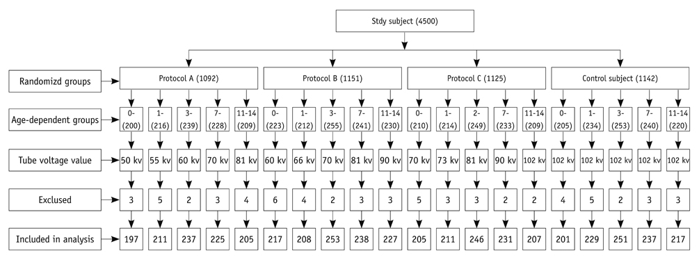
Forty-five hundred children aged 0-14 years (yr) were randomly divided into four groups according to the tube voltage protocols for chest DR: lower kilovoltage potential (kVp) (A), intermediate kVp (B), and higher kVp (C) groups, and the fixed high kVp group (controls). The results were analyzed among five different age groups (0-1 yr, 1-3 yr, 3-7 yr, 7-11 yr and 11-14 yr). The dose area product (DAP) and visual grading analysis score (VGAS) were determined and compared by using one-way analysis of variance.
RESULTS
The mean DAP of protocol C was significantly lower as compared with protocols A, B and controls (p < 0.05). DAP was higher in protocol A than the controls (p <0.001), but it was not statistically significantly different between B and the controls (p = 0.976). Mean VGAS was lower in the controls than all three protocols (p < 0.001 for all). Mean VGAS did not differ between protocols A and B (p = 0.334), but was lower in protocol C than A (p = 0.008) and B (p = 0.049).
CONCLUSION
Protocol C (higher kVp) may help optimize the trade-off between radiation dose and image quality, and it may be acceptable for use in a pediatric age group from these results.
MeSH Terms
Figure
Reference
-
1. Brenner D, Elliston C, Hall E, Berdon W. Estimated risks of radiation-induced fatal cancer from pediatric CT. AJR Am J Roentgenol. 2001. 176:289–296.2. Vano E. ICRP recommendations on 'Managing patient dose in digital radiology'. Radiat Prot Dosimetry. 2005. 114:126–130.3. Busch HP, Faulkner K. Image quality and dose management in digital radiography: a new paradigm for optimisation. Radiat Prot Dosimetry. 2005. 117:143–147.4. Honey ID, Mackenzie A, Evans DS. Investigation of optimum energies for chest imaging using film-screen and computed radiography. Br J Radiol. 2005. 78:422–427.5. Tingberg A, Sjöström D. Optimisation of image plate radiography with respect to tube voltage. Radiat Prot Dosimetry. 2005. 114:286–293.6. Ullman G, Sandborg M, Dance DR, Hunt RA, Alm Carlsson G. Towards optimization in digital chest radiography using Monte Carlo modelling. Phys Med Biol. 2006. 51:2729–2743.7. Uffmann M, Neitzel U, Prokop M, Kabalan N, Weber M, Herold CJ, et al. Flat-panel-detector chest radiography: effect of tube voltage on image quality. Radiology. 2005. 235:642–650.8. Rong XJ, Shaw CC, Liu X, Lemacks MR, Thompson SK. Comparison of an amorphous silicon/cesium iodide flat-panel digital chest radiography system with screen/film and computed radiography systems--a contrast-detail phantom study. Med Phys. 2001. 28:2328–2335.9. Willis CE. Optimizing digital radiography of children. Eur J Radiol. 2009. 72:266–273.10. Sæther HK, Lagesen B, Trægde Martinsen AC, Holsen EP, Øvrebø KM. Dose levels from thoracic and pelvic examinations in two pediatric radiological departments in Norway - a comparison study of dose-area product and radiographic technique. Acta Radiol. 2010. 51:1137–1142.11. Olgar T, Onal E, Bor D, Okumus N, Atalay Y, Turkyilmaz C, et al. Radiation exposure to premature infants in a neonatal intensive care unit in Turkey. Korean J Radiol. 2008. 9:416–419.12. Samei E, Hill JG, Frey GD, Southgate WM, Mah E, Delong D. Evaluation of a flat panel digital radiographic system for low-dose portable imaging of neonates. Med Phys. 2003. 30:601–607.13. Strotzer M, Völk M, Fründ R, Hamer O, Zorger N, Feuerbach S. Routine chest radiography using a flat-panel detector: image quality at standard detector dose and 33% dose reduction. AJR Am J Roentgenol. 2002. 178:169–171.14. Völk M, Hamer OW, Feuerbach S, Strotzer M. Dose reduction in skeletal and chest radiography using a large-area flat-panel detector based on amorphous silicon and thallium-doped cesium iodide: technical background, basic image quality parameters, and review of the literature. Eur Radiol. 2004. 14:827–834.15. European guidelines on quality criteria for diagnostic radiographic images in pediatrics. 1996. Luxembourg, EC: EUR 16261 report;Available at: http://www.e-radiography.net/regsetc/European_guide_children_extract.pdf.16. Yakoumakis EN, Tsalafoutas IA, Aliberti M, Pantos GI, Yakoumakis NE, Karaiskos P, et al. Radiation doses in common X-ray examinations carried out in two dedicated paediatric hospitals. Radiat Prot Dosimetry. 2007. 124:348–352.17. Dougeni ED, Delis HB, Karatza AA, Kalogeropoulou CP, Skiadopoulos SG, Mantagos SP, et al. Dose and image quality optimization in neonatal radiography. Br J Radiol. 2007. 80:807–815.18. Båth M, Månsson LG. Visual grading characteristics (VGC) analysis: a non-parametric rank-invariant statistical method for image quality evaluation. Br J Radiol. 2007. 80:169–176.19. Nickoloff EL, Lu ZF, Dutta AK, So JC. Radiation dose descriptors: BERT, COD, DAP, and other strange creatures. Radiographics. 2008. 28:1439–1450.20. Doherty P, O'Leary D, Brennan PC. Do CEC guidelines under-utilise the full potential of increasing kVp as a dose-reducing tool? Eur Radiol. 2003. 13:1992–1999.21. Geijer H, Norrman E, Persliden J. Optimizing the tube potential for lumbar spine radiography with a flat-panel digital detector. Br J Radiol. 2009. 82:62–68.22. Sandborg M, Tingberg A, Ullman G, Dance DR, Alm Carlsson G. Comparison of clinical and physical measures of image quality in chest and pelvis computed radiography at different tube voltages. Med Phys. 2006. 33:4169–4175.23. Geijer H, Persliden J. Varied tube potential with constant effective dose at lumbar spine radiography using a flat-panel digital detector. Radiat Prot Dosimetry. 2005. 114:240–245.24. Fink C, Hallscheidt PJ, Noeldge G, Kampschulte A, Radeleff B, Hosch WP, et al. Clinical comparative study with a large-area amorphous silicon flat-panel detector: image quality and visibility of anatomic structures on chest radiography. AJR Am J Roentgenol. 2002. 178:481–486.25. Bacher K, Smeets P, Bonnarens K, De Hauwere A, Verstraete K, Thierens H. Dose reduction in patients undergoing chest imaging: digital amorphous silicon flat-panel detector radiography versus conventional film-screen radiography and phosphor-based computed radiography. AJR Am J Roentgenol. 2003. 181:923–929.26. Moore CS, Beavis AW, Saunderson JR. Investigation of optimum X-ray beam tube voltage and filtration for chest radiography with a computed radiography system. Br J Radiol. 2008. 81:771–777.27. Billinger J, Nowotny R, Homolka P. Diagnostic reference levels in pediatric radiology in Austria. Eur Radiol. 2010. 20:1572–1579.
- Full Text Links
- Actions
-
Cited
- CITED
-
- Close
- Share
- Similar articles
-
- A Comparative Study of Image Quality and Radiation Dose with Changes in Tube Voltage and Current for a Digital Chest Radiography
- Digital Tomosynthesis of the Chest: Comparison of Patient Exposure Dose and Image Quality between Standard Default Setting and Low Dose Setting
- Relationship of Image Quality and Radiation Dose for Chest Radiography in the Medical Institutions for Pneumoconiosis: A Comparison to the Korean Diagnostic Reference Level
- Effect of the amount of battery charge on tube voltage in different hand-held dental x-ray systems
- National Survey of Radiation Doses of Pediatric Chest Radiography in Korea: Analysis of the Factors Affecting Radiation Doses


