Korean J Radiol.
2012 Oct;13(5):610-617. 10.3348/kjr.2012.13.5.610.
National Survey of Radiation Doses of Pediatric Chest Radiography in Korea: Analysis of the Factors Affecting Radiation Doses
- Affiliations
-
- 1Department of Radiology, University of Ulsan College of Medicine, Asan Medical Center, Seoul 138-736, Korea. dokh@amc.seoul.kr
- 2National Institute of Food and Drug Safety Evaluation, Radiation Safety Division, Cheongwon 363-700, Korea.
- KMID: 1392940
- DOI: http://doi.org/10.3348/kjr.2012.13.5.610
Abstract
OBJECTIVE
To investigate radiation doses in pediatric chest radiography in a national survey and to analyze the factors that affect radiation doses.
MATERIALS AND METHODS
The study was based on the results of 149 chest radiography machines in 135 hospitals nationwide. For each machine, a chest radiograph was obtained by using a phantom representing a 5-year-old child (ATOM(R) dosimetry phantom, model 705-D, CIRS, Norfolk, VA, USA) with each hospital's own protocol. Five glass dosimeters (M-GD352M, Asahi Techno Glass Corporation, Shizuoka, Japan) were horizontally installed at the center of the phantom to measure the dose. Other factors including machine's radiography system, presence of dedicated pediatric radiography machine, presence of an attending pediatric radiologist, and the use of automatic exposure control (AEC) were also evaluated.
RESULTS
The average protocol for pediatric chest radiography examination in Korea was 94.9 peak kilovoltage and 4.30 milliampere second. The mean entrance surface dose (ESD) during a single examination was 140.4 microgray (microGy). The third quartile, median, minimum and maximum value of ESD were 160.8 microGy, 93.4 microGy, 18.8 microGy, and 2334.6 microGy, respectively. There was no significant dose difference between digital and non-digital radiography systems. The use of AEC significantly reduced radiation doses of pediatric chest radiographs (p < 0.001).
CONCLUSION
Our nationwide survey shows that the third quartile, median, and mean ESD for pediatric chest radiograph is 160.8 microGy, 93.4 microGy, and 140.4 microGy, respectively. No significant dose difference is noticed between digital and non-digital radiography systems, and the use of AEC helps significantly reduce radiation doses.
MeSH Terms
Figure
-

Fig. 1 Dose distribution. A. Distribution of radiation dose and number of machines allocated in each range of dose. B. Radiation dose distribution of machines. Yellow line indicates median value of overall distribution and red line indicates third quartile.
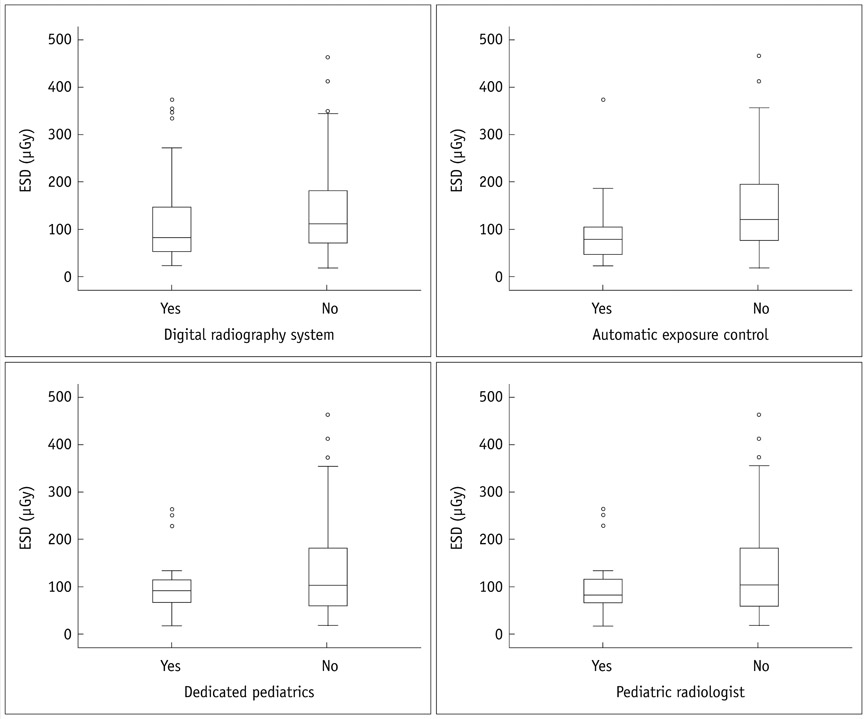
Fig. 2 Boxplots comparing radiography system, AEC, dedicated pediatric radiography machines and pediatric radiologists for ESD (µGy). ESD = entrance surface dose, AEC = automatic exposure control
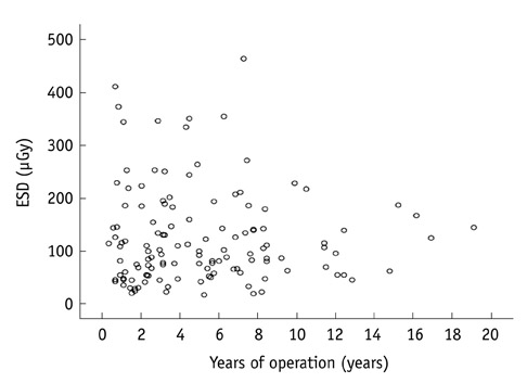
Fig. 3 Scattergram of machine's years of operation and ESD (*r = -0.24, p = 0.783). Pearson correlation coefficient. ESD = entrance surface dose
Reference
-
1. Doody MM, Lonstein JE, Stovall M, Hacker DG, Luckyanov N, Land CE. Breast cancer mortality after diagnostic radiography: findings from the U.S. Scoliosis Cohort Study. Spine (Phila Pa 1976). 2000. 25:2052–2063.2. Tanimura M, Matsui I, Abe J, Ikeda H, Kobayashi N, Ohira M, et al. Increased risk of hepatoblastoma among immature children with a lower birth weight. Cancer Res. 1998. 58:3032–3035.3. Shu XO, Potter JD, Linet MS, Severson RK, Han D, Kersey JH, et al. Diagnostic X-rays and ultrasound exposure and risk of childhood acute lymphoblastic leukemia by immunophenotype. Cancer Epidemiol Biomarkers Prev. 2002. 11:177–185.4. Lee YJ, Chung YE, Lim JS, Kim JH, Kim YJ, Lee HJ, et al. Cumulative radiation exposure during follow-up after curative surgery for gastric cancer. Korean J Radiol. 2012. 13:144–151.5. Goo HW. CT radiation dose optimization and estimation: an update for radiologists. Korean J Radiol. 2012. 13:1–11.6. Goo HW. Pediatric CT: understanding of radiation dose and optimization of imaging techniques. J Korean Radiol Soc. 2005. 52:1–5.7. Yang DH, Goo HW. Pediatric 16-slice CT protocols: radiation dose and image quality. J Korean Radiol Soc. 2008. 59:333–347.8. Brenner DJ. Estimating cancer risks from pediatric CT: going from the qualitative to the quantitative. Pediatr Radiol. 2002. 32:228–231. discussion 242-244.9. Hart D, Wall BF, Shrimpton PC, Bungay DR, Dance CR. Reference doses and patient size in pediatric radiology. 2003. Chilton: National Radiological Protection Board Report 2000 R318, NRPB.10. EC Study Group. European guidelines on quality criteria for diagnostic radiographic images in pediatrics. 1996. Luxemburg: 1996 EUR 16261.11. United Nations Scientific Committee on the Effects of Atomic Radiation (UNSCEAR). Sources and Effects of Ionizing Radiation. 2000. New York: 2000 United Nations.12. Jung YY, Goo HW. The optimal parameter for radiation dose in pediatric low dose abdominal CT: cross-sectional dimensions versus body weight. J Korean Radiol Soc. 2008. 58:169–175.13. Don S. Radiosensitivity of children: potential for overexposure in CR and DR and magnitude of doses in ordinary radiographic examinations. Pediatr Radiol. 2004. 34:Suppl 3. S167–S172. discussion S234-S241.14. Huda W. Assessment of the problem: pediatric doses in screen-film and digital radiography. Pediatr Radiol. 2004. 34:Suppl 3. S173–S182. discussion S234-S241.15. Recommendations of the International Commission on Radiological Protection. ICRP Publication 60. Annals of the ICRP 21, No. 1-3. 1991. Oxford: Pergamon Press.16. Radiological protection and safety in medicine. ICRP Publication 73. Annals of the ICRP 26, No. 2. 1996. Oxford: Pergamon Press.17. Schaefer-Prokop C, Neitzel U, Venema HW, Uffmann M, Prokop M. Digital chest radiography: an update on modern technology, dose containment and control of image quality. Eur Radiol. 2008. 18:1818–1830.18. Uffmann M, Schaefer-Prokop C. Digital radiography: the balance between image quality and required radiation dose. Eur J Radiol. 2009. 72:202–208.19. Japanese Association of Radiological Technologists. "Guideline 2006". Available at: http://www.jart.jp/guideline/index.html.
- Full Text Links
-
- Actions
-
Cited
- CITED
-
- Close
- Share
-
- Similar articles
-
- The survey of the surface doses of the dental x-ray machines
- Scattered Radiation Doses to the Patients and Medical Practitioneer from Extracorporeal Shock Wave Lithotripsy
- Radiation Exposure to Premature Infants in a Neonatal Intensive Care Unit in Turkey
- Reference dose levels for dental periapical radiography in Chonnam Province
- Radiation absorbed doses of cone beam computed tomography
