Lab Med Online.
2018 Apr;8(2):56-61. 10.3343/lmo.2018.8.2.56.
Anaplastic Large Cell Lymphoma with Massive Eosinophilia and Complex Karyotype Initially Misdiagnosed as Chronic Eosinophilic Leukemia
- Affiliations
-
- 1Department of Laboratory Medicine, College of Medicine, Ewha Womans University, Seoul, Korea. JungWonH@ewha.ac.kr
- 2Department of Pathology, College of Medicine, Ewha Womans University, Seoul, Korea.
- 3Department of Internal Medicine, College of Medicine, Ewha Womans University, Seoul, Korea.
- KMID: 2407563
- DOI: http://doi.org/10.3343/lmo.2018.8.2.56
Abstract
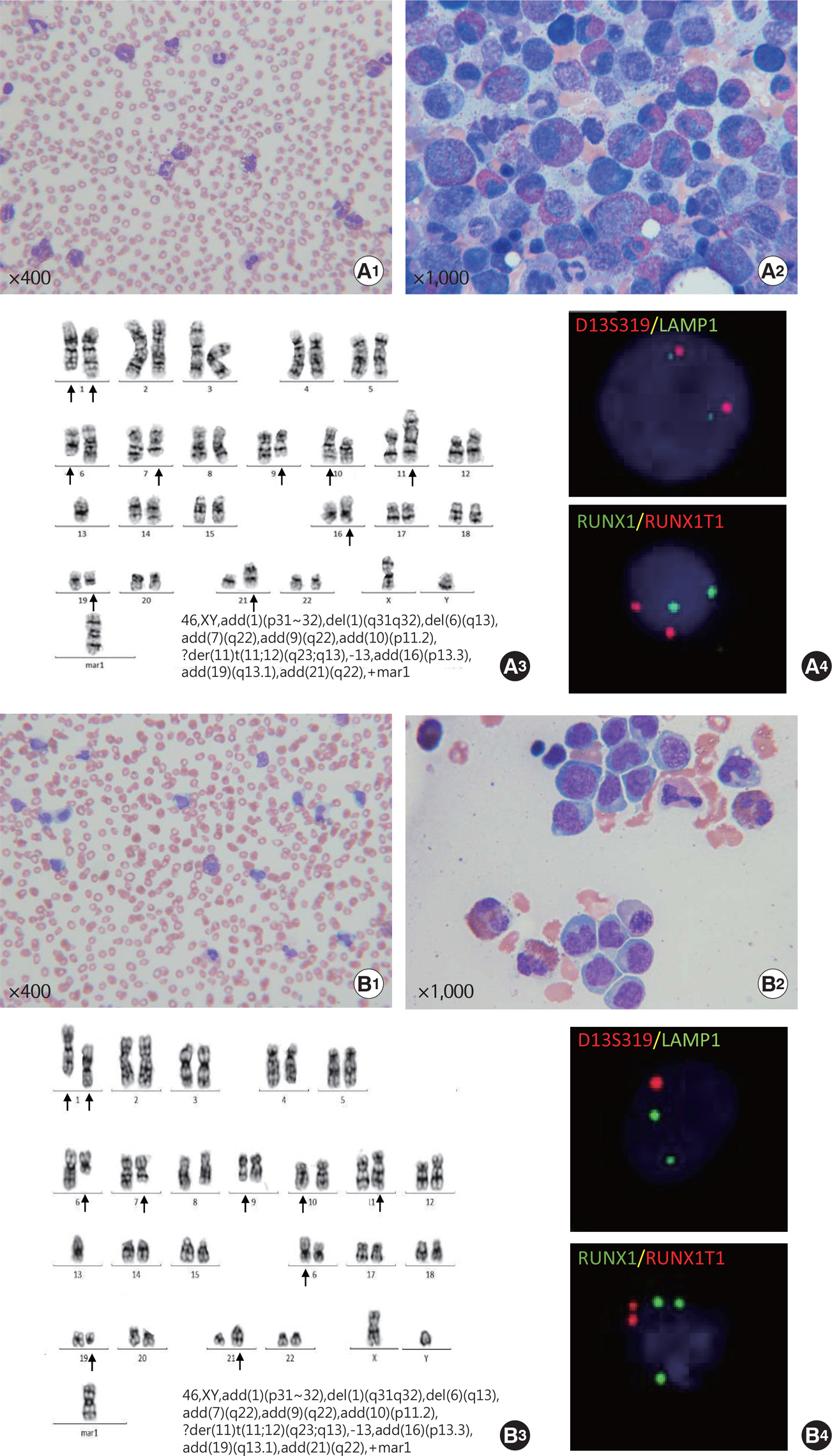
- We report a patient with massive eosinophilia and a complex karyotype that was initially misdiagnosed as chronic eosinophilic leukemia (CEL), but later diagnosed as anaplastic large cell lymphoma (ALCL) masked by massive eosinophilia. The complex karyotype observed at initial diagnosis remained unchanged later, after the evidence of bone marrow involvement of ALCL was obtained. At diagnosis, genetic aberrations corresponding to metaphase cytogenetics were not identified by interphase fluorescence in situ hybridization, although abnormal results were noted at follow-up. Together, these observations indicate that the complex karyotype at initial work-up has been derived from a low proportion of lymphoma cells with high mitotic ability that were not identified by microscopy, rather than from massive eosinophils. These findings suggest that our patient had ALCL with secondary eosinophilia rather than CEL since initial diagnosis.
Keyword
MeSH Terms
Figure
Reference
-
1.Gotlib J. World Health Organization-defned eosinophilic disorders: 2015 update on diagnosis, risk stratifcation, and management. Am J Hematol. 2015. 90:1077–89.2.Ogata M., Ogata Y., Kohno K., Uno N., Ohno E., Ohtsuka E, et al. Eosinophilia associated with adult T-cell leukemia: role of interleukin 5 and granulocyte-macrophage colony-stimulating factor. Am J Hematol. 1998. 59:242–5.
Article3.Montgomery ND., Dunphy CH., Mooberry M., Laramore A., Foster MC., Park SI, et al. Diagnostic complexities of eosinophilia. Arch Pathol Lab Med. 2013. 137:259–69.
Article4.Bain BJ. Hypereosinophilia. Curr Opin Hematol. 2000. 7:21–5.
Article5.Desenne JJ., Acquatella G., Stern R., Muller A., Sanchez M., Somoza R. Blood eosinophilia in Hodgkin's disease. A follow-up of 25 cases in Venezuela. Cancer. 1992. 69:1248–53.
Article6.Utsunomiya A., Ishida T., Inagaki A., Ishii T., Yano H., Komastsu H, et al. Clinical signifcance of a blood eosinophilia in adult T-cell leukemia/lymphoma: a blood eosinophilia is a signifcant unfavorable prognostic factor. Leuk Res. 2007. 31:915–20.7.McKelvie PA., Oon S., Romas E., Nandurkar H., Tam CS. A case of systemic anaplastic lymphoma kinase-negative anaplastic large cell lymphoma associated with hypereosinophilia, granulomatous myositis and vasculitis. Leuk Lymphoma. 2012. 53:2279–82.
Article8.Orofno N., Guidotti F., Cattaneo D., Sciume M., Gianelli U., Cortelezzi A, et al. Marked eosinophilia as initial presentation of breast implant-associated anaplastic large cell lymphoma. Leuk Lymphoma. 2016. 57:2712–5.9.Powers ML., Watson BW., Frater JL., Kreisel F., Hassan A. Peripheral eosinophilia camoufaging anaplastic large cell lymphoma. Int J Surg Pathol. 2011. 19:405–8.10.Gonsalves WI., He R., Pardanani A., Gupta V., Smeltzer JP., Hanson CA, et al. Chronic eosinophilic leukemia-not otherwise specifed (NOS) in the background of a large cell lymphoma. Case Rep Hematol. 2013. 2013:458303.11.Fan YS., Rizkalla K. Comprehensive cytogenetic analysis including multicolor spectral karyotyping and interphase fuorescence in situ hybridization in lymphoma diagnosis. a summary of 154 cases. Cancer Genet Cytogenet. 2003. 143:73–9.12.Wan TS. Cancer cytogenetics: methodology revisited. Ann Lab Med. 2014. 34:413–25.
Article13.Mereu E., Pellegrino E., Scarfo I., Inghirami G., Piva R. The heterogeneous landscape of ALK negative ALCL. Oncotarget. 2017. 8:18525–36.
Article14.Mussolin L., Pillon M., Bonato P., Leszl A., Franceschetto G., Di Meglio A, et al. Cytogenetic analysis of pediatric anaplastic large cell lymphoma. Pediatr Blood Cancer. 2010. 55:446–51.
Article15.Zeng Y., Feldman AL. Genetics of anaplastic large cell lymphoma. Leuk Lymphoma. 2016. 57:21–7.
Article
- Full Text Links
- Actions
-
Cited
- CITED
-
- Close
- Share
- Similar articles
-
- Anaplastic Large Cell Lymphoma with Massive Eosinophilia and Complex Karyotype Initially Misdiagnosed as Chronic Eosinophilic Leukemia
- Dermatofibroma in Patient with Relapsing Primary Cutaneous Anaplastic Large Cell Lymphoma
- Anaplastic large cell lymphoma with marked peripheral eosinophilia misdiagnosed as Kimura disease
- Anaplastic Large Cell Lymphoma Mimicking a Muscle Abscess: A Case Report
- CD30-Positive Anaplastic Lymphoma Kinase-Negative Systemic Anaplastic Large-Cell Lymphoma in a 9-Year-Old Boy


