Diffusion-Weighted MR Imaging of Unusual White Matter Lesion in a Patient with Menkes Disease
- Affiliations
-
- 1Department of Rehabilitation Medicine, Gyeongsang National University College of Medicine, Jinju, Korea. ryoojw@gsnu.ac.kr
- 2Department of Radiology, Gyeongsang National University College of Medicine, Jinju, Korea.
- 3The Institute of Health Science, Gyeongsang National University College of Medicine, Jinju, Korea.
- KMID: 753871
- DOI: http://doi.org/10.3348/kjr.2007.8.1.82
Abstract
- We report here on the diffusion-weighted imaging of unusual white matter lesions in a case of Menkes disease. On the initial MR imaging, the white matter lesions were localized in the deep periventricular white matter in the absence of diffuse cortical atrophy. The lesion showed diffuse high signal on the diffusion-weighted images and diffuse progression and persistent hyperintensity on the follow up imaging. Our case suggests that the white matter lesion may precede diffuse cortical atrophy in a patient with Menkes disease.
MeSH Terms
Figure
-
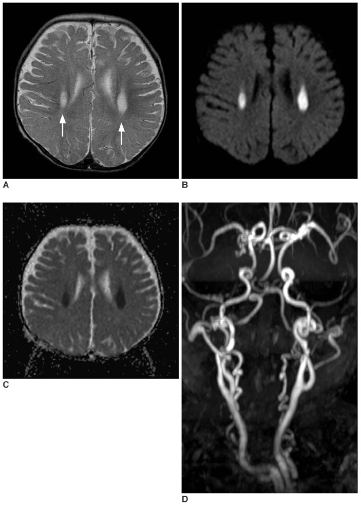
Fig. 1 Initial MR images obtained at age of 10 months. A. T2-wieghted axial image (TR/TE = 4700/104 msec, 5 mm thickness) demonstrates symmetric hyperintense lesion in both deep periventricular white matters (arrows). B. Diffusion weighted image (TR/TE = 3500/92, b maximum = 1000 s/mm2, three directions, 5 mm thickness) shows the bright signal intensity of the lesion. C. On the apparent diffusion coefficient map, the lesion shows diffuse low signal intensities, suggesting restricted diffusion. D. Three dimensional MR angiography shows markedly tortuous intracranial and extracranial vessels, which are characteristics of Menkes disease.
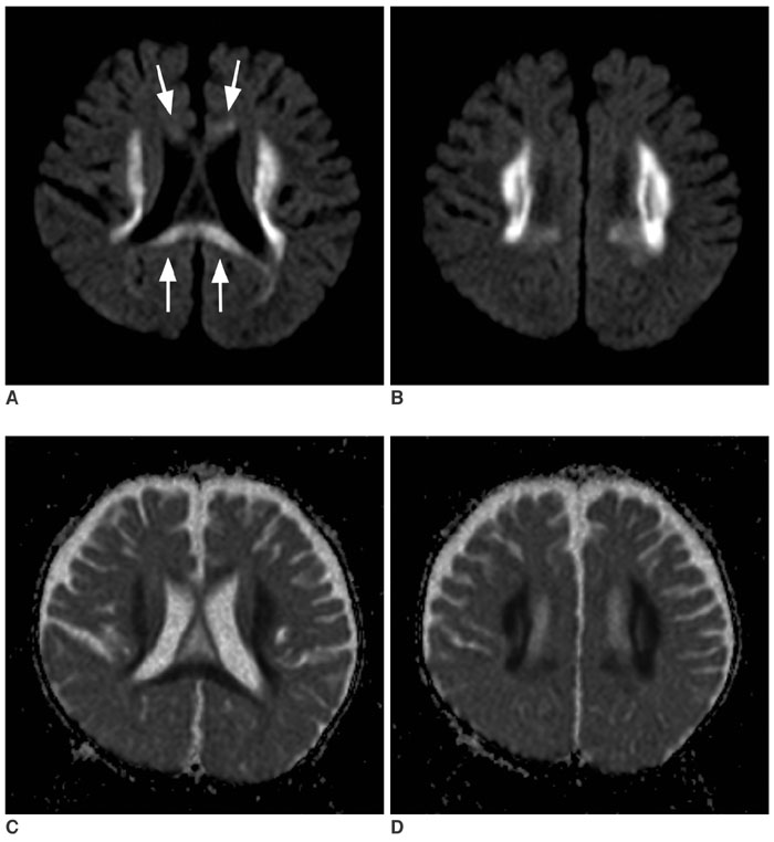
Fig. 2 Follow up diffusion study obtained at the age of 13 months. A, B. Consecutive diffusion-weighted images (TR/TE = 3500/95, b maximum = 1000 s/mm2) demonstrate marked extension of the white matter lesions to the adjacent white matter. The lesion also involves the corpus callosum (arrows). Note there is no definite progression of cortical atrophy. C, D. Apparent diffusion coefficient maps of the lesion show diffusely decreased apparent diffusion coefficient, similar to that of the initial lesions (Fig. 1C).
Reference
-
1. Menkes JH, Alter M, Steigleder GK, Weakley DR, Sung JH. A sex-linked recessive disorder with retardation of growth, peculiar hair, and focal cerebral and cerebellar degeneration. Pediatrics. 1962. 29:764–779.2. Kaler SG. Menkes disease. Adv Pediatr. 1994. 41:263–304.3. Faerber EN, Grover WD, DeFilipp GJ, Capitanio MA, Liu TH, Swartz JD. Cerebral MR of Menkes kinky-hair disease. AJNR Am J Neuroradiol. 1989. 10:190–192.4. Leventer RJ, Kornberg AJ, Phelan EM, Kean MJ. Early magnetic resonance imaging findings in Menkes' disease. J Child Neurol. 1997. 12:222–224.5. Takahashi S, Ishii K, Matsumoto K, Higano S, Ishibashi T, Zuguchi M, et al. Cranial MRI and MR angiography in Menkes' syndrome. Neuroradiology. 1993. 35:556–558.6. Kim OH, Suh JH. Intracranial and extracranial MR angiography in Menkes disease. Pediatr Radiol. 1997. 27:782–784.7. Hsich GE, Robertson RL, Irons M, Soul JS, du Plessis AJ. Cerebral infarction in Menkes' disease. Pediatr Neurol. 2000. 23:425–428.8. Blaser SI, Berns DH, Ross JS, Lanska MJ, Weissman BM. Serial MR studies in Menkes disease. J Comput Assist Tomogr. 1989. 13:113–115.9. Ozawa H, Kodama H, Murata Y, Takashima S, Noma S. Transient temporal lobe changes and a novel mutation in a patient with Menkes disease. Pediatr Int. 2001. 43:437–440.10. Valanne L, Ketonen L, Majander A, Suomalainen A, Pihko H. Neuroradiologic findings in children with mitochondrial disorders. AJNR Am J Neuroradiol. 1998. 19:369–377.
- Full Text Links
-
- Actions
-
Cited
- CITED
-
- Close
- Share
-
- Similar articles
-
- Acute Marchiafava-Bignami Disease With Typical White Matter Involvement on Diffusion Weighted MRI
- Rapid Regression of White Matter Changes in Hypoglycemic Encephalopathy
- Diffusion Weighted MRI Findings in Two Patients with Hypoxic Brain Damage
- Uremic Encephalopathy with Atypical Magnetic Resonance Features on Diffusion-Weighted Images
- The Analysis of Pathogenesis in the Hypertensive Encephalopathy using Diffusion-Weighted MR Imaging
