LncRNA PART1 Attenuates Myocardial Ischemia-Reperfusion Injury by Regulating TFAP2C/DUSP5 Axis via miR-302a-3p
- Affiliations
-
- 1Medical Care Center, Hainan General Hospital, Hainan Affiliated Hospital of Hainan Medical University, Haikou, China
- 2Department of Otorhinolaryngology Head and Neck Surgery, Hainan General Hospital, Hainan Affiliated Hospital of Hainan Medical University, Haikou, China
- 3Hainan Medical University, Haikou, China
- KMID: 2556520
- DOI: http://doi.org/10.4070/kcj.2023.0131
Abstract
- Background and Objectives
Myocardial ischemia-reperfusion injury (MIRI) refers to the damage of cardiac function caused by restoration of blood flow perfusion in ischemic myocardium. However, long non-coding RNA prostate androgen regulated transcript 1 (PART1)’s role in MIRI remain unclear.
Methods
Immunofluorescence detected LC3 expression. Intermolecular relationships were verified by dual luciferase reporter assay. 3-(4,5-dimethylthiazol-2-yl)-2,5-diphenyltetrazolium bromide, flow cytometry and transferase-mediated dUTP nick-end labeling (TUNEL) assays analyzed cell viability and apoptosis. The release of lactate dehydrogenase was tested via enzyme-linked immunosorbent assay (ELISA). Left anterior descending coronary artery surgery induced a MIRI mouse model. Infarct area was detected by 2,3,5-triphenyltetrazolium chloride staining. Hematoxylin and eosin staining examined myocardial injury. ELISA evaluated myocardial marker (creatine kinase MB) level.
Results
PART1 was decreased in hypoxia/reoxygenation (H/R) induced AC16 cells and MIRI mice. PART1 upregulation attenuated the increased levels of Bax, beclin-1 and the ratio of LC3II/I, and enhanced the decrease of Bcl-2 and p62 expression in H/R-treated cells. PART1 upregulation alleviated H/R-triggered autophagy and apoptosis via miR-302a-3p. Mechanically, PART1 targeted miR-302a-3p to upregulate transcription factor activating enhancer-binding protein 2C (TFAP2C). TFAP2C silencing reversed the protected effects of miR-302a-3p inhibitor on H/R treated AC16 cells. We further established TFAP2C combined to dual-specificity phosphatase 5 (DUSP5) promoter and activated DUSP5. TFAP2C upregulation suppressed H/R-stimulated autophagy and apoptosis through upregulating DUSP5. Overexpressed PART1 reduced myocardial infarction area and attenuated MIRI in mice.
Conclusion
PART1 improved the autophagy and apoptosis in H/R-exposed AC16 cells through miR-302a-3p/TFAP2C/DUSP5 axis, which might provide novel targets for MIRI treatment.
Keyword
Figure
-
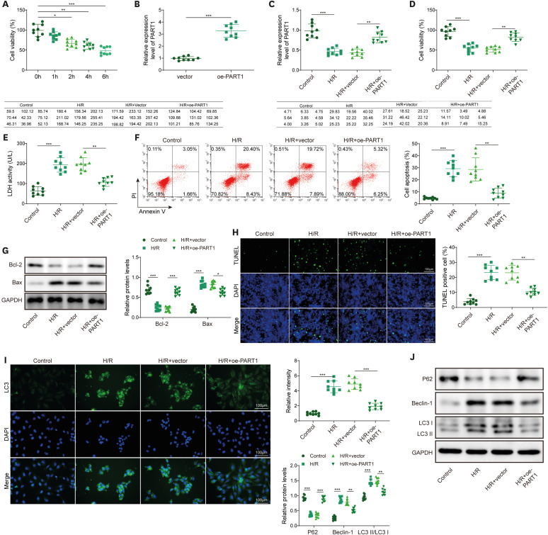
Figure 1 Overexpression of PART1 inhibited autophagy and apoptosis in H/R-challenged cardiomyocytes. (A) MTT assay detected cell viability at different hypoxia time points. (B) PART1 transfection efficiency was verified by RT-qPCR. (C) RT-qPCR detection of PART1 level after overexpression of PART1 in H/R-treated AC16 cells. (D) MTT tested cell viability. (E) ELISA tested LDH concentration. (F) Cells apoptosis was detected utilizing Flow cytometry. (G) TUNEL staining detected apoptosis. (H) Bcl-2 and Bax expressions were measured using Western blot. (I) Immunofluorescence detection of LC3 expression in AC16 cells. (J) Western blot detected p62, LC3II/I, Beclin-1 levels. Data were shown as mean ± SD based on three independent experiments (n=3).ELISA = enzyme-linked immunosorbent assay; H/R = hypoxia/reoxygenation; LDH = lactate dehydrogenase; MTT = 3-(4,5-dimethylthiazol-2-yl)-2,5-diphenyltetrazolium bromide; oe- = overexpression plasmids; PART1 = prostate androgen regulated transcript 1; RT-qPCR = quantitative real-time polymerase chain reaction; SD = standard deviation; TUNEL = transferase-mediated dUTP nick-end labeling.*p<0.05, **p<0.01, ***p<0.001.

Figure 2 PART1 targeted miR-302a-3p and upregulated TFAP2C level. (A) StarBase predicted binding site of miR-302a-3p with PART1 or TFAP2C. (B) Luciferase activity of PART1-WT/MUT (left panel) or TFAP2C-WT/MUT (right panel) in AC16 cells treated with miR-302a-3p mimics or mimics NC. (C, D) RT-qPCR assessed miR-302a-3p (C) and TFAP2C (D) levels after overexpression of PART1 in AC16 cells. (E) Western blot analyzed TFAP2C protein level. (F, G) RT-qPCR detected miR-302a-3p (F) and TFAP2C (G) after miR-302a-3p overexpression or inhibition in AC16 cells. (H) Western blot tested the protein level of TFAP2C. Data were shown as mean ± SD based on three independent experiments (n=3).MUT = mutant type; NC = negative control; PART1 = prostate androgen regulated transcript 1; RT-qPCR = quantitative real-time polymerase chain reaction; SD = standard deviation; TFAP2C = transcription factor activating enhancer-binding protein 2C; WT = wild type.*p<0.05, **p<0.01, ***p<0.001.
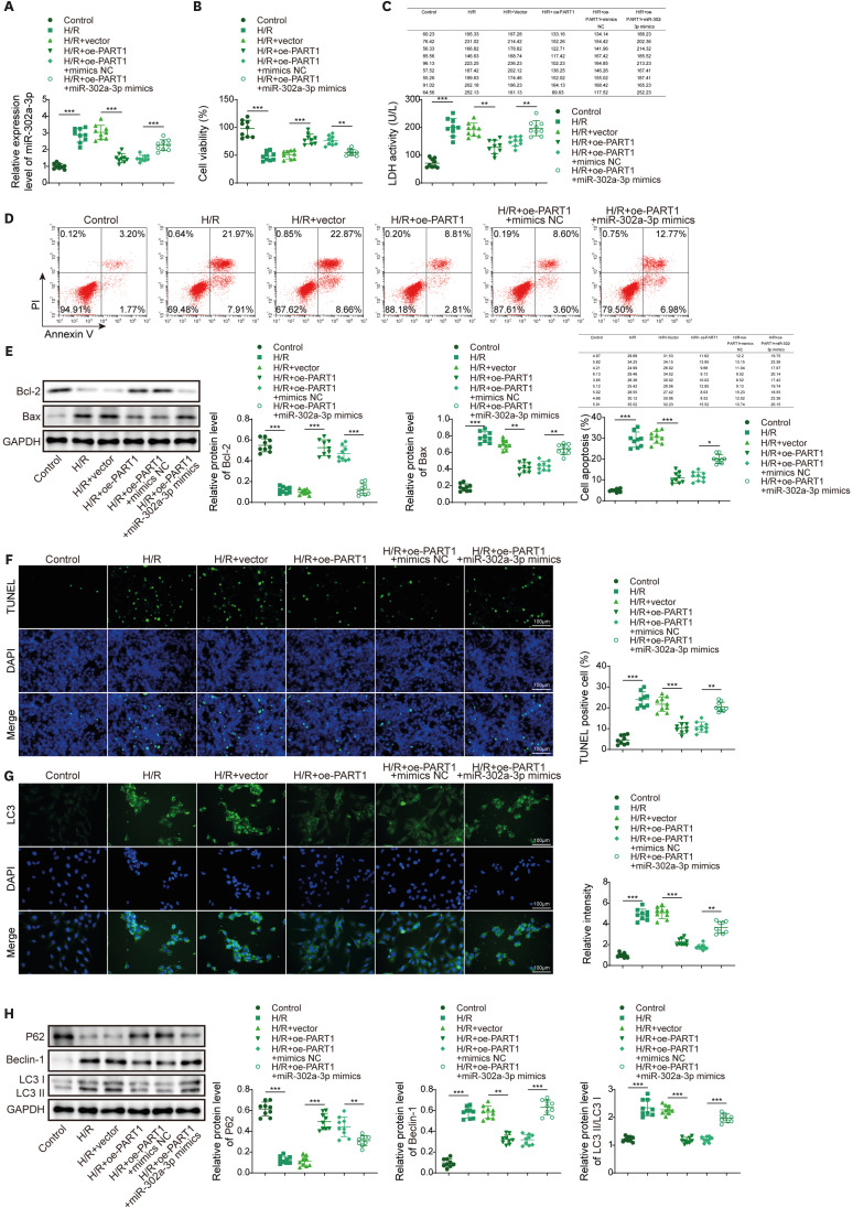
Figure 3 PART1 participated in H/R-mediated cardiomyocyte function injury via regulating miR-302a-3p. (A) RT-qPCR determined miR-302a-3p level. (B) MTT tested cell viability. (C) ELISA tested LDH concentration. (D) Flow cytometry analyzed cell apoptosis. (E) TUNEL staining detected apoptosis. (F) Western blot tested Bcl-2 and Bax levels. (G) Immunofluorescence measured LC3 expression in AC16 cells. (H) Western blot detected the protein levels of p62, LC3II/I, Beclin-1. Data were shown as mean ± SD based on three independent experiments (n=3).ELISA = enzyme-linked immunosorbent assay; LDH = lactate dehydrogenase; MTT = 3-(4,5-dimethylthiazol-2-yl)-2,5-diphenyltetrazolium bromide; PART1 = prostate androgen regulated transcript 1; RT-qPCR = quantitative real-time polymerase chain reaction; SD = standard deviation; TUNEL = transferase-mediated dUTP nick-end labeling.*p<0.05, **p<0.01, ***p<0.001.
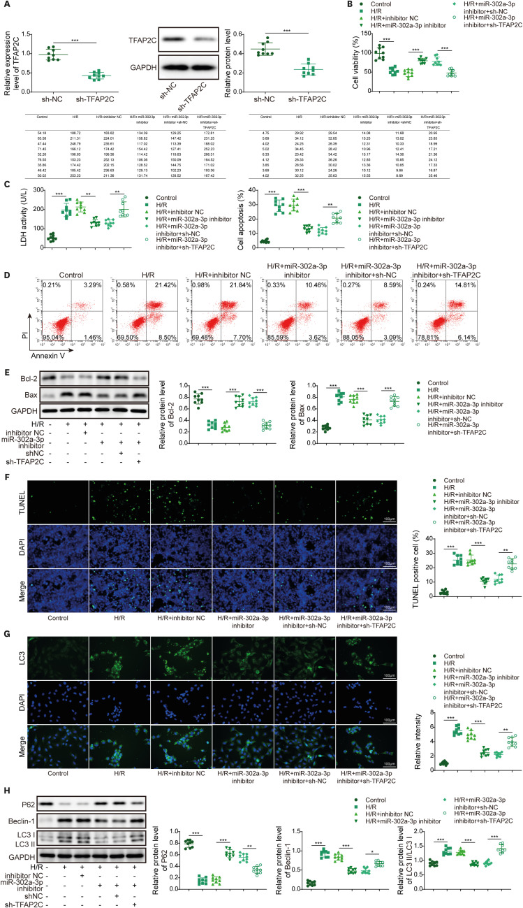
Figure 4 MiR-302a-3p inhibitor attenuated H/R-triggered cardiomyocyte autophagy and apoptosis through TFAP2C. (A) TFAP2C transfection efficiency was verified by RT-qPCR and Western blot. (B) MTT tested cell viability. (C) ELISA tested LDH concentration. (D) Flow cytometry determined apoptosis. (E) TUNEL staining detected apoptosis. (F) Western blot analyzed Bcl-2 and Bax expression levels. (G) Immunofluorescence detected LC3 expression in AC16 cells. (H) Western blot measured p62, LC3II/I, Beclin-1 levels. Data were shown as mean ± SD based on three independent experiments (n=3).ELISA = enzyme-linked immunosorbent assay; H/R = hypoxia/reoxygenation; LDH = lactate dehydrogenase; MTT = 3-(4,5-dimethylthiazol-2-yl)-2,5-diphenyltetrazolium bromide; RT-qPCR = quantitative real-time polymerase chain reaction; SD = standard deviation; TFAP2C = transcription factor activating enhancer-binding protein 2C; TUNEL = transferase-mediated dUTP nick-end labeling.*p<0.05, **p<0.01, ***p<0.001.
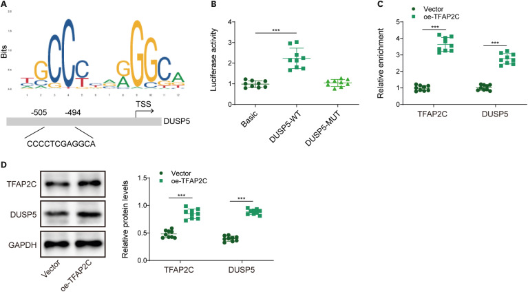
Figure 5 TFAP2C bound to DUSP5 promoter and upregulated DUSP5. (A) Binding site between TFAP2C and DUSP5 by JASPAR. (B) Dual luciferase reporter assay verified the binding of TFAP2C to DUSP5 promoter. (C, D) RT-qPCR and Western blot detected the expression of TFAP2C and DUSP5 after overexpression of TFAP2C. Data were shown as mean ± SD based on three independent experiments (n=3).DUSP5 = dual-specificity phosphatase 5; RT-qPCR = quantitative real-time polymerase chain reaction; SD = standard deviation; TFAP2C = transcription factor activating enhancer-binding protein 2C.*p<0.05, **p<0.01, ***p<0.001.
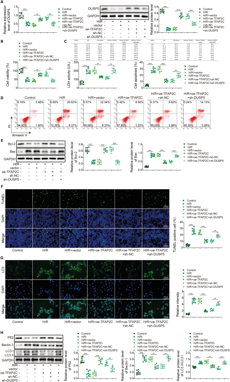
Figure 6 TFAP2C upregulation suppressed H/R-challenged cardiomyocyte autophagy and apoptosis through regulating DUSP5. (A) RT-qPCR and Western blot analysis of DUSP5 expression. (B) MTT tested cell viability. (C) ELISA tested LDH concentration. (D) Flow cytometry detected apoptosis. (E) TUNEL staining detected apoptosis. (F) Western blot analyzed Bcl-2 and Bax expression levels. (G) Immunofluorescence detected LC3 in AC16 cells. (H) Western blot detected p62, LC3II/I, Beclin-1 levels. Data were shown as mean ± SD based on three independent experiments (n=3).DUSP5 = dual-specificity phosphatase 5; ELISA = enzyme-linked immunosorbent assay; H/R = hypoxia/reoxygenation; LDH = lactate dehydrogenase; MTT = 3-(4,5-dimethylthiazol-2-yl)-2,5-diphenyltetrazolium bromide; RT-qPCR = quantitative real-time polymerase chain reaction; SD = standard deviation; TFAP2C = transcription factor activating enhancer-binding protein 2C; TUNEL = transferase-mediated dUTP nick-end labeling.*p<0.05, **p<0.01, ***p<0.001.
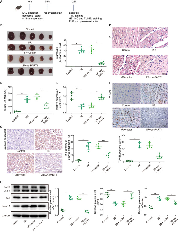
Figure 7 Enforced expression of PART1 relieved I/R-induced MIRI. A mice model of MIRI was prepared and treated with a PART1 overexpression vector. (A) Mouse MIRI model construction timeline. (B) TTC staining detected myocardial infarct volume. (C) H&E staining detected myocardial histopathological changes in mice. (D) Blood level of myocardial marker CK-MB was measured by ELISA. (E) RT-qPCR detection of PART1 expression in mouse myocardial tissue. (F) TUNEL staining detected cardiomyocyte apoptosis. (G) Cleaved caspase3 expression in mouse myocardial tissue was assessed with IHC. (H) Western blot detected p62, LC3II/I, Beclin-1 levels. Data are the means ± SD for three independent experiments (n=5 mice/group).CK-MB = creatine kinase MB; ELISA = enzyme-linked immunosorbent assay; H&E = hematoxylin and eosin; IHC = immunohistochemistry; I/R = ischemia/reperfusion; MIRI = myocardial ischemia-reperfusion injury; PART1 = prostate androgen regulated transcript 1; RT-qPCR = quantitative real-time polymerase chain reaction; SD = standard deviation; TTC = 2,3,5-Triphenyltetrazolium chloride; TUNEL = transferase-mediated dUTP nick-end labeling.*p<0.05, **p<0.01, ***p<0.001.
Reference
-
1. Pagliaro BR, Cannata F, Stefanini GG, Bolognese L. Myocardial ischemia and coronary disease in heart failure. Heart Fail Rev. 2020; 25:53–65. PMID: 31332663.2. Davidson SM, Ferdinandy P, Andreadou I, et al. Multitarget strategies to reduce myocardial ischemia/reperfusion injury: JACC review topic of the week. J Am Coll Cardiol. 2019; 73:89–99. PMID: 30621955.3. Du J, Li Y, Zhao W. Autophagy and myocardial ischemia. Adv Exp Med Biol. 2020; 1207:217–222. PMID: 32671750.4. Niu X, Pu S, Ling C, et al. lncRNA Oip5-as1 attenuates myocardial ischaemia/reperfusion injury by sponging miR-29a to activate the SIRT1/AMPK/PGC1α pathway. Cell Prolif. 2020; 53:e12818. PMID: 32468629.5. Guo Z, Zhao M, Jia G, Ma R, Li M. LncRNA PART1 alleviated myocardial ischemia/reperfusion injury via suppressing miR-503-5p/BIRC5 mediated mitochondrial apoptosis. Int J Cardiol. 2021; 338:176–184. PMID: 34082009.6. Lv XW, He ZF, Zhu PP, Qin QY, Han YX, Xu TT. miR-451-3p alleviates myocardial ischemia/reperfusion injury by inhibiting MAP1LC3B-mediated autophagy. Inflamm Res. 2021; 70:1089–1100. PMID: 34633468.7. Lv W, Jiang J, Li Y, Fu L, Meng F, Li J. MiR-302a-3p aggravates myocardial ischemia-reperfusion injury by suppressing mitophagy via targeting FOXO3. Exp Mol Pathol. 2020; 117:104522. PMID: 32866521.8. Gu Z, Zhou Y, Cao C, Wang X, Wu L, Ye Z. TFAP2C-mediated LINC00922 signaling underpins doxorubicin-resistant osteosarcoma. Biomed Pharmacother. 2020; 129:110363. PMID: 32563982.9. Hammer S, Toenjes M, Lange M, et al. Characterization of TBX20 in human hearts and its regulation by TFAP2. J Cell Biochem. 2008; 104:1022–1033. PMID: 18275040.10. Luo J, Xue D, Song F, Liu X, Li W, Wang Y. DUSP5 (dual-specificity protein phosphatase 5) suppresses BCG-induced autophagy via ERK 1/2 signaling pathway. Mol Immunol. 2020; 126:101–109. PMID: 32795663.11. Zeng M, Wei X, He YL, Chen JX, Lin WT, Xu WX. EGCG protects against myocardial I/RI by regulating lncRNA Gm4419-mediated epigenetic silencing of the DUSP5/ERK1/2 axis. Toxicol Appl Pharmacol. 2021; 433:115782. PMID: 34740634.12. Wang J, Hu X, Fu W, Xie J, Zhou X, Jiang H. Isoproterenol-mediated heme oxygenase-1 induction inhibits high mobility group box 1 protein release and protects against rat myocardial ischemia/reperfusion injury in vivo. Mol Med Rep. 2014; 9:1863–1868. PMID: 24604346.13. Xu Z, Mo Y, Li X, et al. The novel lncRNA AK035396 drives cardiomyocyte apoptosis through Mterf1 in myocardial ischemia/reperfusion injury. Front Cell Dev Biol. 2021; 9:773381. PMID: 34820386.14. Xu S, Wu B, Zhong B, et al. Naringenin alleviates myocardial ischemia/reperfusion injury by regulating the nuclear factor-erythroid factor 2-related factor 2 (Nrf2) /System xc-/ glutathione peroxidase 4 (GPX4) axis to inhibit ferroptosis. Bioengineered. 2021; 12:10924–10934. PMID: 34699317.15. Valikeserlis I, Athanasiou AA, Stakos D. Cellular mechanisms and pathways in myocardial reperfusion injury. Coron Artery Dis. 2021; 32:567–577. PMID: 33471478.16. Liu CY, Zhang YH, Li RB, et al. LncRNA CAIF inhibits autophagy and attenuates myocardial infarction by blocking p53-mediated myocardin transcription. Nat Commun. 2018; 9:29. PMID: 29295976.17. Fu D, Gao T, Liu M, et al. LncRNA TUG1 aggravates cardiomyocyte apoptosis and myocardial ischemia/reperfusion injury. Histol Histopathol. 2021; 36:1261–1272. PMID: 34668176.18. Sun Y, Zhang Y, Ye Z, et al. circRNA-miRNA complex participates in the apoptosis of myocardial cells in myocardial ischemia/reperfusion injury. Discov Med. 2022; 33:13–26. PMID: 35882241.19. Fang YC, Yeh CH. Inhibition of miR-302 suppresses hypoxia-reoxygenation-induced H9c2 cardiomyocyte death by regulating Mcl-1 expression. Oxid Med Cell Longev. 2017; 2017:7968905. PMID: 28491238.20. Yu SY, Dong B, Fang ZF, Hu XQ, Tang L, Zhou SH. Knockdown of lncRNA AK139328 alleviates myocardial ischaemia/reperfusion injury in diabetic mice via modulating miR-204-3p and inhibiting autophagy. J Cell Mol Med. 2018; 22:4886–4898. PMID: 30047214.21. Liang H, Li F, Li H, Wang R, Du M. Overexpression of lncRNA HULC attenuates myocardial ischemia/reperfusion Injury in rat models and apoptosis of hypoxia/reoxygenation cardiomyocytes via targeting miR-377-5p through NLRP3/Caspase-1/IL-1β signaling pathway inhibition. Immunol Invest. 2021; 50:925–938. PMID: 32674625.22. Jiang X, Guo S, Xu M, et al. TFAP2C-mediated lncRNA PCAT1 inhibits ferroptosis in docetaxel-resistant prostate cancer through c-Myc/miR-25-3p/SLC7A11 signaling. Front Oncol. 2022; 12:862015. PMID: 35402284.23. Bai T, Cui Y, Yang X, et al. miR-302a-3p targets FMR1 to regulate pyroptosis of renal tubular epithelial cells induced by hypoxia-reoxygenation injury. Exp Physiol. 2021; 106:2531–2541. PMID: 34605097.24. Fabian MR, Sonenberg N, Filipowicz W. Regulation of mRNA translation and stability by microRNAs. Annu Rev Biochem. 2010; 79:351–379. PMID: 20533884.25. Makkos A, Ágg B, Petrovich B, Varga ZV, Görbe A, Ferdinandy P. Systematic review and network analysis of microRNAs involved in cardioprotection against myocardial ischemia/reperfusion injury and infarction: involvement of redox signalling. Free Radic Biol Med. 2021; 172:237–251. PMID: 33965565.26. Kutty RG, Talipov MR, Bongard RD, et al. Dual specificity phosphatase 5-substrate interaction: a mechanistic perspective. Compr Physiol. 2017; 7:1449–1461. PMID: 28915331.27. Ghasemi M, Turnbull T, Sebastian S, Kempson I. The MTT assay: utility, limitations, pitfalls, and interpretation in bulk and single-cell analysis. Int J Mol Sci. 2021; 22:12827. PMID: 34884632.28. Wallberg F, Tenev T, Meier P. Analysis of apoptosis and necroptosis by fluorescence-activated cell sorting. Cold Spring Harb Protoc. 2016; 2016:pdb.prot087387. PMID: 27037070.29. Brunelle JK, Zhang B. Apoptosis assays for quantifying the bioactivity of anticancer drug products. Drug Resist Updat. 2010; 13:172–179. PMID: 20947411.30. Klein R, Nagy O, Tóthová C, Chovanová F. Clinical and diagnostic significance of lactate dehydrogenase and its isoenzymes in animals. Vet Med Int. 2020; 2020:5346483. PMID: 32607139.
- Full Text Links
-
- Actions
-
Cited
- CITED
-
- Close
- Share
-
- Similar articles
-
- Inhibition of Long Noncoding RNA SNHG15 Ameliorates Hypoxia/Ischemia-Induced Neuronal Damage by Regulating miR-302a-3p/STAT1/NF-κB Axis
- Paper “Inhibition of Long Noncoding RNA SNHG15 Ameliorates Hypoxia/Ischemia-Induced Neuronal Damage by Regulating miR-302a-3p/STAT1/NF-κB Axis” by Hu C, et al.[Yonsei Med J 2021;62(4):325-337]
- CircZNF609 Aggravated Myocardial Ischemia Reperfusion Injury via Mediation of miR-214-3p/PTGS2 Axis
- LncRNA AC005332.7 Inhibited Ferroptosis to Alleviate Acute Myocardial Infarction Through Regulating miR-331-3p/CCND2 Axis
- LncRNA XLOC_006390 facilitates cervical cancer tumorigenesis and metastasis as a ceRNA against miR-331-3p and miR-338-3p
