Enhancement of bioactivity and osseointegration in Ti-6Al-4V orthodontic mini-screws coated with calcium phosphate on the TiO 2 nanotube layer
- Affiliations
-
- 1Department of Dental Biomaterials, Institute of Biodegradable Materials, School of Dentistry, Jeonbuk National University, Jeonju, Korea
- KMID: 2536334
- DOI: http://doi.org/10.4041/kjod22.118
Abstract
Objective
This study evaluated the effect of cyclic pre-calcification treatment on the improvement of bioactivity and osseointegration of Ti-6Al-4V miniscrews.
Methods
The experimental groups were: an untreated group (UT), an anodized and heat-treated group (AH), and an anodized treatment followed by cyclic pre-calcification treatment group (ASPH). A bioactive material with calcium phosphate was coated on the mini-screws, and its effects on bioactivity and osseointegration were evaluated in in vitro and in vivo tests of following implantation in the rat tibia.
Results
As a result of immersing the ASPH group in simulated body fluid for 2 days, protrusions appearing in the initial stage of hydroxyapatite precipitation were observed. On the 3rd day, the protrusions became denser, other protrusions overlapped and grew on it, and the calcium and phosphorus concentrations increased. The removal torque values increased significantly in the following order: UT group (2.08 ± 0.67 N·cm), AH group (4.10 ± 0.72 N·cm), and ASPH group (6.58 ± 0.66 N·cm) with the ASPH group showing the highest value (p < 0.05). In the ASPH group, new bone was observed that was connected to the threads, and it was confirmed that a bony bridge connected to the adjacent bone was formed.
Conclusions
In conclusion, it was found that the surface treatment method used in the ASPH group improved the bioactivity and osseointegration of Ti-6Al-4V orthodontic miniscrews.
Figure
-
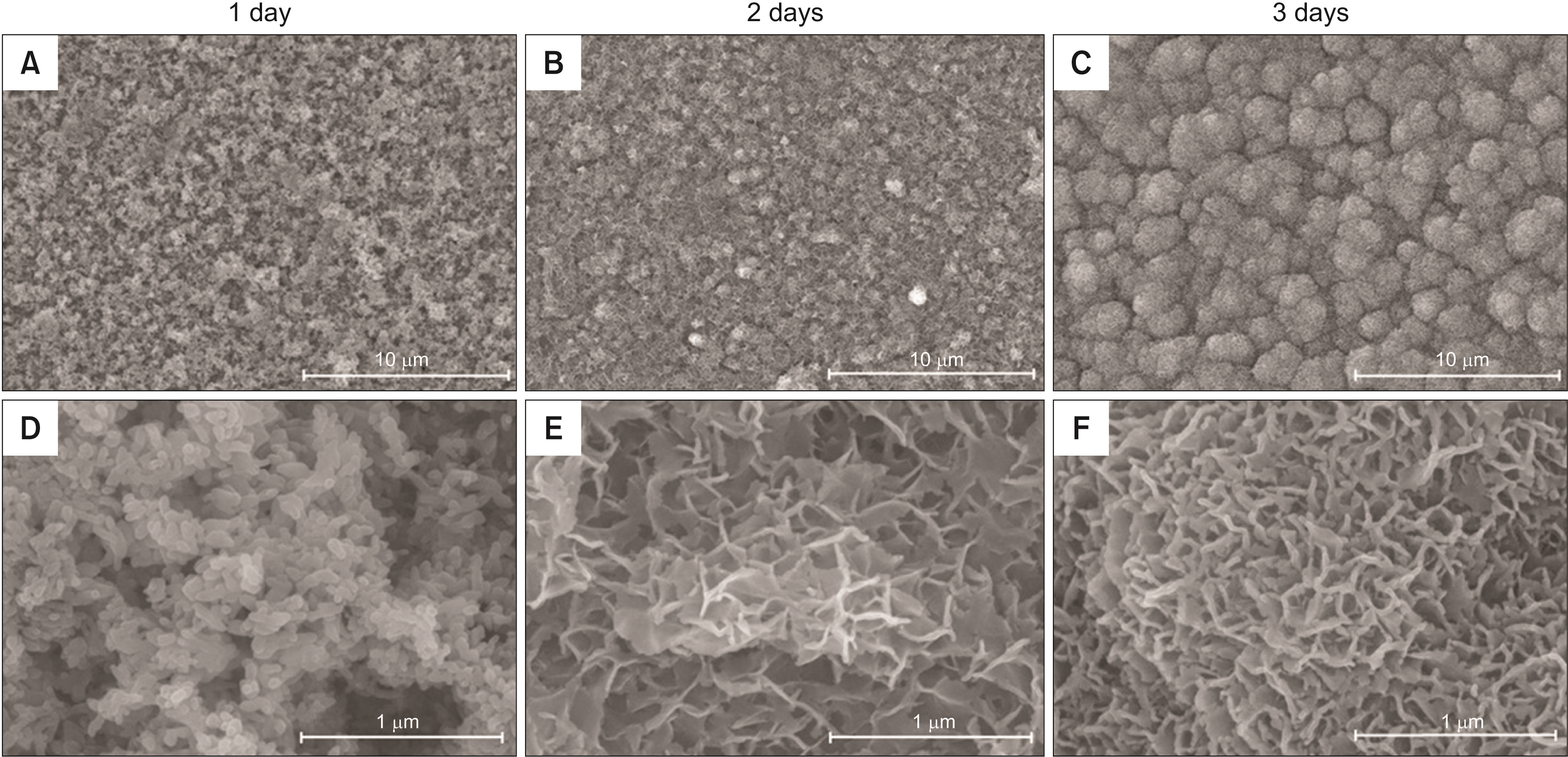
Figure 1 FE-SEM images (×5,000 in top row and ×50,000 in bottom row) of ASPH group immersed in SBF for 1 day (A, D), 2 days (B, E), and 3 days (C, F). FE-SEM, field emission scanning electron microscope; ASPH, anodized treatment followed by cyclic pre-calcification; SBF, simulated body fluid.
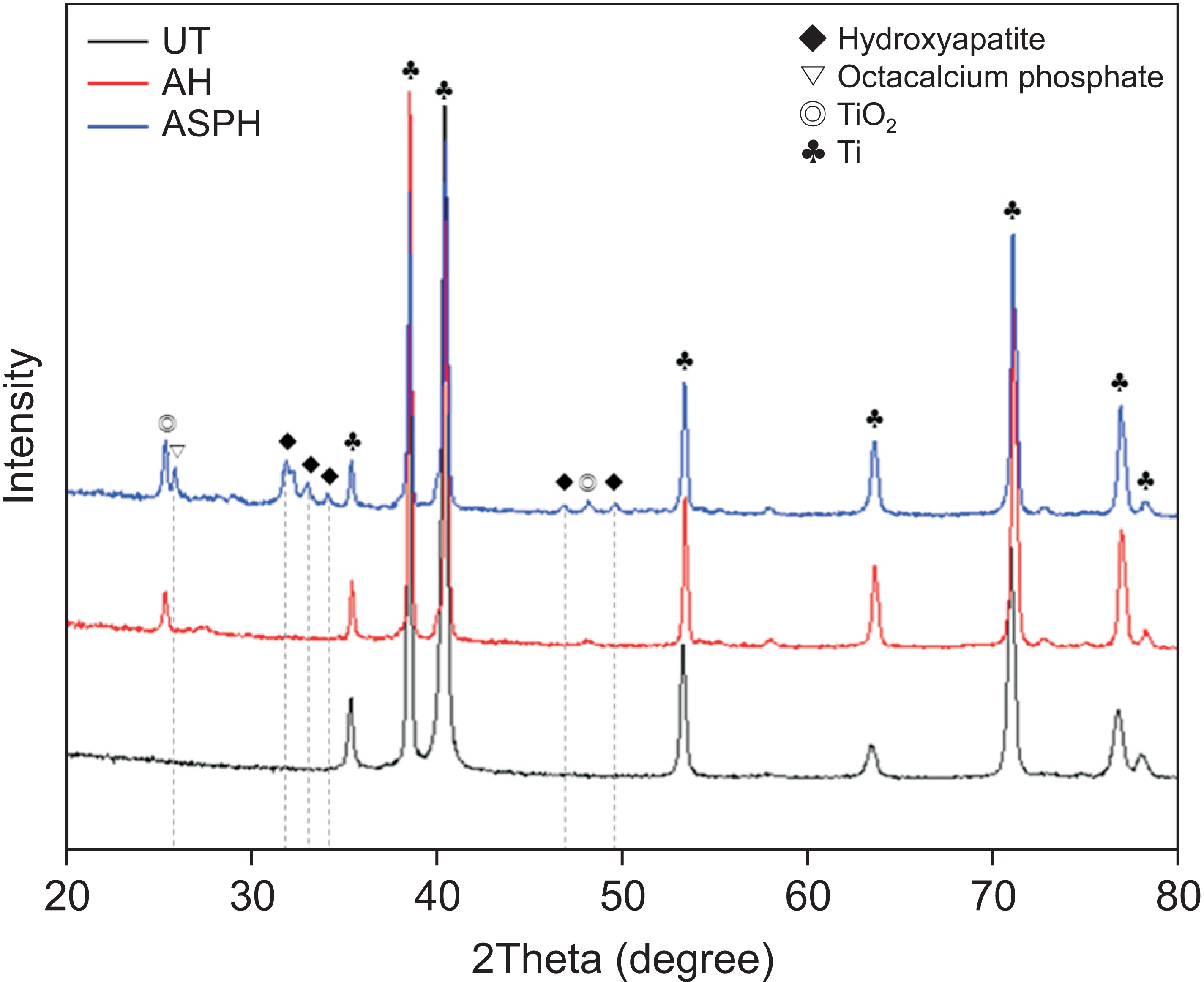
Figure 2 X-ray diffraction analysis of UT, AH, and ASPH groups. UT, untreated group; AH, anodized and heat-treated group; ASPH, anodized treatment followed by cyclic pre-calcification group.
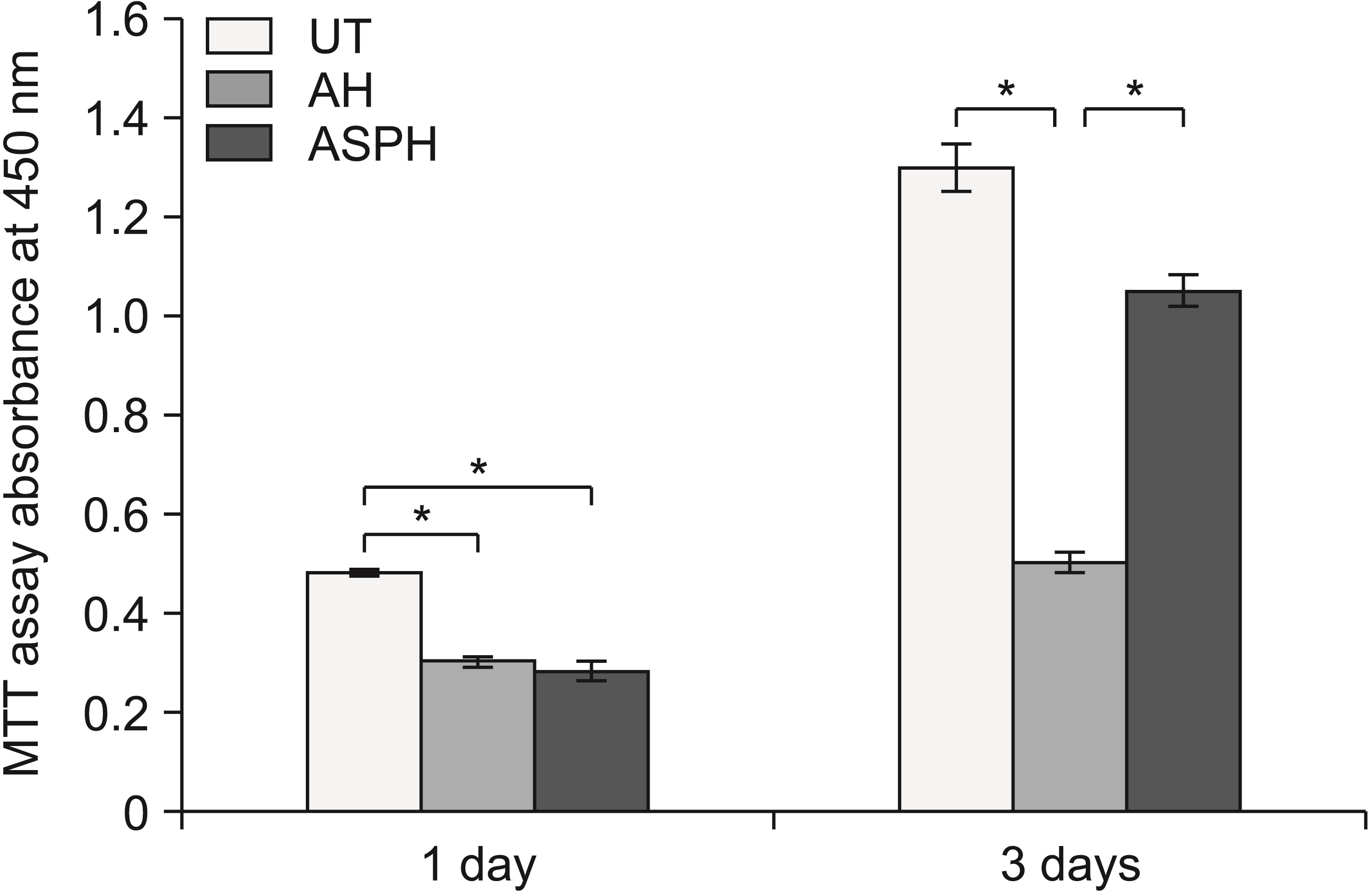
Figure 3 Cytotoxicity test of MC3T3-E1 osteoblast cells in the UT, AH, and ASPH groups for 1 and 3 days (MTT assay). The results are presented as the means ± standard deviation. UT, untreated group; AH, anodized and heat-treated group; ASPH, anodized treatment followed by cyclic pre-calcification group; MTT, methylthiazolyldiphenyl-tetrazolium bromide. *p < 0.05.

Figure 4 Mini-screws removal torque values of the UT, AH, and ASPH groups at 4 weeks after implantation. The results are presented as the means ± standard deviation. UT, untreated group; AH, anodized and heat-treated group; ASPH, anodized treatment followed by cyclic pre-calcification group. *p < 0.05.
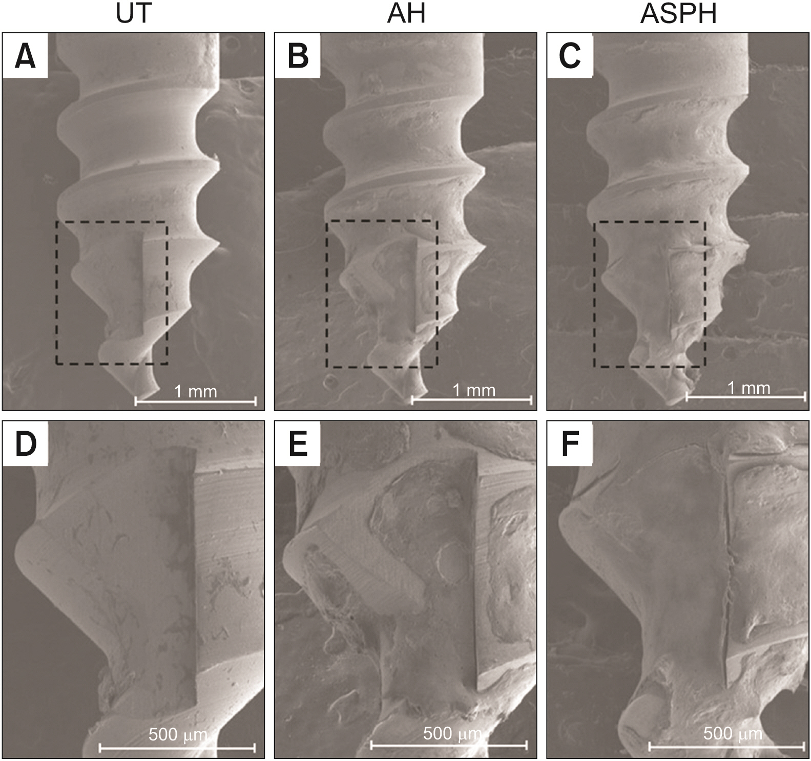
Figure 5 Surface morphology (×40) of mini-screws removed after implantation in rat tibia for 4 weeks. UT (A, D), AH (B, E), and ASPH (C, F) groups. The black squares show high-magnification images (×100). UT, untreated group; AH, anodized and heat-treated group; ASPH, anodized treatment followed by cyclic pre-calcification group.

Figure 6 Optical microscope analysis (×30) after 4 weeks of mini-screws implantation. Surface morphology of UT (A, D), AH (B, E), and ASPH (C, F) group mini-screws removed after implantation in rat tibia for 4 weeks. The black squares show high-magnification images (×100). The red arrow indicates osteogenesis. UT, untreated group; AH, anodized and heat-treated group; ASPH, anodized treatment followed by cyclic pre-calcification group.
Reference
-
1. Kakali L, Alharbi M, Pandis N, Gkantidis N, Kloukos D. 2019; Success of palatal implants or mini-screws placed median or paramedian for the reinforcement of anchorage during orthodontic treatment: a systematic review. Eur J Orthod. 41:9–20. DOI: 10.1093/ejo/cjy015. PMID: 29608666. PMID: https://www.scopus.com/inward/record.uri?partnerID=HzOxMe3b&scp=85060584713&origin=inward.2. Hergel CA, Acar YB, Ates M, Kucukkeles N. 2019; In-vitro evaluation of the effects of insertion and sterilization procedures on the mechanical and surface characteristics of mini screws. Eur Oral Res. 53:25–31. DOI: 10.26650/eor.20197993. PMID: 31309189. PMCID: PMC6612762.3. Oswal S, Agarkar SS, Jethe S, Yerawadekar S, Kawale P, Deshmukh S, et al. 2020; Clinical use of orthodontic mini-implants for intrusion and retraction: a systematic review. Australas Orthod J. 36:87–100. DOI: 10.21307/aoj-2020-011. PMID: 3cf6a92d071b40c78885afe2e9b622af. PMID: https://www.scopus.com/inward/record.uri?partnerID=HzOxMe3b&scp=85134233501&origin=inward.4. Mecenas P, Espinosa DG, Cardoso PC, Normando D. 2020; Stainless steel or titanium mini-implants? Angle Orthod. 90:587–97. DOI: 10.2319/081619-536.1. PMID: 33378494. PMCID: PMC8028470. PMID: https://www.scopus.com/inward/record.uri?partnerID=HzOxMe3b&scp=85088590598&origin=inward.5. Reynders R, Ronchi L, Bipat S. 2009; Mini-implants in orthodontics: a systematic review of the literature. Am J Orthod Dentofacial Orthop. 135:564.e1–19. discussion 564–5. DOI: 10.1016/j.ajodo.2008.09.026. PMID: 19409331. PMID: https://www.scopus.com/inward/record.uri?partnerID=HzOxMe3b&scp=65249139979&origin=inward.6. Moon YY, Lee MH, Song KW, Lee MH, Bae TS. 2008; Characteristics of TiO2 nanotube on Ti-6Al-4V alloy. J Korean Res Soc Dent Mater. 35:339–48.7. El-Zehary RR, Tawfik MA, Mourad SI, Enan ET. 2021; Influence of orthodontic mini-screw surface modifications on implant topography and osseointegration: an in-vivo study. Tang Egypt Dent J. 67:1291–305. DOI: 10.21608/edj.2021.63997.1512.8. Xia L, Xie Y, Fang B, Wang X, Lin K. 2018; In situ modulation of crystallinity and nano-structures to enhance the stability and osseointegration of hydroxyapatite coatings on Ti-6Al-4V implants. Chem Eng J. 347:711–20. DOI: 10.1016/j.cej.2018.04.045. PMID: https://www.scopus.com/inward/record.uri?partnerID=HzOxMe3b&scp=85046146746&origin=inward.9. Mehnath S, Arjama M, Rajan M, Premkumar K, Karthikeyan K, Jeyaraj M. 2020; Mineralization of bioactive marine sponge and electrophoretic deposition on Ti-6Al-4V implant for osteointegration. Surf Coat Technol. 392:125727. DOI: 10.1016/j.surfcoat.2020.125727. PMID: https://www.scopus.com/inward/record.uri?partnerID=HzOxMe3b&scp=85083313122&origin=inward.10. Kim SP, Cho HR, Choe HC. 2021; Bioactive element coatings on nano-mesh formed Ti-6Al-4V alloy surface using plasma electrolytic oxidation. Surf Coat Technol. 406:126649. DOI: 10.1016/j.surfcoat.2020.126649. PMID: https://www.scopus.com/inward/record.uri?partnerID=HzOxMe3b&scp=85096403301&origin=inward.11. Lin Q, Huang D, Du J, Wei Y, Hu Y, Lian X, et al. 2019; Nano-hydroxyapatite crystal formation based on calcified TiO2 nanotube arrays. Appl Surf Sci. 478:237–46. DOI: 10.1016/j.apsusc.2019.01.226. PMID: https://www.scopus.com/inward/record.uri?partnerID=HzOxMe3b&scp=85060768869&origin=inward.12. Kodama A, Bauer S, Komatsu A, Asoh H, Ono S, Schmuki P. 2009; Bioactivation of titanium surfaces using coatings of TiO2 nanotubes rapidly pre-loaded with synthetic hydroxyapatite. Acta Biomater. 5:2322–30. DOI: 10.1016/j.actbio.2009.02.032. PMID: 19332383. PMID: https://www.scopus.com/inward/record.uri?partnerID=HzOxMe3b&scp=67349141019&origin=inward.13. Nguyen TDT, Park IS, Lee MH, Bae TS. 2013; Enhanced biocompatibility of a pre-calcified nanotubular TiO2 layer on Ti-6Al-7Nb alloy. Surf Coat Technol. 236:127–34. DOI: 10.1016/j.surfcoat.2013.09.038. PMID: https://www.scopus.com/inward/record.uri?partnerID=HzOxMe3b&scp=84888848901&origin=inward.14. Zhang L, Zhao Z, Li Y, Wu J, Zheng L, Tang T. 2010; Osseointegration of orthodontic micro-screws after immediate and early loading. Angle Orthod. 80:354–60. DOI: 10.2319/021909-106.1. PMID: 19905862. PMCID: PMC8973229. PMID: https://www.scopus.com/inward/record.uri?partnerID=HzOxMe3b&scp=73349094746&origin=inward.15. Baino F, Yamaguchi S. 2020; The use of simulated body fluid (SBF) for assessing materials bioactivity in the context of tissue engineering: review and challenges. Biomimetics (Basel). 5:57. DOI: 10.3390/biomimetics5040057. PMID: 33138246. PMCID: PMC7709622. PMID: 423061ae851d4d94a7dc9fd870c08ab9. PMID: https://www.scopus.com/inward/record.uri?partnerID=HzOxMe3b&scp=85094632327&origin=inward.16. Li Y, Li B, Song Y, Ma A, Li C, Zhang X, et al. 2019; Improved osteoblast adhesion and osseointegration on TiO2 nanotubes surface with hydroxyapatite coating. Dent Mater J. 38:278–86. DOI: 10.4012/dmj.2018-118. PMID: 30541994. PMID: https://www.scopus.com/inward/record.uri?partnerID=HzOxMe3b&scp=85064003930&origin=inward.17. Zhou Z, Shi Q, Wang J, Chen X, Hao Y, Zhang Y, et al. 2021; The unfavorable role of titanium particles released from dental implants. Nanotheranostics. 5:321–32. DOI: 10.7150/ntno.56401. PMID: 33732603. PMCID: PMC7961127. PMID: https://www.scopus.com/inward/record.uri?partnerID=HzOxMe3b&scp=85103143661&origin=inward.18. Moghaddam SF, Mohammadi A, Behroozian A. 2021; The effect of sandblasting and acid etching on survival rate of orthodontic miniscrews: a split-mouth randomized controlled trial. Prog Orthod. 22:2. DOI: 10.1186/s40510-020-00347-z. PMID: 33409700. PMCID: PMC7788110. PMID: 39aa49823b1b42e99727b65a5d40f704. PMID: https://www.scopus.com/inward/record.uri?partnerID=HzOxMe3b&scp=85098772550&origin=inward.19. Cheng Y, Yang H, Yang Y, Huang J, Wu K, Chen Z, et al. 2018; Progress in TiO2 nanotube coatings for biomedical applications: a review. J Mater Chem B. 6:1862–86. DOI: 10.1039/C8TB00149A. PMID: 32254353. PMID: https://www.scopus.com/inward/record.uri?partnerID=HzOxMe3b&scp=85044770698&origin=inward.20. Stich T, Alagboso F, Křenek T, Kovářík T, Alt V, Docheva D. 2021; Implant-bone-interface: reviewing the impact of titanium surface modifications on osteogenic processes in vitro and in vivo. Bioeng Transl Med. 7:e10239. DOI: 10.1002/btm2.10239. PMID: 35079626. PMCID: PMC8780039. PMID: 5bfc36652be74fd1abd02f70f98d8a6a. PMID: https://www.scopus.com/inward/record.uri?partnerID=HzOxMe3b&scp=85109729182&origin=inward.21. El-Mezayen MA, Gaber KM, Refai WM. 2022; Detection of bone deposition on the surface of immediately removed or left mini-screw after canine retraction (SEM Study). Egypt Orthod J. 61:12–23. DOI: 10.21608/eos.2021.107405.1037.22. Jiman PA, Prodan D, Moldovan M, Muntean A, Sarosi C, Tarmure V, et al. 2021; New and recovered temporary anchorage devices, in vitro assessment of structural and surface properties. Materials (Basel). 14:6271. DOI: 10.3390/ma14216271. PMID: 34771795. PMCID: PMC8584799. PMID: aaaa799a0d1f4ba28d814bbcc8c2d390. PMID: https://www.scopus.com/inward/record.uri?partnerID=HzOxMe3b&scp=85117811723&origin=inward.23. Bucur SM, Vaida LL, Olteanu CD, Checchi V. 2021; A brief review on micro-implants and their use in orthodontics and dentofacial orthopaedics. Appl Sci. 11:10719. DOI: 10.3390/app112210719. PMID: 778f92d47d164116b344e0c7dd70f68f. PMID: https://www.scopus.com/inward/record.uri?partnerID=HzOxMe3b&scp=85119268509&origin=inward.24. Łosiewicz B, Stróż A, Osak P, Maszybrocka J, Gerle A, Dudek K, et al. 2021; Production, characterization and application of oxide nanotubes on Ti-6Al-7Nb alloy as a potential drug carrier. Materials (Basel). 14:6142. DOI: 10.3390/ma14206142. PMID: 34683734. PMCID: PMC8538941. PMID: 422563c12efb43319e63fc84f9c6ec6b. PMID: https://www.scopus.com/inward/record.uri?partnerID=HzOxMe3b&scp=85117573517&origin=inward.25. Wang K, Jin H, Song Q, Huo J, Zhang J, Li P. 2021; Titanium dioxide nanotubes as drug carriers for infection control and osteogenesis of bone implants. Drug Deliv Transl Res. 11:1456–74. DOI: 10.1007/s13346-021-00980-z. PMID: 33942245. PMID: https://www.scopus.com/inward/record.uri?partnerID=HzOxMe3b&scp=85105376065&origin=inward.26. Oh EJ, Nguyen TD, Lee SY, Jeon YM, Bae TS, Kim JG. 2014; Enhanced compatibility and initial stability of Ti6Al4V alloy orthodontic miniscrews subjected to anodization, cyclic precalcification, and heat treatment. Korean J Orthod. 44:246–53. DOI: 10.4041/kjod.2014.44.5.246. PMID: 25309864. PMCID: PMC4192526. PMID: https://www.scopus.com/inward/record.uri?partnerID=HzOxMe3b&scp=84907908008&origin=inward.27. Kim HY, Kim SC. 2016; Bone cutting capacity and osseointegration of surface-treated orthodontic mini-implants. Korean J Orthod. 46:386–94. DOI: 10.4041/kjod.2016.46.6.386. PMID: 27896213. PMCID: PMC5118218. PMID: https://www.scopus.com/inward/record.uri?partnerID=HzOxMe3b&scp=84995662772&origin=inward.28. Nguyen TT, Jang YS, Kim YK, Kim SY, Lee MH, Bae TS. 2019; Osteogenesis-related gene expression and guided bone regeneration of a strontium-doped calcium-phosphate-coated titanium mesh. ACS Biomater Sci Eng. 5:6715–24. DOI: 10.1021/acsbiomaterials.9b01042. PMID: 33423489. PMID: https://www.scopus.com/inward/record.uri?partnerID=HzOxMe3b&scp=85075023799&origin=inward.29. Nguyen TD, Moon SH, Oh TJ, Park IS, Lee MH, Bae TS. 2015; The effect of APH treatment on surface bonding and osseointegration of Ti-6Al-7Nb implants: an in vitro and in vivo study. J Biomed Mater Res B Appl Biomater. 103:641–8. DOI: 10.1002/jbm.b.33210. PMID: 24976109. PMID: https://www.scopus.com/inward/record.uri?partnerID=HzOxMe3b&scp=84925682824&origin=inward.30. Johansson P, Barkarmo S, Hawthan M, Peruzzi N, Kjellin P, Wennerberg A. 2018; Biomechanical, histological, and computed X-ray tomographic analyses of hydroxyapatite coated PEEK implants in an extended healing model in rabbit. J Biomed Mater Res A. 106:1440–7. DOI: 10.1002/jbm.a.36345. PMID: 29341426. PMID: https://www.scopus.com/inward/record.uri?partnerID=HzOxMe3b&scp=85042167153&origin=inward.31. Im C, Park JH, Jeon YM, Kim JG, Jang YS, Lee MH, et al. 2022; Improvement of osseointegration of Ti-6Al-4V ELI alloy orthodontic mini-screws through anodization, cyclic pre-calcification, and heat treatments. Prog Orthod. 23:11. DOI: 10.1186/s40510-022-00405-8. PMID: 35368222. PMCID: PMC8977256. PMID: 9a48620e4c7245dab8337a21f3e64b7e. PMID: https://www.scopus.com/inward/record.uri?partnerID=HzOxMe3b&scp=85127523040&origin=inward.
- Full Text Links
-
- Actions
-
Cited
- CITED
-
- Close
- Share
-
- Similar articles
-
- Osteoblastic behavior to zirconium coating on Ti-6Al-4V alloy
- A HISTOMORPHOMETRIC STUDY OF BONE APPOSITION TO NEWLY DEVELOPED TI-BASED ALLOYS IN RABBIT BONE
- Brazing characteristics of ZrO2 and Ti-6Al-4V brazed joints with increasing temperature
- The effect of Zirconium Nitride coating on shear bond strength with denture base resin in Co-Cr alloy and titanium alloy
- Precalcification Treatment of TiO2 Nanotube on Ti-6Al-4V Alloy
