Orthodontic intrusion treatment of mandibular anterior teeth in a periodontal patient with hyperdivergent skeletal pattern: 8-year follow-up
- Affiliations
-
- 1Dental Clinic Center, Pusan National University Hosptial, Busan, Republic of Korea
- 2(Bio)medical Research Institute, Pusan National University Hospital, Busan, Republic of Korea
- 3Department of Orthodontics, School of Dentistry, Pusan National University, Yangsan, Republic of Korea
- KMID: 2525978
- DOI: http://doi.org/10.14368/jdras.2021.37.1.48
Abstract
- Patients who have extruded anterior teeth and deep bite with pathologic tooth migration, it is necessary not only periodontal treat-ment for reduce inflammation, but also orthodontic treatment for intrusion of anterior teeth. However, it is difficult to place the orthodontic brackets due to the deep bite, and there is a problem that the extrusion of the posterior teeth occurs more easily than the intrusion of the anterior teeth biomechanically. In particular, in patients with long face, relative intrusion of the anterior teeth by extrusion of the posterior teeth causes the clockwise rotation of the mandible and makes the facial profile worse. Therefore the biomechanical consideration and appliance design that can block these problems are required from the treatment plan. This is a patient who had a deep overbite with extruded anterior teeth, treated by treatment and intrusion of mandibular anterior teeth using cute brackes and miniscrews, and resulted in favorable maintenance during 8-year retention.
Figure
-

Fig. 1 (A) Pretreatment extraoral photographs. Gummy smile and excessive tooth exposure were observed. (B) Pretreatment intraoral photographs. Deep bite and gingival recession were observed.

Fig. 2 (A) Pretreatment lateral cephalometric radiograph and analysis. Deep overbite and large overjet were observed. SNA, the angle of sella-nasion-A point; SNB, the angle of sella-nasion-B point; ANB, the angle of A point-nasion-B point; SN-Mn., the angle of SN plane and mandibular plane; FMA, Frankfort mandibular plane angle; U1 to SN, the angle of the long axis of maxillary central incisors between sella-nasion plane; IMPA, the angle of the lowermost tangent to the mandible and the long axis of mandibular incisors; U1-Stmi, the distance of the U1 incisal edge to stomion of upper lip; Interlabial gap, the distance of the stomion of upper lip to lower lip; Overjet, the anterior-posterior distance of the maxillary central incisors over the mandibular central incisors; Overbite, the vertical overlap of the maxillary central incisors over the mandibular central incisors. (B) Pretreatment panoramic (1) & periapical radiographs (2, 3). Angular alveolar bone loss was observed on the distal root surface of # 21. Generalized alveolar bone loss was also observed on the mandibular anterior teeth. *, **, ***, indicates that the value is beyond one, two or three times of the standard deviation.

Fig. 3 Intraoral photos during orthodontic treatment. (A) Brackets (maxillary full arch) and cute brackets (mandibular anterior teeth) were bonded. Anchorage preparations were done using temporary anchorage devices(TADs) on mandible. (B - D) Progression of orthodontic treatment. (during 18 months). (B) Overbite was improved. (D) Vertical positions on maxillary arch were maintained using TADs.
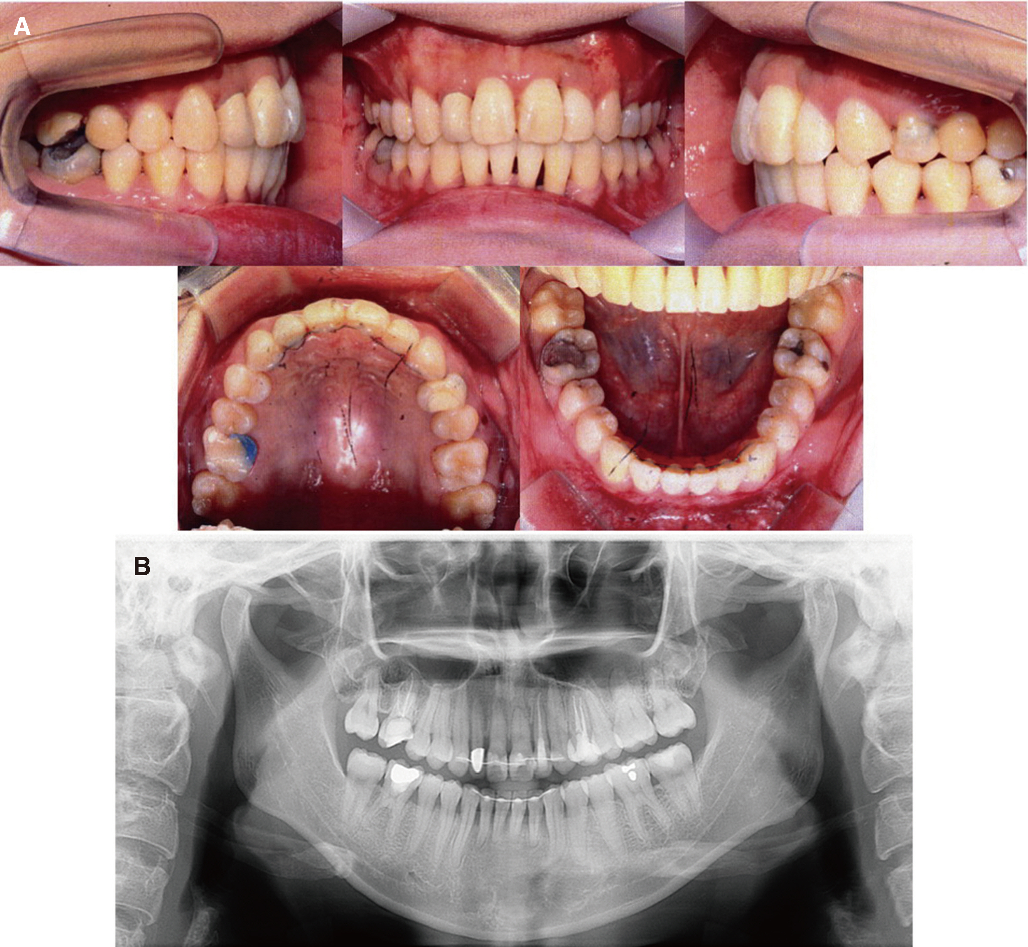
Fig. 4 Posttreatment intraoral photographs (A) and panoramic radiograph (B). Normal overbite and overjet were achieved. Fixed retainer was placed on both anterior teeth.
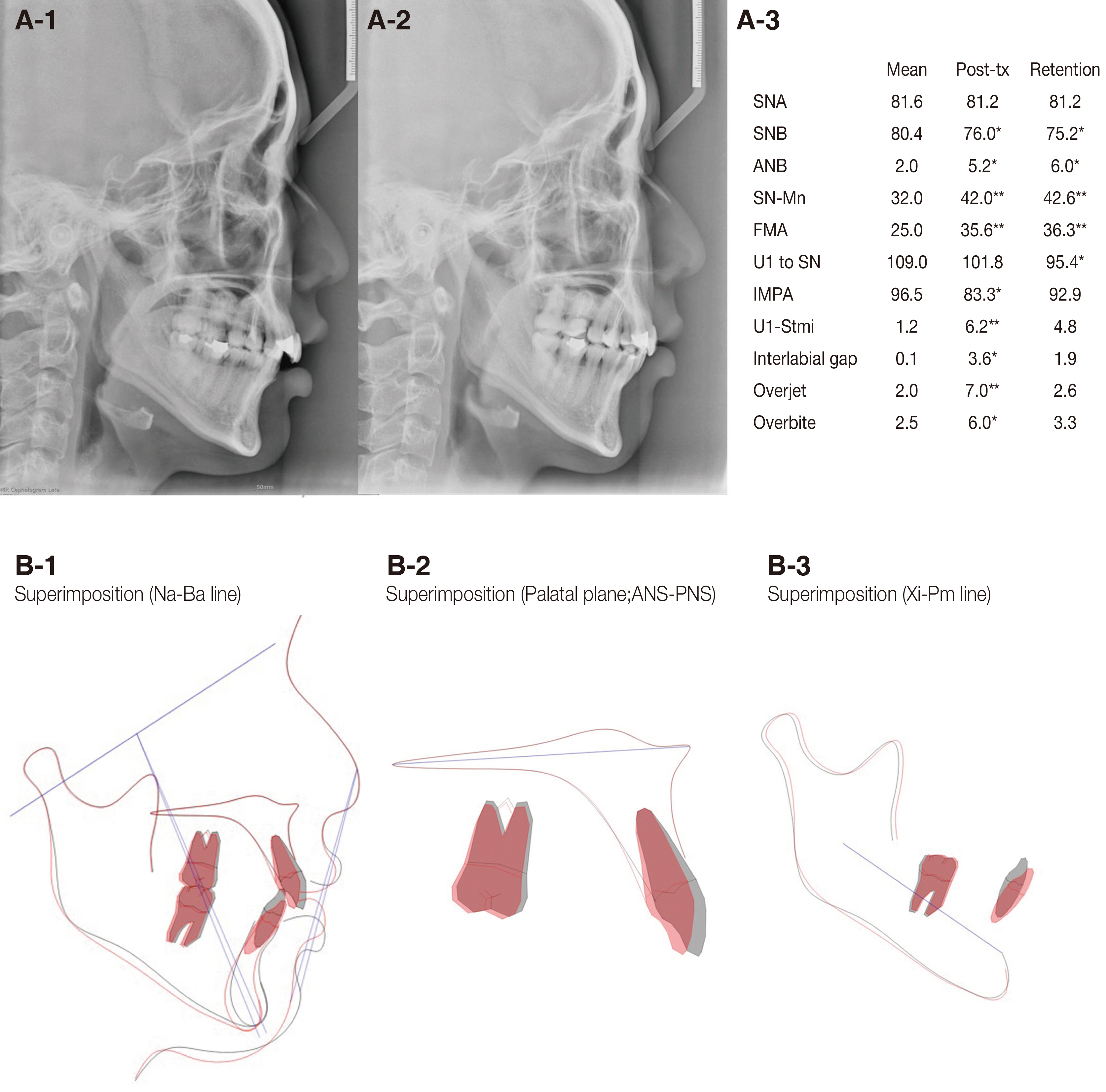
Fig. 5 Pretreatment, posttreatment lateral cephalometric radiographs (A) and superimposition analysis (B). (A) Pretreatment (1), posttreatment (2) radiographs and cephalometric analysis (3). (B) Superimposition of pretreatment and posttreatment. B-1, Superimposition of the maxilla and mandible based on the cranial base (Nasion-Basion line). B-2, Superimposition of the maxilla based on palatal plane (ANS-PNS). B-3, Superimposition of the mandible based on Xi-Pm line (Xi, the point placed in the center of the mandibular ascending ramus; Pm, point where curvature of the anterior contour of the symphysis changes from concave to convex). During orthodontic treatment, the vertical position of the mandibular posterior teeth was maintained and the mandibular anterior teeth were intruded and labioversion. However, the maxillary molar was mildly extruded during the leveling process, and a slight clockwise rotation of the mandible was observed (gray line: pretreatment, red line: posttreatment). *, **, ***, indicates that the value is beyond one, two or three times of the standard deviation.

Fig. 6 Intraoral photographs (A) and panoramic & periapical radiographs (B) for 8-year retention period. Overjet and overbite were maintained. #16 was extracted. Implant fixture was placed on #16 site. Lamina dura was observed on periapical radiographs.
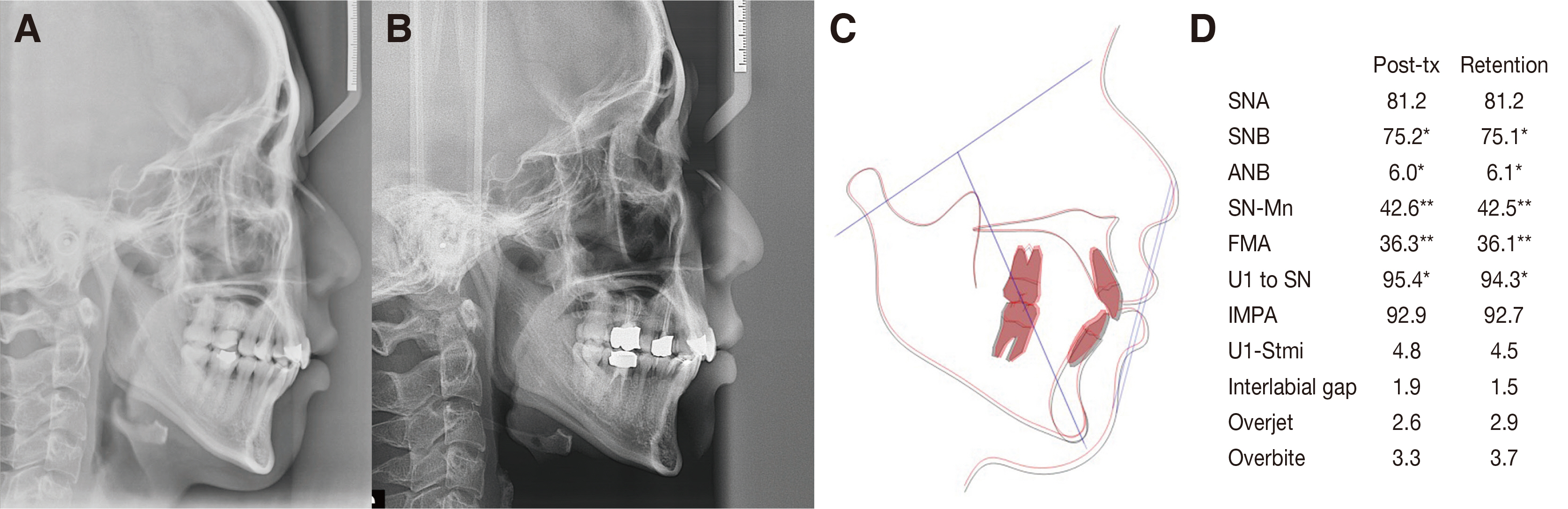
Fig. 7 Lateral cephalometric radiograph of posttreatment (A), 5-year follow-up (B), superimposition of A & B, (C) and analysis (D).
Reference
-
References
1. Fukunaga T, Kuroda S, Kurosaka H, Takano-Yamamoto T. 2006; Skeletal anchorage for orthodontic correction of maxillary protrusion with adult periodontitis. Angle Orthod. 76:148–55. DOI: 10.1043/0003-3219(2006)076[0148:SAFOCO]2.0.CO;2. PMID: 16448285.2. Martinez-Canut P, Carrasquer A, Magán R, Lorca A. 1997; A study on factors associated with pathologic tooth migration. J Clin Periodontol. 24:492–7. DOI: 10.1111/j.1600-051X.1997.tb00217.x. PMID: 9226390.3. Melsen B, Agerbaek N, Eriksen J, Terp S. 1988; New attachment through periodontal treatment and orthodontic intrusion. Am J Orthod Dentofacial Orthop. 94:104–16. DOI: 10.1016/0889-5406(88)90358-7. PMID: 3165238.4. Proffit W, Fields H, Larson B, Sarver D. 2018. Contemporary Orthodontic. 6th ed. Elselvier.5. Burstone CR. 1977; Deep overbite correction by intrusion. Am J Orthod. 72:1–22. DOI: 10.1016/0002-9416(77)90121-X. PMID: 267433.6. Thilander B. Lindhe J, editor. 1989. Orthodontic tooth movement in periodontal therapy. Textbook of Clinical Periodontology. Munksgaard;Copenhagen: p. 450–500.7. Alstad S, Zachrisson BU. 1979; Longitudinal study of periodontal condition associated with orthodontic treatment in adolescents. Am J Orthod. 76:277–86. DOI: 10.1016/0002-9416(79)90024-1. PMID: 290273.8. Artun J, Urbye KS. 1988; The effect of orthodontic treatment on periodontal bone support in patients with advanced loss of marginal periodontium. Am J Orthod Dentofacial Orthop. 93:143–8. DOI: 10.1016/0889-5406(88)90292-2. PMID: 3422529.9. Melsen B, Agerbaek N, Markenstam G. 1989; Intrusion of incisors in adult patients with marginal bone loss. Am J Orthod. 96:232–41. DOI: 10.1016/0889-5406(89)90460-5. PMID: 2773869.10. Gkantidis N, Christou P, Topouzelis N. 2010; The orthodontic-periodontic interrelationship in integrated treatment challenges: a systematic review. J Oral Rehabil. 37:377–90. DOI: 10.1111/j.1365-2842.2010.02068.x. PMID: 20202098.11. Kuroda S, Yamada K, Deguchi T, Hashimoto T, Kyung HM, Takona-Yamamoto T. 2007; Root proximity is a major factor for screw failure in orthodontic anchorage. Am J Orthod Dentofacial Orthop. 131 Suppl 4:S68–73. DOI: 10.1016/j.ajodo.2006.06.017. PMID: 17448389.12. Kuroda S, Sugawara Y, Deguchi T, Kyung HM, Takona-Yamamoto T. 2007; Clinical use of miniscrew implants as orthodontic anchorage: success rates and postoperative discomfort. Am J Orthod Dentofacial Orthop. 131:9–15. DOI: 10.1016/j.ajodo.2005.02.032. PMID: 17208101.13. Baumrind S, Korn EL, Boyd RL. 1996; Apical root resorption in orthodontically treated adults. Am J Orthod Dentofacial Orthop. 110:311–20. DOI: 10.1016/S0889-5406(96)80016-3. PMID: 8814033.14. Han G, Huang S, Von den Hoff JW, Zeng X, Kuijpers-Jagtman AM. 2005; Root resorption after orthodontic intrusion and extrusion: an intraindividual study. Angle Orthod. 75:912–8. DOI: 10.1043/0003-3219(2005)75[912:RRAOIA]2.0.CO;2. PMID: 16448231.15. Carrillo R, Rossouw PE, Franco PF, Opperman LA, Buschang PH. 2007; Intrusion of multiradicular teeth and related root resorption with mini-screw implant anchorage: a radiographic evaluation. Am J Orthod Dentofacial Orthop. 132:647–55. DOI: 10.1016/j.ajodo.2006.08.017. PMID: 18005839.16. Sameshima GT, Sinclair PM. 2001; Predicting and preventing root resorption: part II. Treatment factors. Am J Orthod Dentofacial Orthop. 119:511–5. DOI: 10.1067/mod.2001.113410. PMID: 11343023.17. Erkan M, Pikdoken L, Usumez S. 2007; Gingival response to mandibular incisor intrusion. Am J Orthod Dentofacial Orthop. 132:143.e9–13. DOI: 10.1016/j.ajodo.2006.10.015. PMID: 17693359.18. Hwang HS, Jeon HR, Kim SP, Kim WS, Lee GH. 2011; A new orthodontic appliance for rapid anterior alignment in adults: Mini-tube appliance (MTA). J Korean Dent Assoc. 49:334–45.19. Ericsson I, Thilander B. 1980; Orthodontic relapse in dentitions with reduced periodontal support: an experimental study in dogs. Eur J Orthod. 2:51–7. DOI: 10.1093/ejo/2.1.51. PMID: 6935058.20. Alexander RG. Nanda R, Burstone CJ, editors. 1996. Treatment and retention for longterm stability. Retention and stability in orthodontics. DaehanNarae Publishing Inc.;Seoul: p. 113–23.21. Al-Buraiki H, Sadowsky C, Schneider B. 2005; The effectiveness and long-term stability of overbite correction with incisor intrusion mechanics. Am J Orthod Dentofacial Orthop. 127:47–55. DOI: 10.1016/j.ajodo.2003.10.034. PMID: 15643414.22. Bernstein RL, Preston CB, Lampasso J. 2007; Leveling the curve of Spee with a continuous archwire technique: a long-term cephalometric study. Am J Orthod Dentofacial Orthop. 131:363–71. DOI: 10.1016/j.ajodo.2005.05.056. PMID: 17346592.
- Full Text Links
-
- Actions
-
Cited
- CITED
-
- Close
- Share
-
- Similar articles
-
- 3-D FEA on the intrusion of mandibular anterior segment using orthodontic miniscrews
- Intrusion of the extruded maxillary central incisor using skeletal anchorage system and unilateral segmental intrusion arch
- Intrusion Of The Severe Extruded Maxillary Post.Teeth Using Post.Segmental Osteotomy And Orthodontic Treatment With Skeletal Anchorage System: Cases Report
- Non-surgical treatment and retention of open bite in adult patients with orthodontic mini-implants
- Surgical orthodontic treatment of skeletal Class III malocclusion using mini-implant: correction of horizontal and vertical dental compensation
