Non-surgical treatment and retention of open bite in adult patients with orthodontic mini-implants
- Affiliations
-
- 1Department of Orthodontics, Gachon University Dental Hospital. orthodm@gilhospital.com
- 2Public Health Doctor, Korea.
- 3Private Practice, Korea.
- 4Department of Dentistry, Chung-Ang University College of Medicine, Korea.
- KMID: 1975784
- DOI: http://doi.org/10.4041/kjod.2009.39.6.402
Abstract
- Successful treatment of the non-growing patient with an open bite of either dental skeletal pattern often presents a difficult challenge. The morphologic pattern in anterior open bite is characterized by longer vertical dimensions, an increase in development of the maxillary posterior dento-alveolar structure and a steep mandibular plane. In such cases, molar intrusion would be a good remedy for treatment. This article reports the successful treatment and retention of two anterior open-bite cases. We used orthodontic mini-implants for treatment and a circumferential retainer with posterior bite block or skeletal fixed retainer for retention. The diagnostic criteria and mechanics for appropriate treatment are discussed. Our results suggest that open bite can be reduced successfully with intrusion of molars using orthodontic mini-implants (OMI) without orthognathic surgery, and that circumferential retainer with posterior bite block and skeletal fixed retainer are effective for retention.
MeSH Terms
Figure
-
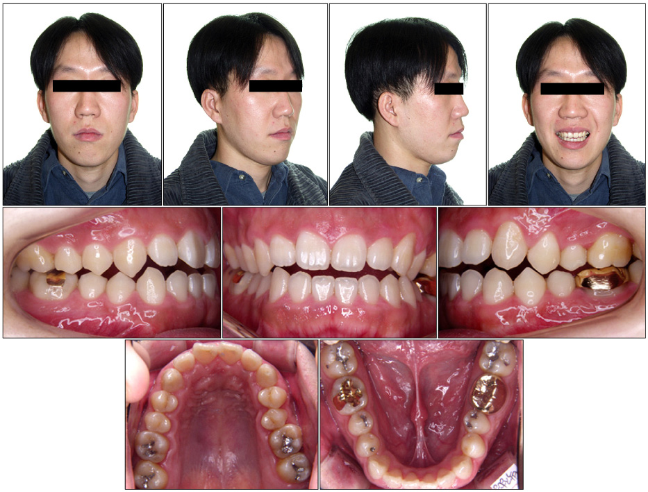
Fig. 1 Pretreatment facial and intraoral photographs.
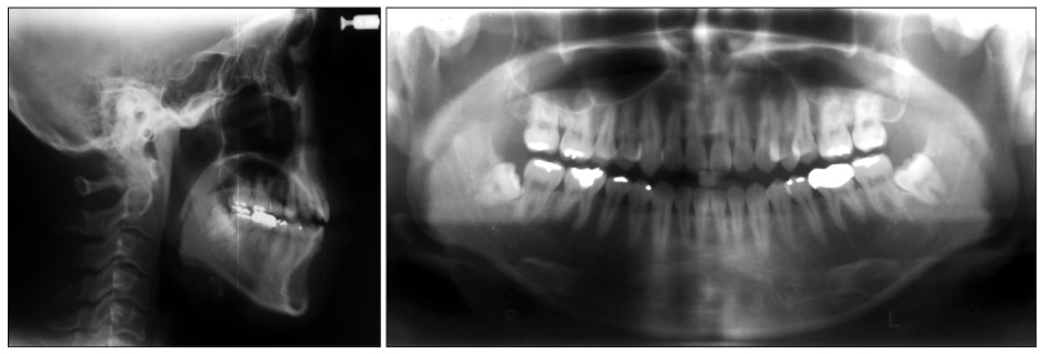
Fig. 2 Pretreatment lateral cephalometric and panoramic radiograph.
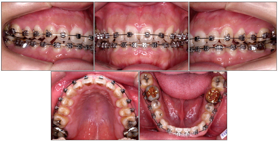
Fig. 3 Intraoral photographs (6 months after start of treatment). Only intrusion of molars has allowed a positive overbite.
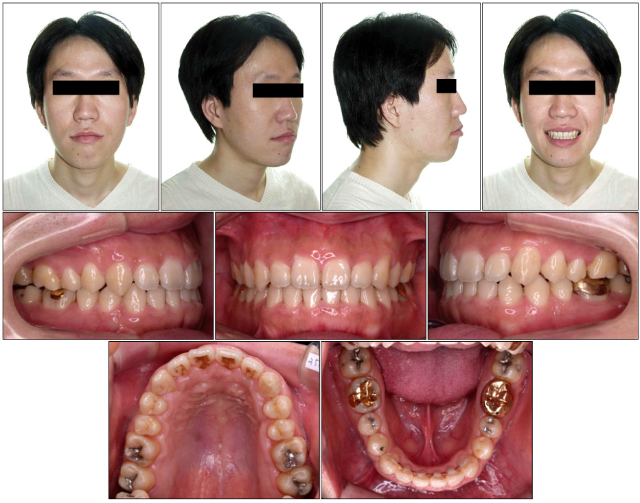
Fig. 4 Posttreatment facial and intraoral photographs (15 months after start of treatment).
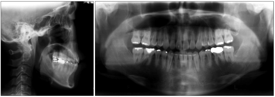
Fig. 5 Posttreatment lateral cephalometric and panoramic radiograph.
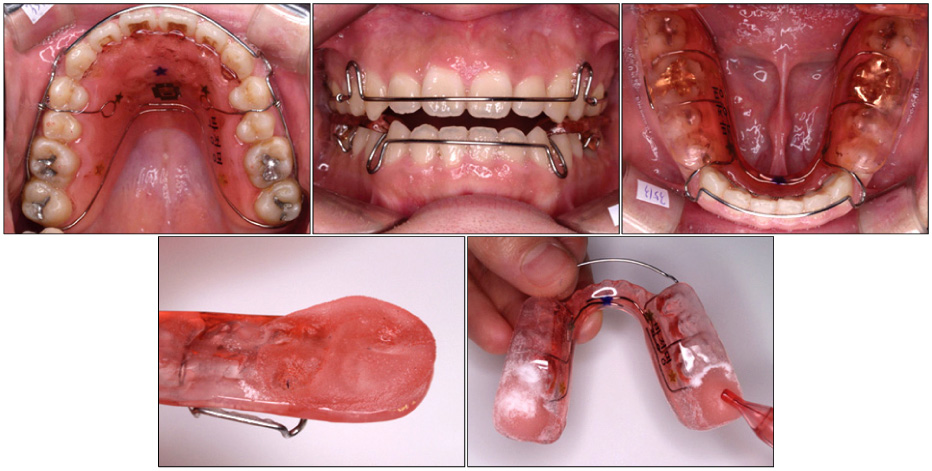
Fig. 6 Retainer photographs (Mx. arch; circumferential retainer, Mn. arch; circumferential retainer with posterior bite block).
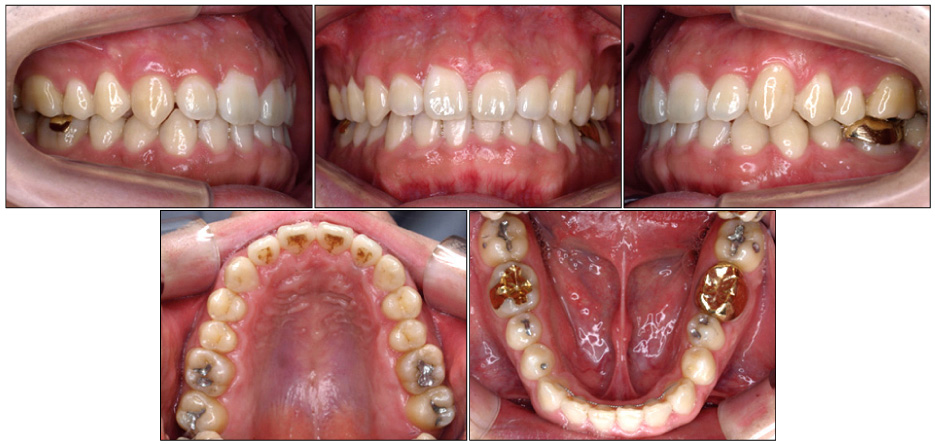
Fig. 7 20 months-retention intraoral photographs (8 months after circumferential retainer with posterior bite block removal).
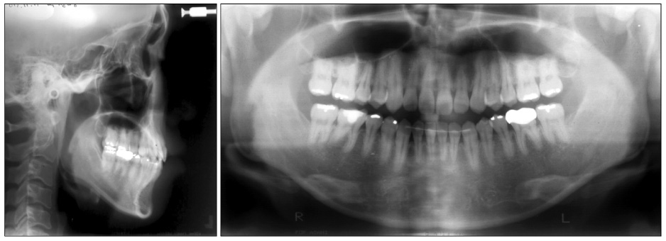
Fig. 8 20 months-retention lateral cephalometric and panoramic radiograph.
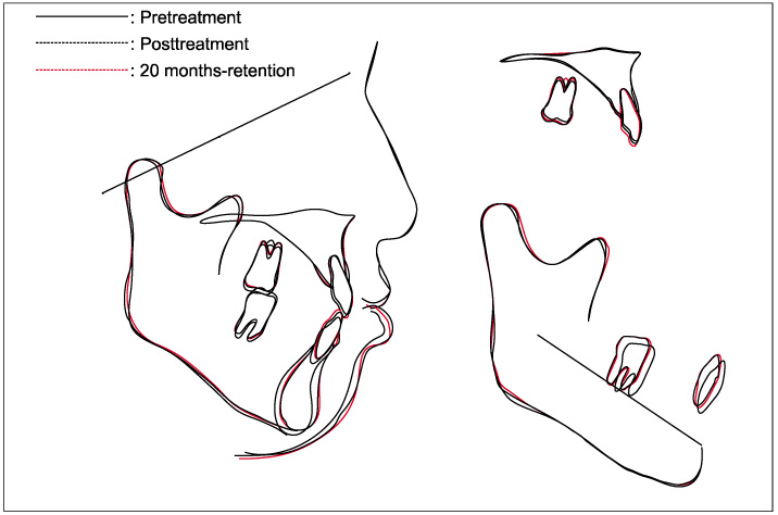
Fig. 9 Superimposition of cephalometric tracings.
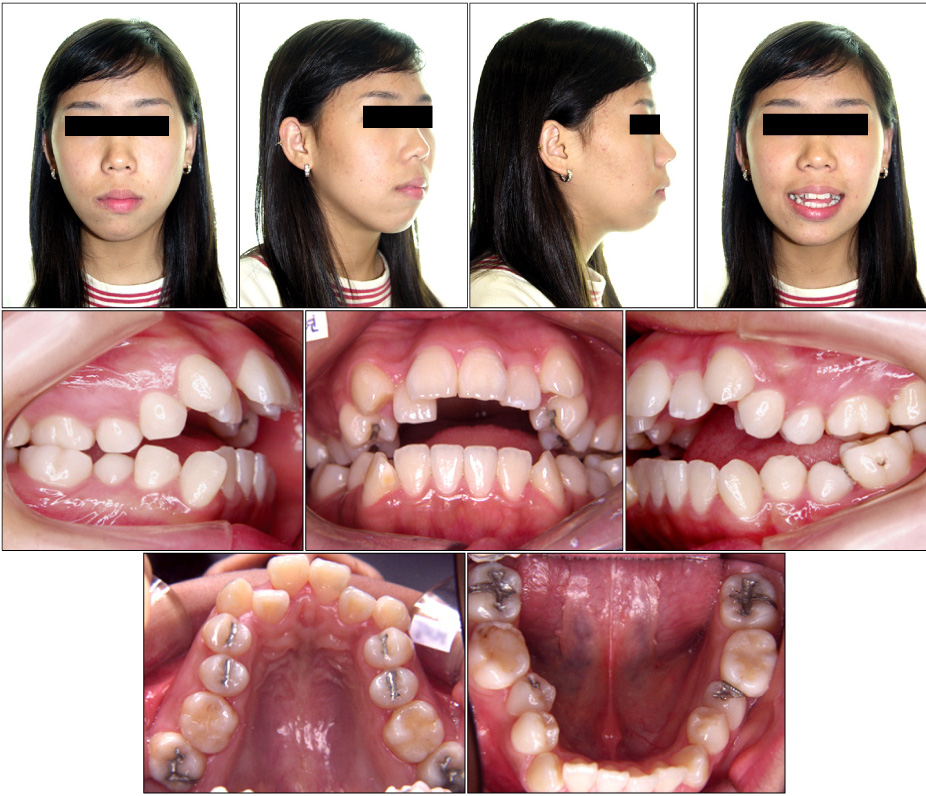
Fig. 10 Pretreatment facial and intraoral photographs.
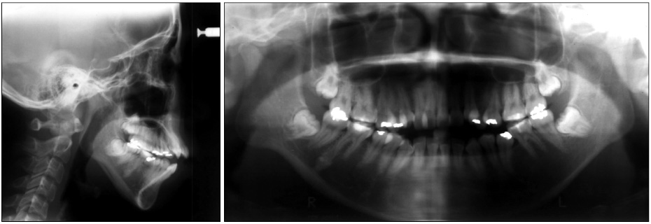
Fig. 11 Pretreatment lateral cephalometric and panoramic radiograph.
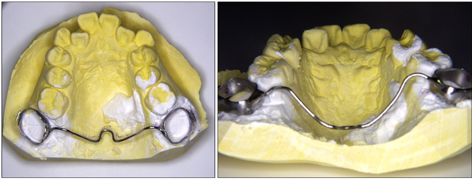
Fig. 12 Transpalatal arch for intrusion of maxillary 2nd molar (TPA is kept apart from the palatal surface for intrusion of the maxillary 2nd molar).

Fig. 13 Intraoral photographs (5 months after start of treatment).
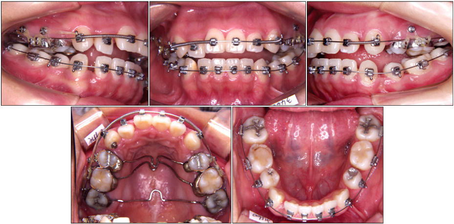
Fig. 14 Intraoral photographs (8 months after start of treatment).
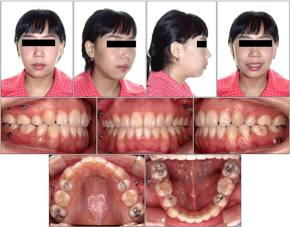
Fig. 15 Posttreatment facial and intraoral photographs (28 months after start of treatment).
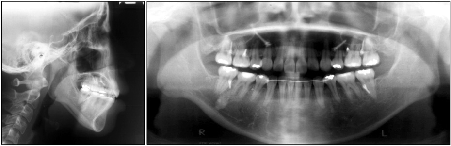
Fig. 16 Posttreatment lateral cephalometric and panoramic radiograph.
Fig. 17 Skeletal fixed retainer (SFR).
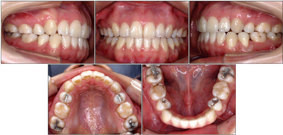
Fig. 18 13 months-retention intraoral photographs (1 month after SFR removal).
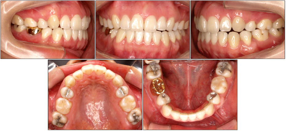
Fig. 19 27 months-retention intraoral photographs (15 months after SFR removal).
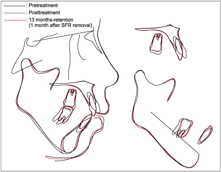
Fig. 20 Superimposition of cephalometric tracings.
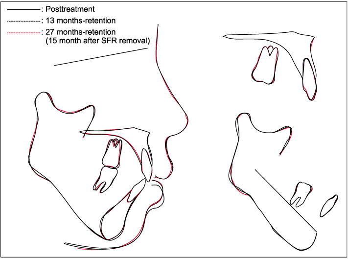
Fig. 21 Superimposition of cephalometric tracings.
Cited by 3 articles
-
Severe bidentoalveolar protrusion treated with lingual Biocreative therapy using palatal miniplate
Kyu-Rhim Chung, Do-Min Jeong, Hyun-Jung Park, Seong-Hun Kim, Gerald Nelson
Korean J Orthod. 2010;40(4):276-287. doi: 10.4041/kjod.2010.40.4.276.Three-dimensional finite element analysis for determining the stress distribution after loading the bone surface with two-component mini-implants of varying length
Bohm Choi, Dong-Ok Lee, Sung-Seo Mo, Seong-Hun Kim, Ki-Ho Park, Kyu-Rhim Chung, Gerald Nelson, Seong Ho Han
Korean J Orthod. 2011;41(6):423-430. doi: 10.4041/kjod.2011.41.6.423.Correction of dental Class III with posterior open bite by simple biomechanics using an anterior C-tube miniplate
Hyo-Won Ahn, Kyu-Rhim Chung, Suk-Man Kang, Lu Lin, Gerald Nelson, Seong-Hun Kim
Korean J Orthod. 2012;42(5):270-278. doi: 10.4041/kjod.2012.42.5.270.
Reference
-
1. Epker BN, Fish L. Surgical-orthodontic collection of open-bite deformity. Am J Orthod. 1977. 71:278–299.2. Proffit WR, Phillips C, Dann C 4th. Who seeks surgical-orthodontic treatment? Int J Adult Orthodon Orthognath Surg. 1990. 5:153–160.3. Proffit WR, Fields HW Jr. Contemporary Orthodontics. 1993. 2nd ed. St Louis: Mosby-Year Book Inc;464–506.4. Isaacson JR, Isaacson RJ, Speidel TM, Worms FW. Extreme variation in vertical facial growth and associated variation in skeletal and dental relations. Angle Orthod. 1971. 41:219–229.5. Sassouni V. A classification of skeletal facial types. Am J Orthod. 1969. 55:109–123.
Article6. Schudy FF. The rotation of the mandible resulting from growth: its implications in orthodontic treatment. Angle Orthod. 1965. 35:36–50.7. Chae JM, Chang NY, Cho JH, Kang KH, Kim SC. Treatment of skeletal Class II adult patient with vertical and transverse problems caused by nasal airway obstruction using microimplant anchorage. Korean J Orthod. 2009. 39:257–272.
Article8. Freudenthaler JW, Haas R, Bantleon HP. Bicortical titanium screws for critical orthodontic anchorage in the mandible: a preliminary report on clinical applications. Clin Oral Implants Res. 2001. 12:358–363.
Article9. Bernhart T, Vollgruber A, Gahleitner A, Dörtbudak O, Haas R. Alternative to median region of the palate for placement of an orthodontic implant. Clin Oral Implants Res. 2000. 11:595–601.
Article10. Tosun T, Keles A, Erverdi N. Method for the placement of palatal implants. Int J Oral Maxillofac Implants. 2002. 17:95–100.11. Kim SJ, Lim SH. Anatomic study of the incisive canal in relation to midpalatal placement of mini-implant. Korean J Orthod. 2009. 39:146–158.
Article12. Higuchi KW, Slack JM. The use of titanium fixtures for intraoral anchorage to facilitate orthodontic tooth movement. Int J Oral Maxillofac Implants. 1991. 6:338–344.13. Kuroda S, Katayama A, Takano-Yamamoto T. Severe anterior open-bite case treated using titanium screw anchorage. Angle Orthod. 2004. 74:558–567.14. Lee YS, Kim JC. A cephalometric study on the airway size according to the types of the malocclusion. Korean J Orthod. 1995. 25:19–29.15. Kim JH, Lee KS. Relations between posture and size of the tongue and dentoalveolar pattern. Korean J Orthod. 1987. 17:33–45.16. Subtenly JD, Sakuda M. Open bite: diagnosis and treatment. Am J Orthod. 1964. 50:337–358.17. Proffit WR, Fields HW, Nixon WL. Occlusal forces in normal- and long-face adults. J Dent Res. 1983. 62:566–570.
Article18. Chun YS, Row J. Cephalometric analysis in Children and Adolescent. 1999. Seoul: JeeSeung Publishing Co;163–166.19. Kim YH. Anterior open bite and its treatment with multiloop edgewise archwire. Angle Orthod. 1987. 57:290–321.20. Proffit WR, Fields HW Jr, Sarver DM. Contemporary Orthodontics. 2007. 4th ed. St Louis: Mosby;695–699.21. Umemori M, Sugawara J, Mitani H, Nagasaka H, Kawamura H. Skeletal anchorage system for open-bite correction. Am J Orthod Dentofacial Orthop. 1999. 115:166–174.
Article22. Moon CH. Clinical use and failure of skeletal anchorage system. J Korean Dental Assoc. 2002. 40:68–74.23. Janson G, Valarelli FP, Henriques JF, de Freitas MR, Cançado RH. Stability of anterior open bite nonextraction treatment in the permanent dentition. Am J Orthod Dentofacial Orthop. 2003. 124:265–276.
Article24. de Freitas MR, Beltrão RT, Janson G, Henriques JF, Cançado RH. Long-term stability of anterior open bite extraction treatment in the permanent dentition. Am J Orthod Dentofacial Orthop. 2004. 125:78–87.
Article25. Sugawara J, Baik UB, Umemori M, Takahashi I, Nagasaka H, Kawamura H, et al. Treatment and posttreatment dentoalveolar changes following intrusion of mandibular molars with application of a skeletal anchorage system (SAS) for open bite correction. Int J Adult Orthodon Orthognath Surg. 2002. 17:243–253.26. Kuroda S, Sakai Y, Tamamura N, Deguchi T, Takano-Yamamoto T. Treatment of severe anterior open bite with skeletal anchorage in adults: comparison with orthognathic surgery outcomes. Am J Orthod Dentofacial Orthop. 2007. 132:599–605.
Article27. Lee HA, Park YC. Treatment and posttreatment changes following intrusion of maxillary posterior teeth with miniscrew implants for open bite correction. Korean J Orthod. 2008. 38:31–40.
Article
- Full Text Links
-
- Actions
-
Cited
- CITED
-
- Close
- Share
-
- Similar articles
-
- Treatment and posttreatment changes following intrusion of maxillary posterior teeth with miniscrew implants for open bite correction
- Management of acquired open bite associated with temporomandibular joint osteoarthritis using miniscrew anchorage
- Treatment of anterior open bite with bimaxillary anterior segmental osteotomy and genioplasty
- Clinical study on success rate of microscrew implants for orthodontic anchorage
- Management of open bite that developed during treatment for internal derangement and osteoarthritis of the temporomandibular joint
