Management of open bite that developed during treatment for internal derangement and osteoarthritis of the temporomandibular joint
- Affiliations
-
- 1Department of Orthodontics, School of Dental Medicine, Tsurumi University, Yokohama, Japan. arai-chihiro@tsurumi-u.ac.jp
- 2Private Practice, Seoul, Korea.
- 3Department of Oral and Maxillofacial Surgery, School of Dental Medicine, Tsurumi University, Yokohama, Japan.
- KMID: 1974805
- DOI: http://doi.org/10.4041/kjod.2015.45.3.136
Abstract
- This case report describes the orthodontic treatment performed for open bite caused by internal derangement (ID) and osteoarthritis (OA) of the temporomandibular joint (TMJ). A Japanese woman, aged 31 years and 11 months, referred to our department by an oral surgeon had an open bite with clockwise rotation of the mandible and degeneration of the condyle. The overbite was corrected through intrusion of the maxillary and mandibular molars using mini-screw implants to induce counterclockwise rotation of the mandible. Then, the mandibular second premolars were extracted and comprehensive orthodontic treatment was performed to establish a Class I molar relationship with distalization of the maxillary arch and to eliminate anterior crowding. Following treatment, her facial profile improved and a functional and stable occlusion was achieved without recurrence of the TMJ symptoms. These results suggest that orthodontic intrusion of the molars is one of the safer and less stressful alternatives for the management of open bite due to degeneration of the condyles caused by ID and OA of TMJ.
MeSH Terms
Figure
-
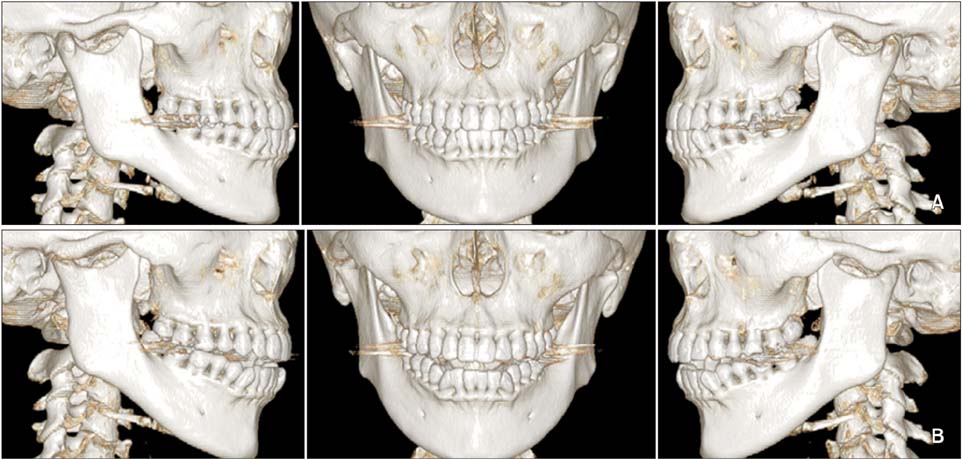
Figure 1 Computed tomography images. A, Before condylar degeneration at the age of 28 years and 9 months. B, After condylar degeneration at the age of 31 years and 10 months.

Figure 2 Computed tomography images of the left condyle. The white arrow illustrates osteophytes.

Figure 3 Pretreatment facial and intraoral photographs.
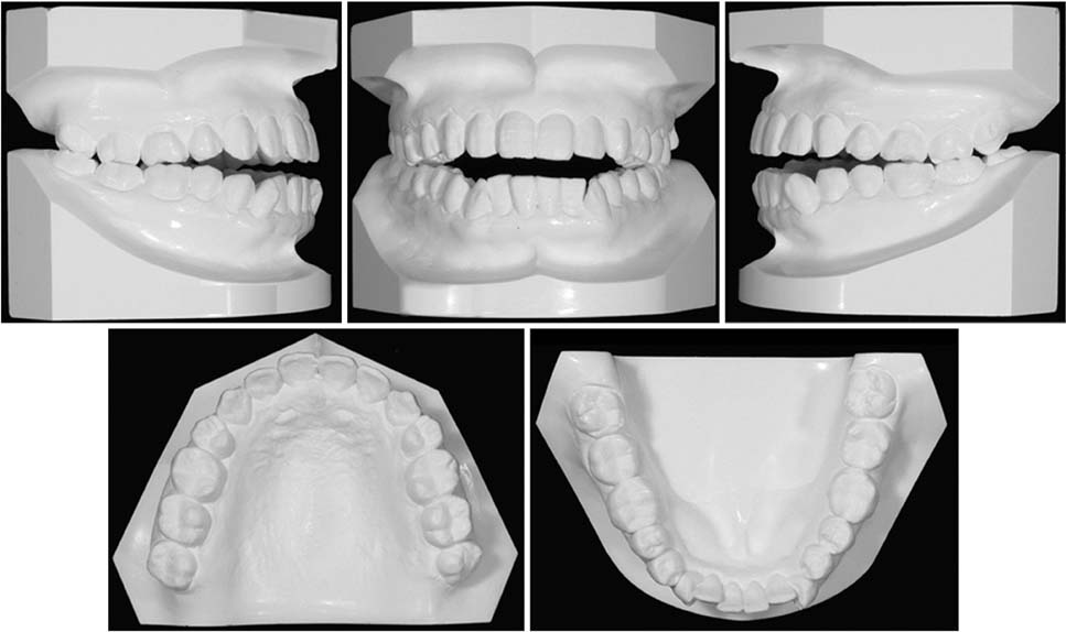
Figure 4 Pretreatment dental casts.

Figure 5 Pretreatment radiographic examination. A, Pretreatment panoramic radiograph. B, Pretreatment lateral cephalogram.
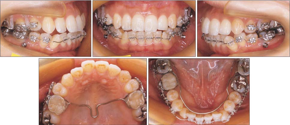
Figure 6 Intraoral photographs obtained after molar intrusion.

Figure 7 Intraoral photographs at distalization of the maxillary arch.

Figure 8 Post-treatment facial and intraoral photographs.

Figure 9 Post-treatment dental casts.

Figure 10 Post-treatment radiographic examination. A, Post-treatment panoramic radiograph. B, Post-treatment lateral cephalogram.

Figure 11 Superimposition of pre- (solid lines) and post-treatment (dashed lines) lateral cephalometric tracings.
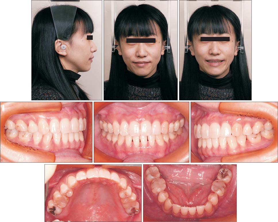
Figure 12 Facial and intraoral photographs obtained 2 years after treatment.
Cited by 1 articles
-
Total intrusion and distalization of the maxillary arch to improve smile esthetics
Eui Seon Baek, Soonshin Hwang, Kyung-Ho Kim, Chooryung J. Chung
Korean J Orthod. 2017;47(1):59-73. doi: 10.4041/kjod.2017.47.1.59.
Reference
-
1. Dimitroulis G. Temporomandibular disorders: a clinical update. BMJ. 1998; 317:190–194.2. McNeill C. Temporomandibular disorders: guidelines for classification, assessment, and management. 2nd ed. Chicago, IL: Quintessence Books;1993.3. Nitzan DW, Dolwick MF, Martinez GA. Temporomandibular joint arthrocentesis: a simplified treatment for severe, limited mouth opening. J Oral Maxillofac Surg. 1991; 49:1163–1167. discussion 1168-70.
Article4. Dimitroulis G, Dolwick MF, Martinez A. Temporomandibular joint arthrocentesis and lavage for the treatment of closed lock: a follow-up study. Br J Oral Maxillofac Surg. 1995; 33:23–26.
Article5. Kondoh T, Dolwick MF, Hamada Y, Seto K. Visually guided irrigation for patients with symptomatic internal derangement of the temporomandibular joint: a preliminary report. Oral Surg Oral Med Oral Pathol Oral Radiol Endod. 2003; 95:544–551.
Article6. Dimitroulis G. A review of 56 cases of chronic closed lock treated with temporomandibular joint arthroscopy. J Oral Maxillofac Surg. 2002; 60:519–524.
Article7. Bertram S, Rudisch A, Innerhofer K, Pümpel E, Grubwieser G, Emshoff R. Diagnosing TMJ internal derangement and osteoarthritis with magnetic resonance imaging. J Am Dent Assoc. 2001; 132:753–761.
Article8. Arnett GW, Milam SB, Gottesman L. Progressive mandibular retrusion-idiopathic condylar resorption. Part II. Am J Orthod Dentofacial Orthop. 1996; 110:117–127.
Article9. Arnett GW, Milam SB, Gottesman L. Progressive mandibular retrusion--idiopathic condylar resorption. Part I. Am J Orthod Dentofacial Orthop. 1996; 110:8–15.
Article10. Wolford LM, Cardenas L. Idiopathic condylar resorption: diagnosis, treatment protocol, and outcomes. Am J Orthod Dentofacial Orthop. 1999; 116:667–677.
Article11. Arnett GW, Tamborello JA. Progressive Class II development: female idiopathic condylar resorption. Oral Maxillofac Surg Clin North Am. 1990; 2:699–716.12. Crawford JG, Stoelinga PJ, Blijdorp PA, Brouns JJ. Stability after reoperation for progressive condylar resorption after orthognathic surgery: report of seven cases. J Oral Maxillofac Surg. 1994; 52:460–466.
Article13. Merkx MA, Van Damme PA. Condylar resorption after orthognathic surgery. Evaluation of treatment in 8 patients. J Craniomaxillofac Surg. 1994; 22:53–58.14. Huang YL, Pogrel MA, Kaban LB. Diagnosis and management of condylar resorption. J Oral Maxillofac Surg. 1997; 55:114–119.
Article15. Bailey LJ, Proffit WR. Combined surgical and orthodontic treatment. In : Proffit WR, Fields HW, editors. Contemporary orthodontics. 3rd ed. St. Louis: Mosby;2000. p. 679–682.16. Paik CH, Woo YJ, Boyd RL. Treatment of an adult patient with vertical maxillary excess using miniscrew fixation. J Clin Orthod. 2003; 37:423–428.17. Kuroda S, Katayama A, Takano-Yamamoto T. Severe anterior open-bite case treated using titanium screw anchorage. Angle Orthod. 2004; 74:558–567.18. Baek MS, Choi YJ, Yu HS, Lee KJ, Kwak J, Park YC. Long-term stability of anterior open-bite treatment by intrusion of maxillary posterior teeth. Am J Orthod Dentofacial Orthop. 2010; 138:396.e1–396.e9.
Article19. Sugawara J, Baik UB, Umemori M, Takahashi I, Nagasaka H, Kawamura H, et al. Treatment and posttreatment dentoalveolar changes following intrusion of mandibular molars with application of a skeletal anchorage system (SAS) for open bite correction. Int J Adult Orthodon Orthognath Surg. 2002; 17:243–253.
- Full Text Links
-
- Actions
-
Cited
- CITED
-
- Close
- Share
-
- Similar articles
-
- Use of Intermaxillary Traction Appliances and Exercises to Strengthen the Masticatory Muscles of Patients with Anterior Open Bite Caused by Temporomandibular Joint Osteoarthritis: Case Reports
- Anterior Open Bite with Temporomandibular Joint Osteoarthritis Treated with Skeletal Anchorage Device: A Case Report
- Evaluation of osseous changes of TMJ in internal derangement and osteoarthritis patients using MRI
- Approach to prosthetic treatment for patients with open bite due to mandibular displacement: Case report
- Management of acquired open bite associated with temporomandibular joint osteoarthritis using miniscrew anchorage
