Projected lifetime cancer risk from cone-beam computed tomography for orthodontic treatment
- Affiliations
-
- 1Department of Dentistry, University of Ulsan College of Medicine, Seoul, Korea
- 2Department of Orthodontics, Asan Medical Center, University of Ulsan College of Medicine, Seoul, Korea
- 3Private Practice, Seoul, Korea
- 4Private Practice, Suwon, Korea
- 5Department of Preventive Medicine, Korea University College of Medicine, Seoul, Korea
- KMID: 2515822
- DOI: http://doi.org/10.4041/kjod.2021.51.3.189
Abstract
Objective
To estimate the projected cancer risk attributable to diagnostic cone-beam computed tomography (CBCT) performed under different exposure settings for orthodontic purposes in children and adults.
Methods
We collected a list of CBCT machines and their specifications from 38 orthodontists. Organ doses were estimated using median and maximum exposure settings of 105 kVp/156.8 mAs and 130 kVp/200 mAs, respectively. The projected cancer risk attributable to CBCT procedures performed 1–3 times within 2 years was calculated for children (aged 5 and 10 years) and adult (aged 20, 30, and 40 years) male and female patients.
Results
For maximum exposure settings, the mean lifetime fractional ratio (LFR) was 14.28% for children and 0.91% for adults; this indicated that the risk to children was 16 times the risk to adults. For median exposure settings, the mean LFR was 5.25% and 0.58% for children and adults, respectively. The risk of cancer decreased with increasing age. For both median and maximum exposure settings, females showed a higher risk of cancer than did males in all age groups. Cancer risk increased with an increase in the frequency of CBCT procedures within a given period.
Conclusions
The projected dental CBCT-associated cancer risk spans over a wide range depending on the machine parameters and image acquisition settings. Children and female patients are at a higher risk of developing cancer associated with diagnostic CBCT. Therefore, the use of diagnostic CBCT should be justified, and protective measures should be taken to minimize the harmful biological effects of radiation.
Figure
-

Figure 1 ALARA-Dental software (Kyung Hee University, Seoul, Korea, and Korea Centers for Disease Control and Prevention, Cheongju, Korea)13 for estimating organ doses delivered for the acquisition of dental radiographs, including intraoral radiographs, panoramic radiographs, and cone-beam computed tomography images.
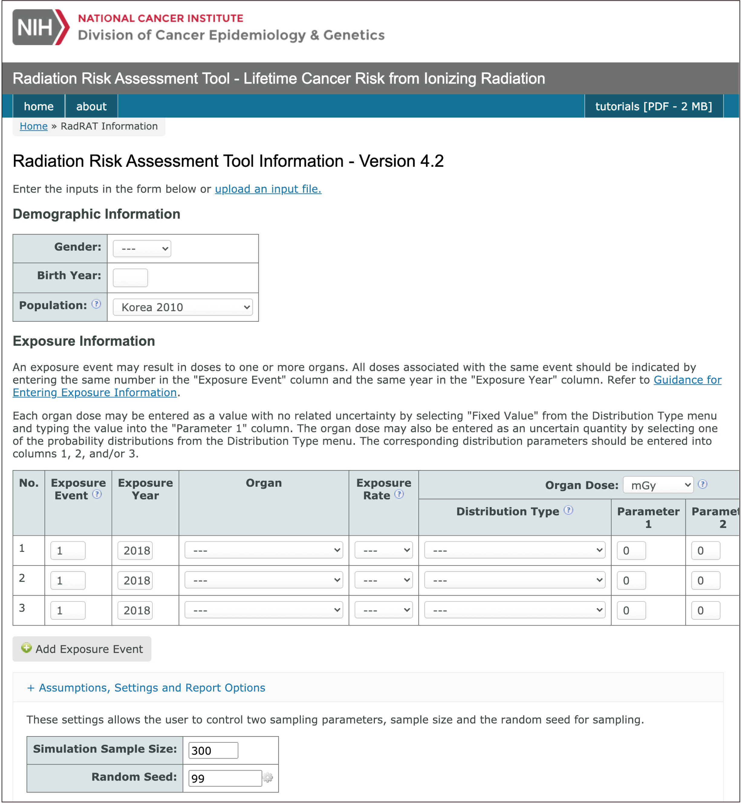
Figure 2 Estimation of cancer risk from cone-beam computed tomography for orthodontic purposes using the radiation risk assessment tool (RadRAT version 4.1.1; National Institutes of Health, Bethesda, MD, USA).18
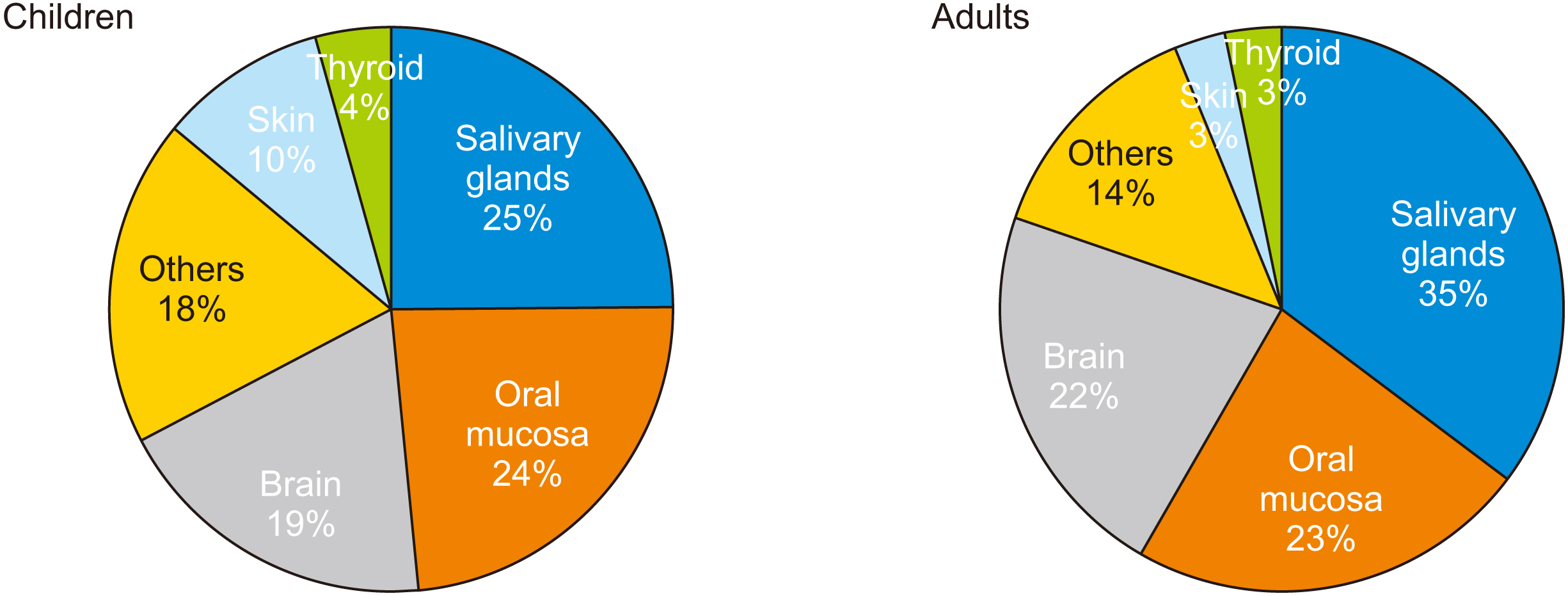
Figure 3 Distribution of mean organ doses (mGy) delivered by cone-beam computed tomography performed for orthodontic purposes in children and adults.
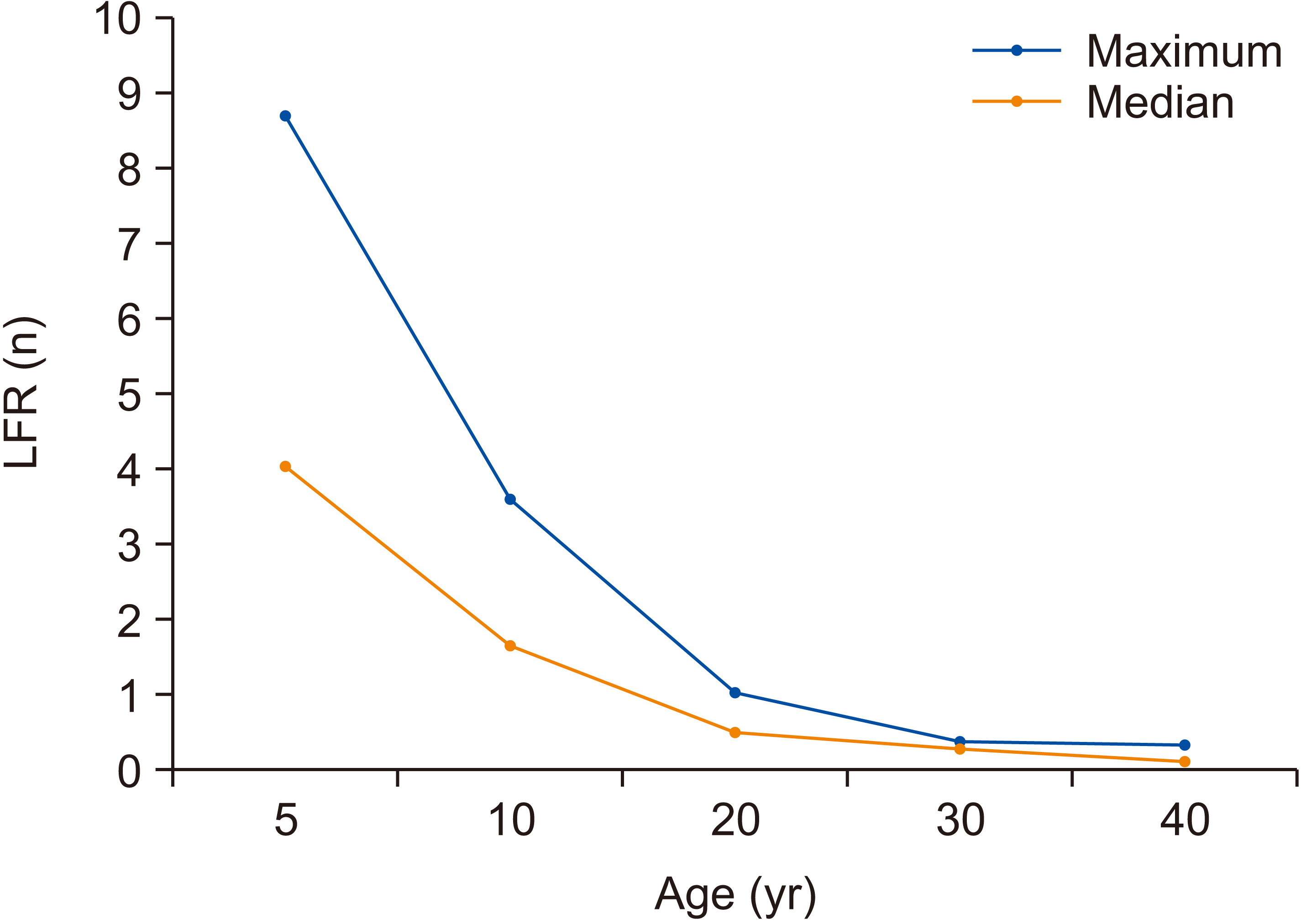
Figure 4 Mean lifetime fractional ratio (LFR) after a single exposure under maximum and median exposure settings, according to age.
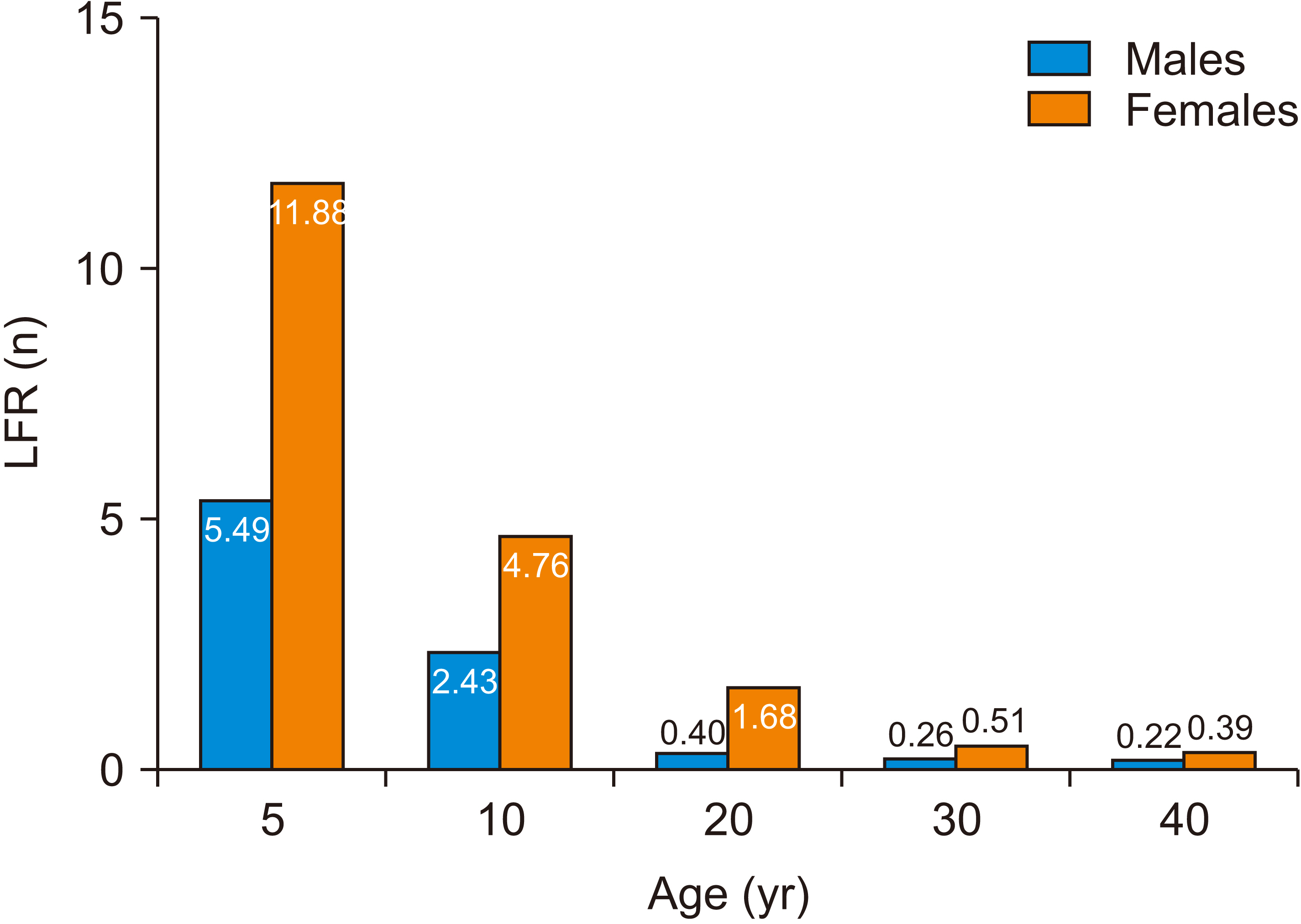
Figure 5 Comparison of the mean lifetime fractional ratio (LFR) after a single exposure under maximum exposure settings between male and female patients in different age groups.
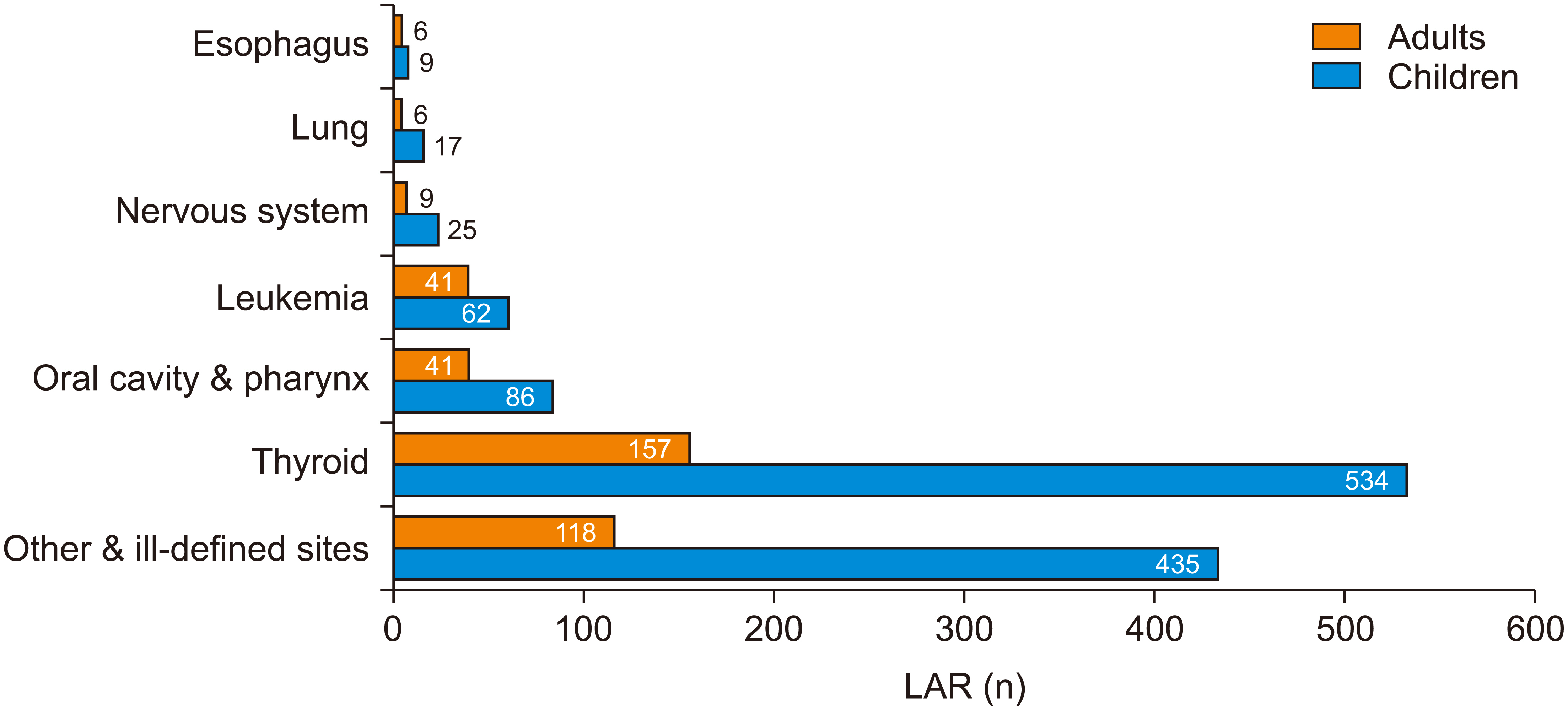
Figure 6 Comparison of mean lifetime attributable risk (LAR) values after a single exposure under maximum exposure parameters between adults and children, according to the cancer site.
Reference
-
1. Adams GL, Gansky SA, Miller AJ, Harrell WE Jr, Hatcher DC. 2004; Comparison between traditional 2-dimensional cephalometry and a 3-dimensional approach on human dry skulls. Am J Orthod Dentofacial Orthop. 126:397–409. DOI: 10.1016/j.ajodo.2004.03.023. PMID: 15470343.
Article2. Tronje G, Welander U, McDavid WD, Morris CR. 1981; Image distortion in rotational panoramic radiography. I. General considerations. Acta Radiol Diagn (Stockh). 22(3A):295–9. DOI: 10.1177/028418518102203A14. PMID: 7315508.3. Loubele M, Bogaerts R, Van Dijck E, Pauwels R, Vanheusden S, Suetens P, et al. 2009; Comparison between effective radiation dose of CBCT and MSCT scanners for dentomaxillofacial applications. Eur J Radiol. 71:461–8. DOI: 10.1016/j.ejrad.2008.06.002. PMID: 18639404.
Article4. Suomalainen A, Vehmas T, Kortesniemi M, Robinson S, Peltola J. 2008; Accuracy of linear measurements using dental cone beam and conventional multislice computed tomography. Dentomaxillofac Radiol. 37:10–7. DOI: 10.1259/dmfr/14140281. PMID: 18195249.
Article5. Kapila SD, Nervina JM. 2015; CBCT in orthodontics: assessment of treatment outcomes and indications for its use. Dentomaxillofac Radiol. 44:20140282. DOI: 10.1259/dmfr.20140282. PMID: 25358833. PMCID: PMC4277443.
Article6. Wrzesień M, Olszewski J. 2017; Absorbed doses for patients undergoing panoramic radiography, cephalometric radiography and CBCT. Int J Occup Med Environ Health. 30:705–13. DOI: 10.13075/ijomeh.1896.00960. PMID: 28584324.
Article7. Ludlow JB, Timothy R, Walker C, Hunter R, Benavides E, Samuelson DB, et al. 2015; Effective dose of dental CBCT-a meta analysis of published data and additional data for nine CBCT units. Dentomaxillofac Radiol. 44:20140197. DOI: 10.1259/dmfr.20140197. PMID: 25224586. PMCID: PMC4277438.
Article8. Ludlow JB, Ivanovic M. 2008; Comparative dosimetry of dental CBCT devices and 64-slice CT for oral and maxillofacial radiology. Oral Surg Oral Med Oral Pathol Oral Radiol Endod. 106:106–14. DOI: 10.1016/j.tripleo.2008.03.018. PMID: 18504152.
Article9. Sezgin ÖS, Kayipmaz S, Yasar D, Yilmaz AB, Ozturk MH. 2012; Comparative dosimetry of dental cone beam computed tomography, panoramic radiography, and multislice computed tomography. Oral Radiol. 28:32–7. DOI: 10.1007/s11282-011-0078-5.
Article10. United Nations. 2013. UNSCEAR 2013 Report to the general assembly, with scientific annexes. Effect of ionizing radiations. United Nations Publications;New York:11. Pearce MS, Salotti JA, Little MP, McHugh K, Lee C, Kim KP, et al. 2012; Radiation exposure from CT scans in childhood and subsequent risk of leukaemia and brain tumours: a retrospective cohort study. Lancet. 380:499–505. DOI: 10.1016/S0140-6736(12)60815-0. PMID: 22681860. PMCID: PMC3418594.
Article12. Hong JY, Han K, Jung JH, Kim JS. 2019; Association of exposure to diagnostic low-dose ionizing radiation with risk of cancer among youths in South Korea. JAMA Netw Open. 2:e1910584. DOI: 10.1001/jamanetworkopen.2019.10584. PMID: 31483470. PMCID: PMC6727680.
Article13. Kim HJ, Lee JY, Lee HK, Kim KP. 2020. Public health weekly report. Computer programs (ALARA) for calculation of diagnostic radiation dose. Korea Disease Control and Prevention Agency;Cheongju: p. 1023–6.14. Lee C, Kim KP, Long DJ, Bolch WE. 2012; Organ doses for reference pediatric and adolescent patients undergoing computed tomography estimated by Monte Carlo simulation. Med Phys. 39:2129–46. DOI: 10.1118/1.3693052. PMID: 22482634. PMCID: PMC3326072.
Article15. Servomaa A, Tapiovaara M. 1998; Organ dose calculation in medical X-ray examinations by the program PCXMC. Radiat Protect Dosim. 80:213–9. DOI: 10.1093/oxfordjournals.rpd.a032509.16. Solberg TD, DeMarco JJ, Chetty IJ, Mesa AV, Cagnon CH, Li AN, et al. 2001; A review of radiation dosimetry applications using the MCNP Monte Carlo code. Radiochim Acta. 89:337–55. DOI: 10.1524/ract.2001.89.4-5.337.
Article17. Long DJ, Lee C, Tien C, Fisher R, Hoerner MR, Hintenlang D, et al. 2013; Monte Carlo simulations of adult and pediatric computed tomography exams: validation studies of organ doses with physical phantoms. Med Phys. 40:013901. DOI: 10.1118/1.4771934. PMID: 23298124. PMCID: PMC3537704.
Article18. Berrington de Gonzalez A, Iulian Apostoaei A, Veiga LH, Rajaraman P, Thomas BA, Owen Hoffman F, et al. 2012; RadRAT: a radiation risk assessment tool for lifetime cancer risk projection. J Radiol Prot. 32:205–22. DOI: 10.1088/0952-4746/32/3/205. PMID: 22810503. PMCID: PMC3816370.19. National Research Council. 2006. Health risks from exposure to low levels of ionizing radiation: BEIR VII phase 2. National Academies Press;Washington, D.C.:20. Kellerer AM, Nekolla EA, Walsh L. 2001; On the conversion of solid cancer excess relative risk into lifetime attributable risk. Radiat Environ Biophys. 40:249–57. DOI: 10.1007/s004110100106. PMID: 11820733.
Article21. Journy NM, Lee C, Harbron RW, McHugh K, Pearce MS, Berrington de González A. 2017; Projected cancer risks potentially related to past, current, and future practices in paediatric CT in the United Kingdom, 1990-2020. Br J Cancer. 116:109–16. DOI: 10.1038/bjc.2016.351. PMID: 27824812. PMCID: PMC5220140.
Article22. Lee WJ, Choi Y, Ko S, Cha ES, Kim J, Kim YM, et al. 2018; Projected lifetime cancer risks from occupational radiation exposure among diagnostic medical radiation workers in South Korea. BMC Cancer. 18:1206. DOI: 10.1186/s12885-018-5107-x. PMID: 30514249. PMCID: PMC6278159.
Article23. Walsh L, Zhang W, Shore RE, Auvinen A, Laurier D, Wakeford R, et al. 2014; A framework for estimating radiation-related cancer risks in Japan from the 2011 Fukushima nuclear accident. Radiat Res. 182:556–72. DOI: 10.1667/RR13779.1. PMID: 25251702.
Article24. Douple EB, Mabuchi K, Cullings HM, Preston DL, Kodama K, Shimizu Y, et al. 2011; Long-term radiation-related health effects in a unique human population: lessons learned from the atomic bomb survivors of Hiroshima and Nagasaki. Disaster Med Public Health Prep. 5 Suppl 1:S122–33. DOI: 10.1001/dmp.2011.21. PMID: 21402804. PMCID: PMC3907953.
Article25. Pauwels R, Cockmartin L, Ivanauskaité D, Urbonienė A, Gavala S, Donta C, et al. 2014; Estimating cancer risk from dental cone-beam CT exposures based on skin dosimetry. Phys Med Biol. 59:3877–91. DOI: 10.1088/0031-9155/59/14/3877. PMID: 24957710.
Article26. Yeh JK, Chen CH. 2018; Estimated radiation risk of cancer from dental cone-beam computed tomography imaging in orthodontics patients. BMC Oral Health. 18:131. DOI: 10.1186/s12903-018-0592-5. PMID: 30075771. PMCID: PMC6091080.
Article27. Stratis A, Zhang G, Jacobs R, Bogaerts R, Bosmans H. 2019; The growing concern of radiation dose in paediatric dental and maxillofacial CBCT: an easy guide for daily practice. Eur Radiol. 29:7009–18. DOI: 10.1007/s00330-019-06287-5. PMID: 31264018.
Article28. Wu TH, Lin WC, Chen WK, Chang YC, Hwang JJ. 2015; Predicting cancer risks from dental computed tomography. J Dent Res. 94:27–35. DOI: 10.1177/0022034514554226. PMID: 25359782.
Article29. Peterson E, De P, Nuttall R. 2012; BMI, diet and female reproductive factors as risks for thyroid cancer: a systematic review. PLoS One. 7:e29177. DOI: 10.1371/journal.pone.0029177. PMID: 22276106. PMCID: PMC3261873.
Article30. Shore RE, Beck HL, Boice JD, Caffrey EA, Davis S, Grogan HA, et al. 2018; Implications of recent epidemiologic studies for the linear nonthreshold model and radiation protection. J Radiol Prot. 38:1217–33. DOI: 10.1088/1361-6498/aad348. PMID: 30004025.
Article
- Full Text Links
-
- Actions
-
Cited
- CITED
-
- Close
- Share
-
- Similar articles
-
- Three-dimensional structural analysis of the morphological condition of the alveolar bone before and after orthodontic treatment
- Management of root canal perforation by using cone-beam computed tomography
- Three-dimensional imaging modalities in endodontics
- An alternative approach to extruding a vertically impacted lower third molar using an orthodontic miniscrew: A case report with cone-beam CT follow-up
- Detection of maxillary second molar with two palatal roots using cone beam computed tomography: a case report
