An alternative approach to extruding a vertically impacted lower third molar using an orthodontic miniscrew: A case report with cone-beam CT follow-up
- Affiliations
-
- 1Department of Oral Radiology, School of Dentistry, University of Sao Paulo, Sao Paulo, Brazil. arthuro@usp.br
- 2Orthodontic Clinic, Sao Paulo, Brazil.
- KMID: 1799601
- DOI: http://doi.org/10.5624/isd.2014.44.2.171
Abstract
- One of the most common oral surgical procedures is the extraction of the lower third molar (LTM). Postoperative complications such as paresthesia due to inferior alveolar nerve (IAN) injury are commonly observed in cases of horizontal and vertical impaction. The present report discusses a case of a vertically impacted LTM associated with a dentigerous cyst. An intimate contact between the LTM roots and the mandibular canal was observed on a panoramic radiograph and confirmed with cone-beam computed tomographic (CBCT) cross-sectional cuts. An orthodontic miniscrew was then used to extrude the LTM prior to its surgical removal in order to avoid the risk of inferior alveolar nerve injury. CBCT imaging follow-up confirmed the success of the LTM orthodontic extrusion.
MeSH Terms
Figure
-
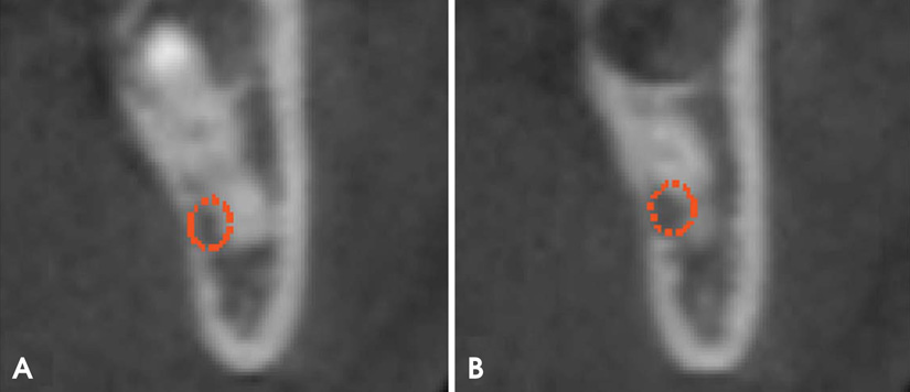
Fig. 1 Initial cross-sectional cone-beam computed tomographic (CBCT) images of the site show the contact between the mandibular canal and the curvatures of the mesial (A) and distal (B) roots, depicted in different CBCT cuts.
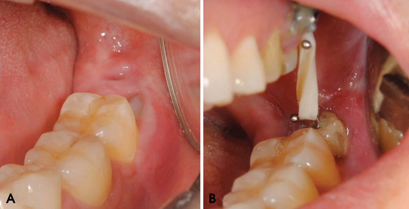
Fig. 2 A clinical photograph shows the lower third molar before (A) and after (B) orthodontic extrusion.
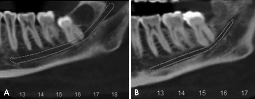
Fig. 3 Initial (A) and final (B) coronal panoramic CBCT reconstruction images of the site show the relationship between the lower third molar roots and the mandibular inferior alveolar canal.
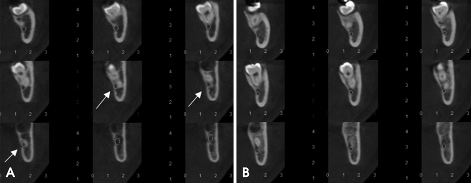
Fig. 4 Initial (A) and final (B) cross-sectional CBCT images of the site show the relationship between the lower third molar roots and the mandibular inferior alveolar canal.

Fig. 5 Mesial (A) and lingual (B) views of the extracted lower third molar show the root curvature (arrow), which comes into contact with the mandibular canal before orthodontic extrusion with the miniscrew.
Reference
-
1. Valmaseda-Castellón E, Berini-Aytés L, Gay-Escoda C. Inferior alveolar nerve damage after lower third molar surgical extraction: a prospective study of 1117 surgical extractions. Oral Surg Oral Med Oral Pathol Oral Radiol Endod. 2001; 92:377–383.
Article2. Susarla SM, Blaeser BF, Magalnick D. Third molar surgery and associated complications. Oral Maxillofac Surg Clin North Am. 2003; 15:177–186.
Article3. Bui CH, Seldin EB, Dodson TB. Types, frequencies, and risk factors for complications after third molar extraction. J Oral Maxillofac Surg. 2003; 61:1379–1389.
Article4. Alessandri Bonetti G, Bendandi M, Laino L, Checchi V, Checchi L. Orthodontic extraction: riskless extraction of impacted lower third molars close to the mandibular canal. J Oral Maxillofac Surg. 2007; 65:2580–2586.
Article5. Wang Y, He D, Yang C, Wang B, Qian W. An easy way to apply orthodontic extraction for impacted lower third molar compressing to the inferior alveolar nerve. J Craniomaxillofac Surg. 2012; 40:234–237.
Article6. Park W, Park JS, Kim YM, Yu HS, Kim KD. Orthodontic extrusion of the lower third molar with an orthodontic mini implant. Oral Surg Oral Med Oral Pathol Oral Radiol Endod. 2010; 110:e1–e6.
Article7. Pell GJ, Gregory BT. Impacted mandibular third molars: classification and modified techniques for removal. Dent Digest. 1933; 39:330–338.8. Tantanapornkul W, Okouchi K, Fujiwara Y, Yamashiro M, Maruoka Y, Ohbayashi N, et al. A comparative study of cone-beam computed tomography and conventional panoramic radiography in assessing the topographic relationship between the mandibular canal and impacted third molars. Oral Surg Oral Med Oral Pathol Oral Radiol Endod. 2007; 103:253–259.
Article9. Scarfe WC, Farman AG, Sukovic P. Clinical applications of cone-beam computed tomography in dental practice. J Can Dent Assoc. 2006; 72:75–80.10. Roberts JA, Drage NA, Davies J, Thomas DW. Effective dose from cone beam CT examinations in dentistry. Br J Radiol. 2009; 82:35–40.
Article11. Guida L, Cuccurullo GP, Lanza A, Tedesco M, Guida A, Annunziata M. Orthodontic-aided extraction of impacted third molar to improve the periodontal status of the neighboring tooth. J Craniofac Surg. 2011; 22:1922–1924.
Article12. Itro A, Lupo G, Marra A, Carotenuto A, Cocozza E, Filipi M, et al. The piezoelectric osteotomy technique compared to the one with rotary instruments in the surgery of included third molars. A clinical study. Minerva Stomatol. 2012; 61:247–253.
- Full Text Links
-
- Actions
-
Cited
- CITED
-
- Close
- Share
-
- Similar articles
-
- Cone beam computed tomography findings of ectopic mandibular third molar in the mandibular condyle: report of a case
- Assessment of the relationship between the mandibular third molar and the mandibular canal using panoramic radiograph and cone beam computed tomography
- Detection of maxillary second molar with two palatal roots using cone beam computed tomography: a case report
- Orthodontic traction of a horizontally impacted mandibular second premolar
- Orthodontic Traction of the Permanent Molar Using Skeletal Anchorage: A Case Report
