Detection of maxillary second molar with two palatal roots using cone beam computed tomography: a case report
- Affiliations
-
- 1Department of Dentistry, Seoul Veterans Hospital, Seoul, Republic of Korea. endo95@naver.com
- KMID: 2162383
- DOI: http://doi.org/10.14368/jdras.2016.32.1.87
Abstract
- The purpose of this clinical report was to show anatomical variations in permanent maxillary second molar using computed tomography (CT). This case report describes the application of CT to detect the unusual root anatomy of maxillary second molar with 2 separate palatal roots for successful endodontic treatment procedures. The use of cone beam computed tomography (CBCT) can overcome the limitation of the periapical standard radiography caused by the overlap of buccal and secondary palatal roots.
Figure
-
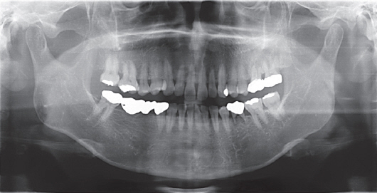
Fig. 1 Preoperative radiograph.
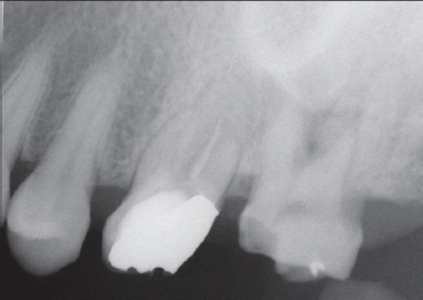
Fig. 2 Radiograph showing severe secondary decay after old bridge removal.
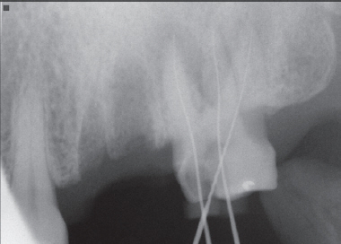
Fig. 3 Working length with 3 roots.
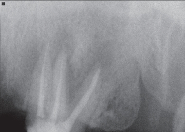
Fig. 4 Postoperative radiograph of maxillary second molar filled with guttapercha and sealer.
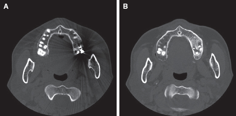
Fig. 5 Cone beam computed tomography showing secondary palatal root. (A, B) Revealed a 2 palatal roots two separated foramina at the apex of maxillary second molar.
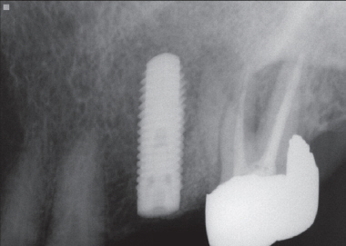
Fig. 6 Periapical standard radiograph was taken at different angles to confirm two separated.
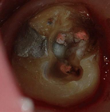
Fig. 7 4th root canal orifice was clearly identified.
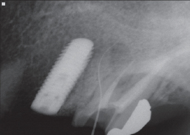
Fig. 8 Working length of 4th canal.
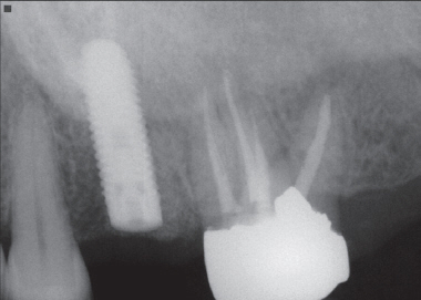
Fig. 9 4th canal filled with guttapercha and sealer.
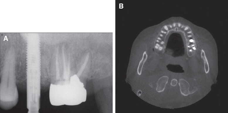
Fig. 10 (A) Periapical standard radiograph: 5-month follow-up after 4th canal filling, (B) Cone beam computed tomography: 5-month follow-up after 4th canal filling.
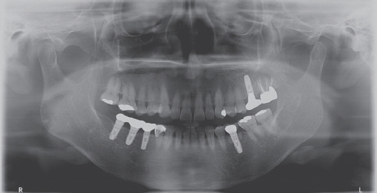
Fig. 11 5-years follow-up after 4th canal filling.
Reference
-
References
1. Stone LH, Stroner WF. Maxillary molars demonstrating more than one palatal root canal. Oral Surg Oral Med Oral Pathol. 1981; 51:649–52. DOI: 10.1016/S0030-4220(81)80017-5.2. Diamond M. Dental anatomy including anatomy of the head and neck. 1952. 3rd ed. New York: MacMillan;p. 203–5.3. Slowey RR. Radiographic aids in the detection of extra root canals. Oral Surg Oral Med Oral Pathol. 1974; 37:762–72. DOI: 10.1016/0030-4220(74)90142-X.4. Libfeld H, Rotstein I. Incidence of four rooted maxillary second molars: literature review and radiographic survey of 1200 teeth. J Endod. 1989; 15:129–31. DOI: 10.1016/S0099-2399(89)80134-7.5. Christie WH, Peikoff MD, Fogel HM. Maxillary molar with two palatal roots: a retrospective clinical study. J Endod. 1991; 17:80–4. DOI: 10.1016/S0099-2399(06)81613-4.6. Shin SJ, Park JW, Lee JK, Hwang SW. Unusual root canal anatomy in maxillay second molar: two case reports. Oral Surg Oral Med Oral Pathol Oral Radiol Endod. 2007; 104:e61–5. DOI: 10.1016/j.tripleo.2007.07.014. PMID: 17942346.7. Barbizam JV, Ribeiro RG, Tanomaru Filho M. Unusual anatomy of permanent maxillary molars. J Endod. 2004; 30:668–71. DOI: 10.1097/01.DON.0000121618.45515.5A. PMID: 15329575.8. Tomazinho FS, Baratto-Filho F, Zaitter S, Leonardi DP, Gonzaga CC. Unusual anatomy of a maxillary first molar with two palatal roots: a case report. J Oral Science. 2010; 52:149–53. DOI: 10.2334/josnusd.52.149.9. Patel S, Dawood A. The use of cone beam computed tomography in the management of external cervical resorption lesions. Int Endod J. 2007; 40:730–7. DOI: 10.1111/j.1365-2591.2007.01247.x. PMID: 17608680.10. Cotton TP, Geisler TM, Holden DT, Schwartz SA, Schindler WG. Endodontic applications of conebeam volumetric tomography. J Endod. 2007; 33:1121–32. DOI: 10.1016/j.joen.2007.06.011. PMID: 17931947.
- Full Text Links
-
- Actions
-
Cited
- CITED
-
- Close
- Share
-
- Similar articles
-
- Assessment of the relationship between the maxillary molars and adjacent structures using cone beam computed tomography
- Endodontic management of a maxillary first molar with three roots and seven root canals with the aid of cone-beam computed tomography
- Comparison of panoramic radiography and cone beam computed tomography for assessing the relationship between the maxillary sinus floor and maxillary molars
- Radix mesiolingualis and radix distolingualis: a case report of a tooth with an unusual morphology
- Maxillary sinus pneumatization after maxillary molar extraction assessed with cone beam computed tomography
