Three-dimensional structural analysis of the morphological condition of the alveolar bone before and after orthodontic treatment
- Affiliations
-
- 1Division of Oral Health Sciences, Department of Orofacial Development and Function, Graduate School, Tokyo Medical and Dental University, Tokyo, Japan. y.shimizu.orts@tmd.ac.jp
- KMID: 2392186
- DOI: http://doi.org/10.4041/kjod.2017.47.6.394
Abstract
- Assessing the condition of the alveolar bone before and after orthodontic treatment is important. Recently, cone-beam computed tomography has been widely accepted as a useful tool for orthodontic treatment. Moreover, using a three-dimensional (3D) structural analysis software enables gathering detailed information and quantifying data. The aim of this study was to introduce various quantitative analyses performed before and after orthodontic treatment by using a 3D structural analysis software for evaluating the morphological condition of the alveolar bone of a patient with gingival recession around the canines.
Figure
-
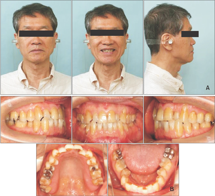
Figure 1 A, Pretreatment frontal/lateral profiles of the patient aged 60 years and 1 month. B, Pretreatment intraoral photographs.
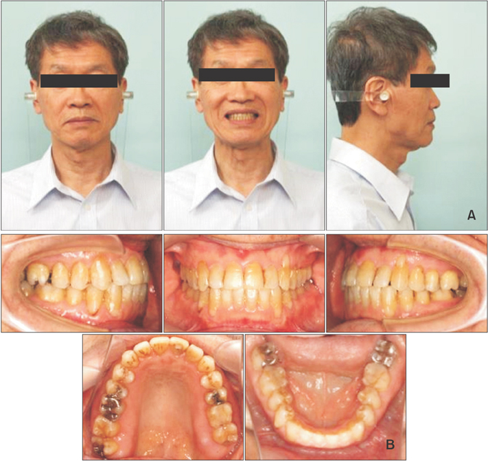
Figure 2 A, Posttreatment frontal/lateral profiles of the patient aged 62 years and 9 months. B, Posttreatment intraoral photographs.
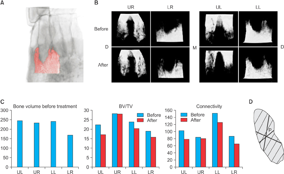
Figure 3 A, Cortical bone surrounding the lower canine (red area). B, Three-dimensional reconstructed images of the cortical bone surrounding the canines. C, The bone volume before treatment, bone volume per tissue volume (BV/TV), and connectivity of the cortical bones before/after treatment. Blue bar, before treatment; red bar, after treatment. D, Diagram of “connectivity,” which is defined as the average volume from the point in the trabecular bone to the end of the trabecular bone in all directions. The shaded area, bone; P, a point. UL, Upper left; UR, upper right; LL, lower left; LR, lower right; M, medial; D, distal.
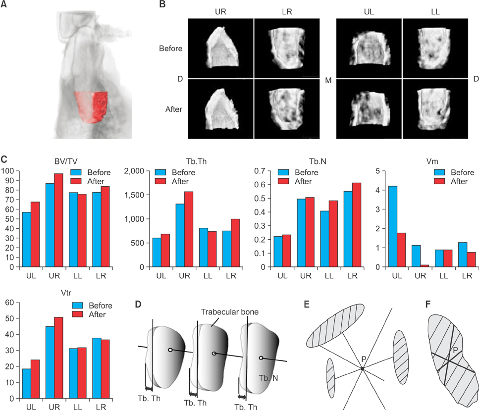
Figure 4 A, Trabecular bone surrounding the lower canine (red area). B, Three-dimensional reconstructed images of the trabecular bone surrounding the canines. C, The bone volume per tissue volume (BV/TV), trabecular thickness (Tb.Th, mm), trabecular number (Tb.N, per mm), marrow space star volume (Vm, mm3), and trabecular star volume (Vtr, mm3) before/after treatment, and a diagram of those parameters. Blue bar, before treatment; red bar, after treatment. D, Diagram of Tb.Th and Tb.N. E, Diagram of Vm. F, Diagram of Vtr. The shaded area, bone; P, a point. UL, Upper left; UR, upper right; LL, lower left; LR, lower right; M, medial; D, distal.
Reference
-
1. Newman WG. Possible etiologic factors in external root resorption. Am J Orthod. 1975; 67:522–539.
Article2. Fuss Z, Tsesis I, Lin S. Root resorption--diagnosis, classification and treatment choices based on stimulation factors. Dent Traumatol. 2003; 19:175–182.
Article3. Ishida Y, Kanno Z, Soma K. Occlusal hypofunction induces atrophic changes in rat gingiva. Angle Orthod. 2008; 78:1015–1022.
Article4. Enokida M, Kaneko S, Yanagishita M, Soma K. Inference of occlusal stimuli on the remodelling of alveolar bone in a rat hypofunction-recovery model. J Oral Biosci. 2005; 47:321–334.
Article5. Shimomoto Y, Chung CJ, Iwasaki-Hayashi Y, Muramoto T, Soma K. Effects of occlusal stimuli on alveolar/jaw bone formation. J Dent Res. 2007; 86:47–51.
Article6. Shimizu Y, Hosomichi J, Kaneko S, Shibutani N, Ono T. Effect of sympathetic nervous activity on alveolar bone loss induced by occlusal hypofunction in rats. Arch Oral Biol. 2011; 56:1404–1411.
Article7. Shimizu Y, Hosomichi J, Nakamura S, Ono T. Micro-computed tomography analysis of changes in the periodontal ligament and alveolar bone proper induced by occlusal hypofunction of rat molars. Korean J Orthod. 2014; 44:263–267.
Article8. Shimizu Y, Ishida T, Hosomichi J, Kaneko S, Hatano K, Ono T. Soft diet causes greater alveolar osteopenia in the mandible than in the maxilla. Arch Oral Biol. 2013; 58:907–911.
Article9. Castro LO, Castro IO, de Alencar AH, Valladares-Neto J, Estrela C. Cone beam computed tomography evaluation of distance from cementoenamel junction to alveolar crest before and after nonextraction orthodontic treatment. Angle Orthod. 2016; 86:543–549.
Article10. Ann HR, Jung YS, Lee KJ, Baik HS. Evaluation of stability after pre-orthodontic orthognathic surgery using cone-beam computed tomography: A comparison with conventional treatment. Korean J Orthod. 2016; 46:301–309.
Article11. Choi JH, Park CH, Yi SW, Lim HJ, Hwang HS. Bone density measurement in interdental areas with simulated placement of orthodontic miniscrew implants. Am J Orthod Dentofacial Orthop. 2009; 136:766.e1–766.e12.
Article12. Renkema AM, Sips ET, Bronkhorst E, Kuijpers-Jagtman AM. A survey on orthodontic retention procedures in The Netherlands. Eur J Orthod. 2009; 31:432–437.
Article13. van Leeuwen EJ, Maltha JC, Kuijpers-Jagtman AM, van't Hof MA. The effect of retention on orthodontic relapse after the use of small continuous or discontinuous forces. An experimental study in beagle dogs. Eur J Oral Sci. 2003; 111:111–116.
Article14. Reitan K. Tissue rearrangement during retention of orthodontically rotated teeth. Angle Orthod. 1959; 29:105–113.15. Edwards JG. A long-term prospective evaluation of the circumferential supracrestal fiberotomy in alleviating orthodontic relapse. Am J Orthod Dentofacial Orthop. 1988; 93:380–387.
Article16. Bouxsein ML, Boyd SK, Christiansen BA, Guldberg RE, Jepsen KJ, Müller R. Guidelines for assessment of bone microstructure in rodents using microcomputed tomography. J Bone Miner Res. 2010; 25:1468–1486.
Article17. Vesterby A. Marrow space star volume can reveal change of trabecular connectivity. Bone. 1993; 14:193–197.
Article18. Vesterby A. Star volume of marrow space and trabeculae in iliac crest: sampling procedure and correlation to star volume of first lumbar vertebra. Bone. 1990; 11:149–155.
Article19. Kazama JJ, Koda R, Yamamoto S, Narita I, Gejyo F, Tokumoto A. Cancellous bone volume is an indicator for trabecular bone connectivity in dialysis patients. Clin J Am Soc Nephrol. 2010; 5:292–298.
Article20. Aoki K, Saito H, Itzstein C, Ishiguro M, Shibata T, Blanque R, et al. A TNF receptor loop peptide mimic blocks RANK ligand-induced signaling, bone resorption, and bone loss. J Clin Invest. 2006; 116:1525–1534.
Article21. Sato K, Suematsu A, Nakashima T, Takemoto-Kimura S, Aoki K, Morishita Y, et al. Regulation of osteoclast differentiation and function by the CaMK-CREB pathway. Nat Med. 2006; 12:1410–1416.
Article22. Kiper POS, Saito H, Gori F, Unger S, Hesse E, Yamana K, et al. Cortical-bone fragility--insights from sFRP4 deficiency in Pyle's disease. N Engl J Med. 2016; 374:2553–2562.23. Oishi S, Shimizu Y, Hosomichi J, Kuma Y, Nagai H, Maeda H, et al. Intermittent hypoxia induces disturbances in craniofacial growth and defects in craniofacial morphology. Arch Oral Biol. 2016; 61:115–124.
Article24. Oishi S, Shimizu Y, Hosomichi J, Kuma Y, Maeda H, Nagai H, et al. Intermittent hypoxia influences alveolar bone proper microstructure via hypoxia-inducible factor and VEGF expression in periodontal ligaments of growing rats. Front Physiol. 2016; 7:416.
Article25. Ito K, Gomi Y, Sato S, Arai Y, Shinoda K. Clinical application of a new compact CT system to assess 3-D images for the preoperative treatment planning of implants in the posterior mandible A case report. Clin Oral Implants Res. 2001; 12:539–542.
Article26. Nomura Y, Watanabe H, Honda E, Kurabayashi T. Reliability of voxel values from cone-beam computed tomography for dental use in evaluating bone mineral density. Clin Oral Implants Res. 2010; 21:558–562.
Article
- Full Text Links
-
- Actions
-
Cited
- CITED
-
- Close
- Share
-
- Similar articles
-
- A study on the morphological changes of lower incisor and symphysis during surgical-orthodontic treatment in skeletal class III malocclusion
- Changes of root length and crestal bone height before and after the orthodontic treatment in nail biting patients
- Alveolar Bone Preservation Using an Orthodontic Mini-implant in an Adolescent Patient with Multiple Missing Teeth
- A study about alveolar crest bone height before and after orthodontic treatment by using bitewing film
- A study on the change of alveolar crest height following orthodontic treatment
