L-histidine and L-carnosine exert anti-brain aging effects in D-galactose-induced aged neuronal cells
- Affiliations
-
- 1Department of Nutritional Science and Food Management, Ewha Womans University, Seoul 03760, Korea
- KMID: 2502480
- DOI: http://doi.org/10.4162/nrp.2020.14.3.188
Abstract
- BACKGROUND/OBJECTIVES
Brain aging is a major risk factor for severe neurodegenerative diseases. Conversely, L-histidine and L-carnosine are known to exhibit neuroprotective effects. The aim of this study was to examine the potential for L-histidine, L-carnosine, and their combination to mediate anti-brain aging effects in neuronal cells subjected to D-galactose-induced aging.
MATERIALS/METHODS
The neuroprotective potential of L-histidine, L-carnosine, and their combination was examined in a retinoic acid-induced neuronal differentiated SH-SY5Y cell line exposed to D-galactose (200 mM) for 48 h. Neuronal cell proliferation, differentiation, and expression of anti-oxidant enzymes and apoptosis markers were subsequently evaluated.
RESULTS
Treatment with L-histidine (1 mM), L-carnosine (10 mM), or both for 48 h efficiently improved the proliferation, neurogenesis, and senescence of D-galactose-treated SH-SY5Y cells. In addition, protein expression levels of both neuronal markers (β tubulin-III and neurofilament heavy protein) and anti-oxidant enzymes, glutathione peroxidase-1 and superoxide dismutase-1 were up-regulated. Conversely, protein expression levels of amyloid β (1-42) and cleaved caspase-3 were down-regulated. Levels of mRNA for the pro-inflammatory cytokines, interleukin (IL)-8, IL-1β, and tumor necrosis factor-α were also down-regulated.
CONCLUSIONS
To the best of our knowledge, we provide the first evidence that L-histidine, L-carnosine, and their combination mediate anti-aging effects in a neuronal cell line subjected to D-galactose-induced aging. These results suggest the potential benefits of L-histidine and L-carnosine as anti-brain aging agents and they support further research of these amino acid molecules.
Keyword
Figure
-
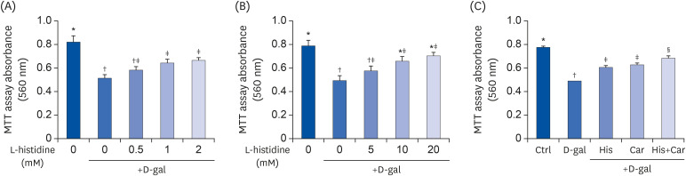
Fig. 1 Effects of L-histidine, L-carnosine, and their combination on proliferation.The neuronal proliferation was assessed by MTT assay. (A) L-histidine and (B) L-carnosine were treated with various concentrations for 48 h. (C) His, Car, and His+Car were treated for 48 h and assessed by MTT assay. The values shown are the mean ± standard error of the mean (n = 3–4).Ctrl, Control; D-gal, 200 mM D-galactose; His, 1 mM L-histidine; Car, 10 mM L-carnosine; His+Car, 1 mM L-histidine + 10 mM L-carnosine.*,†,‡,§Different superscript marks indicate significant differences between groups (P < 0.05).

Fig. 2 Effects of L-histidine, L-carnosine, and their combination on neuronal cell regenerations.Representative pictures for each group (100× magnification). (A) Ctrl, (B) D-gal, (C) His, (D) Car, and (E) His+Car. (F) The average length of the neurites of differentiated SH-SY5Y cells for each group was analyzed. ImageJ software was used to measure the individual neurite length. The protein expressions of an axonal marker, NEFH, and a neuronal marker, β-tubulin III were determined by Western blotting and β-actin was used as a loading control. (G) Representative blots are shown. Quantification of NEFH (H) and β-tubulin III (I) levels to β-actin are shown. The values shown are the mean ± standard error of the mean (n = 3–4).NEFH, Neurofilament heavy polypeptide; Ctrl, Control; D-gal, 200 mM D-galactose; His, 1 mM L-histidine; Car, 10 mM L-carnosine; His+Car, 1 mM L-histidine + 10 mM L-carnosine.*,†,‡,§Different superscript marks indicate significant differences between groups (P < 0.05).
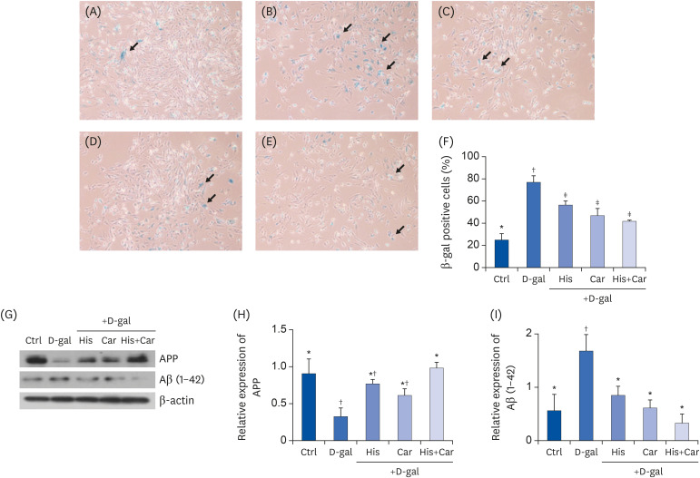
Fig. 3 Effects of L-histidine, L-carnosine, and their combination on cellular senescence and levels of APP and Aβ (1-42) in neuronal cells.SA-β-gal assay was performed to investigate cellular senescence. Representative images for each group at 100× magnification were shown. (A) Ctrl, (B) D-gal, (C) His, (D) Car (E) His+Car. (F) % of SA-β-gal positive cells in total counted cells. The protein expressions of APP and Aβ (1-42) were determined by Western blotting and β-actin was used as a loading control. (G) Representative blots are represented. Quantification of APP (H) and Aβ (1-42) (I) levels to β-actin are shown. The values shown are the mean ± standard error of the mean (n = 3–4).APP, Amyloid β precursor protein; Aβ (1-42), Amyloid β (1-42); SA-β-gal, Senescence-associated β-galactosidase; Ctrl, Control; D-gal, 200 mM D-galactose; His, 1 mM L-histidine; Car, 10 mM L-carnosine; His+Car, 1 mM L-histidine + 10 mM L-carnosine.*,†,‡Different superscript marks indicate significant differences between groups (P < 0.05).
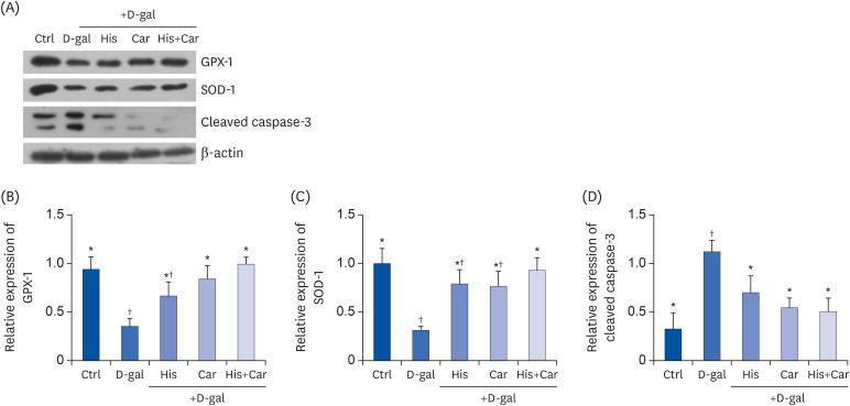
Fig. 4 Effects of L-histidine, L-carnosine, and their combination on anti-oxidant enzymes and caspase-3.The protein expressions of GPX-1, SOD-1, and cleaved caspase-3 were determined by Western blotting and β-actin was used as a loading control. (A) Representative blots are represented (left panels). Quantification of (B) GPX-1, (C) SOD-1, and (D) Cleaved caspase-3 levels to β-actin are shown. The values shown are the mean ± standard error of the mean (n = 3–4).GPX-1, glutathione peroxidase-1; SOD-1, superoxide dismutase-1; Ctrl, Control; D-gal, 200 mM D-galactose; His, 1 mM L-histidine; Car, 10 mM L-carnosine; His+Car, 1 mM L-hisitidne + 10 mM L-carnosine.*,†Different superscript marks indicate significant differences between groups (P < 0.05).
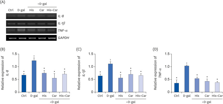
Fig. 5 Effects of L-histidine, L-carnosine, and their combination on pro-inflammatory cytokines in neuronal cells.The mRNA expressions of IL-8, IL-1β, and TNF-α were determined by RT-PCR and GAPDH was used as a loading control. (A) Representative blots are shown. Quantification of (B) IL-8, (C) IL-1β, and (D) TNF-α levels to GAPDH are shown. The values shown are the mean ± SE of the mean (n = 3–4).Ctrl, Control; D-gal, 200 mM D-galactose; His, 1 mM L-histidine; Car, 10 mM L-carnosine; His+Car, 1 mM L-hisitidne + 10 mM L-carnosine.*,†Different superscript marks indicate significant differences between groups (P < 0.05).
Reference
-
1. Gandhi S, Abramov AY. Mechanism of oxidative stress in neurodegeneration. Oxid Med Cell Longev. 2012; 2012:428010. PMID: 22685618.
Article2. Mattson MP, Magnus T. Ageing and neuronal vulnerability. Nat Rev Neurosci. 2006; 7:278–294. PMID: 16552414.
Article3. Ohnuma S, Harris WA. Neurogenesis and the cell cycle. Neuron. 2003; 40:199–208. PMID: 14556704.
Article4. Khodosevich K, Monyer H. Signaling involved in neurite outgrowth of postnatally born subventricular zone neurons in vitro. BMC Neurosci. 2010; 11:18. PMID: 20146799.
Article5. Park MH, Son DJ, Nam KT, Kim SY, Oh SY, Song MJ, Chun HO, Lee TH, Hong JT. 2016.6. Finkel T, Holbrook NJ. Oxidants, oxidative stress and the biology of ageing. Nature. 2000; 408:239–247. PMID: 11089981.
Article7. Zhang YW, Thompson R, Zhang H, Xu H. APP processing in Alzheimer's disease. Mol Brain. 2011; 4:3. PMID: 21214928.
Article8. Yin F, Sancheti H, Patil I, Cadenas E. Energy metabolism and inflammation in brain aging and Alzheimer's disease. Free Radic Biol Med. 2016; 100:108–122. PMID: 27154981.
Article9. Tha KK, Okuma Y, Miyazaki H, Murayama T, Uehara T, Hatakeyama R, Hayashi Y, Nomura Y. Changes in expressions of proinflammatory cytokines IL-1beta, TNF-alpha and IL-6 in the brain of senescence accelerated mouse (SAM) P8. Brain Res. 2000; 885:25–31. PMID: 11121526.10. McLarnon JG. Chemokine interleukin-8 (IL-8) in Alzheimer's and other neurodegenerative diseases. J Alzheimers Dis Parkinsonism. 2016; 6:273.
Article11. Song X, Bao M, Li D, Li YM. Advanced glycation in D-galactose induced mouse aging model. Mech Ageing Dev. 1999; 108:239–251. PMID: 10405984.
Article12. Lu J, Zheng YL, Luo L, Wu DM, Sun DX, Feng YJ. Quercetin reverses D-galactose induced neurotoxicity in mouse brain. Behav Brain Res. 2006; 171:251–260. PMID: 16707173.
Article13. Shwe T, Pratchayasakul W, Chattipakorn N, Chattipakorn SC. Role of D-galactose-induced brain aging and its potential used for therapeutic interventions. Exp Gerontol. 2018; 101:13–36. PMID: 29129736.
Article14. Shen Y, Gao H, Shi X, Wang N, Ai D, Li J, Ouyang L, Yang J, Tian Y, Lu J. Glutamine synthetase plays a role in D-galactose-induced astrocyte aging in vitro and in vivo. Exp Gerontol. 2014; 58:166–173. PMID: 25128847.
Article15. Hsieh HM, Wu WM, Hu ML. Genistein attenuates D-galactose-induced oxidative damage through decreased reactive oxygen species and NF-κB binding activity in neuronal PC12 cells. Life Sci. 2011; 88:82–88. PMID: 21056584.
Article16. Liu YY, Nagpure BV, Wong PT, Bian JS. Hydrogen sulfide protects SH-SY5Y neuronal cells against d-galactose induced cell injury by suppression of advanced glycation end products formation and oxidative stress. Neurochem Int. 2013; 62:603–609. PMID: 23274001.
Article17. Heidari S, Mehri S, Shariaty V, Hosseinzadeh H. Preventive effects of crocin on neuronal damages induced by D-galactose through AGEs and oxidative stress in human neuroblastoma cells (SH-SY5Y). J Pharmacopuncture. 2018; 21:18–25. PMID: 30151301.18. Rassin DK. Essential and non-essential amino acids in neonatal nutrition. Nestle Nutr Workshop Ser. 1994; 33:183–195.19. Cai Q, Takemura G, Ashraf M. Antioxidative properties of histidine and its effect on myocardial injury during ischemia/reperfusion in isolated rat heart. J Cardiovasc Pharmacol. 1995; 25:147–155. PMID: 7723345.
Article20. Matheson IB, Lee J. Chemical reaction rates of amino acids with singlet oxygen. Photochem Photobiol. 1979; 29:879–881.
Article21. Edwards C, Canfield J, Copes N, Brito A, Rehan M, Lipps D, Brunquell J, Westerheide SD, Bradshaw PC. Mechanisms of amino acid-mediated lifespan extension in Caenorhabditis elegans. BMC Genet. 2015; 16:8. PMID: 25643626.
Article22. Adachi N, Liu K, Arai T. Prevention of brain infarction by postischemic administration of histidine in rats. Brain Res. 2005; 1039:220–223. PMID: 15781067.
Article23. Liao RJ, Jiang L, Wang RR, Zhao HW, Chen Y, Li Y, Wang L, Jie LY, Zhou YD, Zhang XN, Chen Z, Hu WW. Histidine provides long-term neuroprotection after cerebral ischemia through promoting astrocyte migration. Sci Rep. 2015; 5:15356. PMID: 26481857.
Article24. Rama Rao KV, Reddy PV, Tong X, Norenberg MD. Brain edema in acute liver failure: inhibition by L-histidine. Am J Pathol. 2010; 176:1400–1408. PMID: 20075201.25. Farshid AA, Tamaddonfard E, Najafi S. Effects of histidine and n-acetylcysteine on experimental lesions induced by doxorubicin in sciatic nerve of rats. Drug Chem Toxicol. 2015; 38:436–441. PMID: 25427688.26. Ruszkiewicz J, Albrecht J. Changes of the thioredoxin system, glutathione peroxidase activity and total antioxidant capacity in rat brain cortex during acute liver failure: modulation by L-histidine. Neurochem Res. 2015; 40:293–300. PMID: 25161077.
Article27. Pichili VB, Rao KV, Jayakumar AR, Norenberg MD. Inhibition of glutamine transport into mitochondria protects astrocytes from ammonia toxicity. Glia. 2007; 55:801–809. PMID: 17357151.
Article28. Kohen R, Yamamoto Y, Cundy KC, Ames BN. Antioxidant activity of carnosine, homocarnosine, and anserine present in muscle and brain. Proc Natl Acad Sci U S A. 1988; 85:3175–3179. PMID: 3362866.
Article29. Yuneva AO, Kramarenko GG, Vetreshchak TV, Gallant S, Boldyrev AA. Effect of carnosine on Drosophila melanogaster lifespan. Bull Exp Biol Med. 2002; 133:559–561. PMID: 12447464.30. Gallant S, Semyonova M, Yuneva M. Carnosine as a potential anti-senescence drug. Biochemistry (Mosc). 2000; 65:866–868. PMID: 10951107.31. Shen Y, Hu WW, Fan YY, Dai HB, Fu QL, Wei EQ, Luo JH, Chen Z. Carnosine protects against NMDA-induced neurotoxicity in differentiated rat PC12 cells through carnosine-histidine-histamine pathway and H(1)/H(3) receptors. Biochem Pharmacol. 2007; 73:709–717. PMID: 17169331.
Article32. Mizuno D, Konoha-Mizuno K, Mori M, Sadakane Y, Koyama H, Ohkawara S, Kawahara M. Protective activity of carnosine and anserine against zinc-induced neurotoxicity: a possible treatment for vascular dementia. Metallomics. 2015; 7:1233–1239. PMID: 25846004.
Article33. Zhang L, Yao K, Fan Y, He P, Wang X, Hu W, Chen Z. Carnosine protects brain microvascular endothelial cells against rotenone-induced oxidative stress injury through histamine H₁ and H₂ receptors in vitro. Clin Exp Pharmacol Physiol. 2012; 39:1019–1025. PMID: 23127196.34. Preston JE, Hipkiss AR, Himsworth DT, Romero IA, Abbott JN. Toxic effects of beta-amyloid(25-35) on immortalised rat brain endothelial cell: protection by carnosine, homocarnosine and beta-alanine. Neurosci Lett. 1998; 242:105–108. PMID: 9533405.35. Zhang ZY, Sun BL, Yang MF, Li DW, Fang J, Zhang S. Carnosine attenuates early brain injury through its antioxidative and anti-apoptotic effects in a rat experimental subarachnoid hemorrhage model. Cell Mol Neurobiol. 2015; 35:147–157. PMID: 25179154.
Article36. Herculano B, Tamura M, Ohba A, Shimatani M, Kutsuna N, Hisatsune T. β-alanyl-L-histidine rescues cognitive deficits caused by feeding a high fat diet in a transgenic mouse model of Alzheimer's disease. J Alzheimers Dis. 2013; 33:983–997. PMID: 23099816.
Article37. Flancbaum L, Fitzpatrick JC, Brotman DN, Marcoux AM, Kasziba E, Fisher H. The presence and significance of carnosine in histamine-containing tissues of several mammalian species. Agents Actions. 1990; 31:190–196. PMID: 2085137.
Article38. Park S, Kim J, Kim Y. Mulberry leaf extract inhibits cancer cell stemness in neuroblastoma. Nutr Cancer. 2012; 64:889–898. PMID: 22860924.
Article39. Teunissen CE, Khalil M. Neurofilaments as biomarkers in multiple sclerosis. Mult Scler. 2012; 18:552–556. PMID: 22492131.
Article40. Dráberová E, Del Valle L, Gordon J, Marková V, Smejkalová B, Bertrand L, de Chadarévian JP, Agamanolis DP, Legido A, Khalili K, Dráber P, Katsetos CD. Class III β-tubulin is constitutively coexpressed with glial fibrillary acidic protein and nestin in midgestational human fetal astrocytes: implications for phenotypic identity. J Neuropathol Exp Neurol. 2008; 67:341–354. PMID: 18379434.
Article41. Cho S, Hwang ES. Fluorescence-based detection and quantification of features of cellular senescence. Methods Cell Biol. 2011; 103:149–188. PMID: 21722803.
Article42. Mattson MP, Arumugam TV. Hallmarks of brain aging: adaptive and pathological modification by metabolic states. Cell Metab. 2018; 27:1176–1199. PMID: 29874566.
Article43. Godbout JP, Johnson RW. Age and neuroinflammation: a lifetime of psychoneuroimmune consequences. Immunol Allergy Clin North Am. 2009; 29:321–337. PMID: 19389585.
Article44. Rea IM, Gibson DS, McGilligan V, McNerlan SE, Alexander HD, Ross OA. Age and age-related diseases: role of inflammation triggers and cytokines. Front Immunol. 2018; 9:586. PMID: 29686666.
Article45. Pardridge WM. Blood-brain barrier transport of nutrients. Nutr Rev. 1986; 44(Suppl):15–25. PMID: 2980844.
Article46. Ohtsuki S, Terasaki T. Contribution of carrier-mediated transport systems to the blood-brain barrier as a supporting and protecting interface for the brain; importance for CNS drug discovery and development. Pharm Res. 2007; 24:1745–1758. PMID: 17619998.
Article47. Keep RF, Smith DE. Chapter 198-oligopeptide transport at the blood-brain and blood-CSF barriers. In : Kastin AJ, editor. Handbook of Biologically Active Peptides. Amsterdam: Academic Press;2006. p. 1423–1428.48. Zlokovic BV. Cerebrovascular permeability to peptides: manipulations of transport systems at the blood-brain barrier. Pharm Res. 1995; 12:1395–1406. PMID: 8584471.49. Sasahara I, Fujimura N, Nozawa Y, Furuhata Y, Sato H. The effect of histidine on mental fatigue and cognitive performance in subjects with high fatigue and sleep disruption scores. Physiol Behav. 2015; 147:238–244. PMID: 25921948.
Article50. Yoshikawa T, Nakamura T, Shibakusa T, Sugita M, Naganuma F, Iida T, Miura Y, Mohsen A, Harada R, Yanai K. Insufficient intake of L-histidine reduces brain histamine and causes anxiety-like behaviors in male mice. J Nutr. 2014; 144:1637–1641. PMID: 25056690.
Article51. Aydın AF, Çoban J, Doğan-Ekici I, Betül-Kalaz E, Doğru-Abbasoğlu S, Uysal M. Carnosine and taurine treatments diminished brain oxidative stress and apoptosis in D-galactose aging model. Metab Brain Dis. 2016; 31:337–345. PMID: 26518192.
Article52. Banerjee S, Poddar MK. Carnosine: effect on aging-induced increase in brain regional monoamine oxidase-A activity. Neurosci Res. 2015; 92:62–70. PMID: 25450310.
Article53. Davinelli S, Di Marco R, Bracale R, Quattrone A, Zella D, Scapagnini G. Synergistic effect of L-Carnosine and EGCG in the prevention of physiological brain aging. Curr Pharm Des. 2013; 19:2722–2727. PMID: 23092324.
Article54. Boldyrev AA, Aldini G, Derave W. Physiology and pathophysiology of carnosine. Physiol Rev. 2013; 93:1803–1845. PMID: 24137022.
Article55. Decker EA, Livisay SA, Zhou S. A re-evaluation of the antioxidant activity of purified carnosine. Biochemistry (Mosc). 2000; 65:766–770. PMID: 10951093.56. Babizhayev MA, Seguin MC, Gueyne J, Evstigneeva RP, Ageyeva EA, Zheltukhina GA. L-carnosine (beta-alanyl-L-histidine) and carcinine (beta-alanylhistamine) act as natural antioxidants with hydroxyl-radical-scavenging and lipid-peroxidase activities. Biochem J. 1994; 304:509–516. PMID: 7998987.57. Canonaco M, Madeo M, Alò R, Giusi G, Granata T, Carelli A, Canonaco A, Facciolo RM. The histaminergic signaling system exerts a neuroprotective role against neurodegenerative-induced processes in the hamster. J Pharmacol Exp Ther. 2005; 315:188–195. PMID: 15976014.
Article58. Hiraga N, Adachi N, Liu K, Nagaro T, Arai T. Suppression of inflammatory cell recruitment by histamine receptor stimulation in ischemic rat brains. Eur J Pharmacol. 2007; 557:236–244. PMID: 17169356.
Article59. Fu Q, Dai H, Hu W, Fan Y, Shen Y, Zhang W, Chen Z. Carnosine protects against Abeta42-induced neurotoxicity in differentiated rat PC12 cells. Cell Mol Neurobiol. 2008; 28:307–316. PMID: 18027086.60. Bae ON, Majid A. Role of histidine/histamine in carnosine-induced neuroprotection during ischemic brain damage. Brain Res. 2013; 1527:246–254. PMID: 23850642.
Article61. Attanasio F, Convertino M, Magno A, Caflisch A, Corazza A, Haridas H, Esposito G, Cataldo S, Pignataro B, Milardi D, Rizzarelli E. Carnosine inhibits Aβ(42) aggregation by perturbing the H-bond network in and around the central hydrophobic cluster. ChemBioChem. 2013; 14:583–592. PMID: 23440928.62. Thinakaran G, Koo EH. Amyloid precursor protein trafficking, processing, and function. J Biol Chem. 2008; 283:29615–29619. PMID: 18650430.
Article63. Gralle M, Botelho MG, Wouters FS. Neuroprotective secreted amyloid precursor protein acts by disrupting amyloid precursor protein dimers. J Biol Chem. 2009; 284:15016–15025. PMID: 19336403.
Article64. Yang WN, Han H, Hu XD, Feng GF, Qian YH. The effects of perindopril on cognitive impairment induced by d-galactose and aluminum trichloride via inhibition of acetylcholinesterase activity and oxidative stress. Pharmacol Biochem Behav. 2013; 114-115:31–36. PMID: 24201055.
Article65. Li JJ, Zhu Q, Lu YP, Zhao P, Feng ZB, Qian ZM, Zhu L. Ligustilide prevents cognitive impairment and attenuates neurotoxicity in D-galactose induced aging mice brain. Brain Res. 2015; 1595:19–28. PMID: 25446001.
Article66. Wade AM, Tucker HN. Antioxidant characteristics of L-histidine. J Nutr Biochem. 1998; 9:308–315.67. Ruszkiewicz J, Fręśko I, Hilgier W, Albrecht J. Decrease of glutathione content in the prefrontal cortical mitochondria of rats with acute hepatic encephalopathy: prevention by histidine. Metab Brain Dis. 2013; 28:11–14. PMID: 23086200.
Article68. Han CH, Lin YS, Lee TL, Liang HJ, Hou WC. Asn-Trp dipeptides improve the oxidative stress and learning dysfunctions in D-galactose-induced BALB/c mice. Food Funct. 2014; 5:2228–2236. PMID: 25055965.69. Hawkins RA, O'Kane RL, Simpson IA, Viña JR. Structure of the blood-brain barrier and its role in the transport of amino acids. J Nutr. 2006; 136:218S–226S. PMID: 16365086.
Article70. Margolis FL. Carnosine in the primary olfactory pathway. Science. 1974; 184:909–911. PMID: 4825893.
Article
- Full Text Links
-
- Actions
-
Cited
- CITED
-
- Close
- Share
-
- Similar articles
-
- Curcumin and hesperetin attenuate D-galactose-induced brain senescence in vitro and in vivo
- Kinetin inhibits apoptosis of aging spleen cells induced by D-galactose in rats
- Biological functions of histidine-dipeptides and metabolic syndrome
- The Neuroprotective Effects of Carnosine in Early Stage of Focal Ischemia Rodent Model
- Bitter taste receptors protect against skin aging by inhibiting cellular senescence and enhancing wound healing
