J Clin Neurol.
2019 Apr;15(2):256-258. 10.3988/jcn.2019.15.2.256.
A Little-Known Brain Imaging Feature in Neuromyelitis Optica Spectrum Disorder: A Leukodystrophy-Like Pattern
- Affiliations
-
- 1Department of Neurology, Dongsan Medical Center, Keimyung University School of Medicine, Daegu, Korea. shy2354@gmail.com
- KMID: 2467742
- DOI: http://doi.org/10.3988/jcn.2019.15.2.256
Abstract
- No abstract available.
MeSH Terms
Figure
-
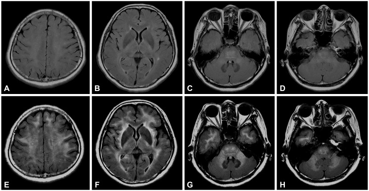
Fig. 1 MRI scans of the patient. Brain MRI performed 9 years previously (A-D) revealed only subtle hyperintensities in the left temporal deep white matter, pons, and middle cerebellar peduncle in axial T2-weighted FLAIR imaging. The present axial FLAIR images (E-H) showed bilateral symmetrical extensive hyperintensities in the subcortical and periventricular white matter, external and internal capsules, temporal pole white matter, pons, middle cerebellar peduncles, and deep cerebellar white matter. FLAIR: fluid-attenuated inversion recovery, MRI: magnetic resonance imaging.
Reference
-
1. Kim HJ, Paul F, Lana-Peixoto MA, Tenembaum S, Asgari N, Palace J, et al. MRI characteristics of neuromyelitis optica spectrum disorder: an international update. Neurology. 2015; 84:1165–1173. PMID: 25695963.2. Ayrignac X, Dalière CC, Nerrant E, Vincent T, De Seze J, Labauge P. Extensive cerebral white matter involvement in a patient with NMO spectrum disorder. Mult Scler. 2014; 20:1401–1403. PMID: 24852925.
Article3. Tajima Y, Yaguchi H, Mito Y. Unique central nervous system involvement and leukoencephalopathy-like magnetic resonance imaging findings in a patient with neuromyelitis optica spectrum disorder with Sjögren’s syndrome. Clin Exp Neuroimmunol. 2017; 8:255–257.
Article4. Ciron J, Colin O, Rosier MP, Lapeyrie S, Biotti D, Brassat D, et al. Neuromyelitis optica spectrum disorder mimicking extensive leukodystrophy. Mult Scler. 2018; 24:1256–1258. PMID: 29676204.
Article5. Stojanov D, Vojinovic S, Aracki-Trenkic A, Tasic A, Benedeto-Stojanov D, Ljubisavljevic S, et al. Imaging characteristics of cerebral autosomal dominant arteriopathy with subcortical infarcts and leucoencephalopathy (CADASIL). Bosn J Basic Med Sci. 2015; 15:1–8.
Article6. Akasbi M, Berenguer J, Saiz A, Brito-Zerón P, Pérez-De-Lis M, Bové A, et al. White matter abnormalities in primary Sjögren syndrome. QJM. 2012; 105:433–443. PMID: 22156707.7. Tzarouchi LC, Zikou AK, Tsifetaki N, Astrakas LG, Konitsiotis S, Voulgari P, et al. White matter water diffusion changes in primary Sjögren syndrome. AJNR Am J Neuroradiol. 2014; 35:680–685. PMID: 24184520.
- Full Text Links
-
- Actions
-
Cited
- CITED
-
- Close
- Share
-
- Similar articles
-
- Postpartum Relapse of Neuromyelitis Optica Spectrum Disorder after a Long Period of Spontaneous Remission
- Narcolepsy Followed by Intractable Vomiting Caused by Recurrent Brain Involvement in Neuromyelitis Optica Spectrum Disorder
- Differential Diagnosis between Multiple Sclerosis and Neuromyelitis Optica Spectrum Disorder
- Early relapse after rituximab treatment in a patient with seronegative neuromyelitis optica spectrum disorder: a case report
- Neuromyelitis Optica Spectrum Disorder Presenting with Pseudoathetosis
