Imaging Sci Dent.
2019 Dec;49(4):295-299. 10.5624/isd.2019.49.4.295.
A clinical pilot study of jawbone mineral density measured by the newly developed dual-energy cone-beam computed tomography method compared to calibrated multislice computed tomography
- Affiliations
-
- 1Department of Dental Anesthesiology and Dental Research Institute, School of Dentistry, Seoul National University, Seoul, Korea. dentane@snu.ac.kr
- 2SMDsolution Co., Ltd., Seoul, Korea.
- 3Ray Co., Ltd., Seongnam, Korea.
- 4Dental Research Institute, School of Dentistry, Seoul National University, Seoul, Korea.
- 5Department of Dental Anesthesiology, Seoul National University Dental Hospital, Seoul, Korea.
- 6Department of Periodontology and Dental Research Institute, School of Dentistry, Seoul National University, Seoul, Korea.
- 7Department of Oral and Maxillofacial Surgery and Dental Research Institute, School of Dentistry, Seoul National University, Seoul, Korea.
- 8Department of Oral and Maxillofacial Radiology and Dental Research Institute, School of Dentistry, Seoul National University, Seoul, Korea. hmslsh@snu.ac.kr
- KMID: 2466552
- DOI: http://doi.org/10.5624/isd.2019.49.4.295
Abstract
- PURPOSE
This clinical pilot study was performed to determine the effectiveness of dual-energy cone-beam computed tomography (DE-CBCT) in measuring bone mineral density (BMD).
MATERIALS AND METHODS
The BMD values obtained using DE-CBCT were compared to those obtained using calibrated multislice computed tomography (MSCT). After BMD calibration with specially designed phantoms, both DE-CBCT and MSCT scanning were performed in 15 adult dental patients. Three-dimensional (3D) Digital Imaging and Communications in Medicine data were imported into a dental software program, and the defined regions of interest (ROIs) on the 3-dimensional surface-rendered images were identified. The automatically-measured BMD values of the ROIs (g/cm³), the differences in the measured BMD values of the matched ROIs obtained by DE-CBCT and MSCT 3D images, and the correlation between the BMD values obtained by the 2 devices were statistically analyzed.
RESULTS
The mean BMD values of the ROIs for the 15 patients as assessed using DE-CBCT and MSCT were 1.09±0.07 g/cm³ and 1.13±0.08 g/cm³, respectively. The mean of the differences between the BMD values of the matched ROIs as assessed using DE-CBCT and calibrated MSCT images was 0.04±0.02 g/cm³. The Pearson correlation coefficient between the BMD values of DE-CBCT and MSCT images was 0.982 (r=0.982, P<0.001).
CONCLUSION
The newly developed DE-CBCT technique could be used to measure jaw BMD in dentistry and may soon replace MSCT, which is expensive and requires special facilities.
MeSH Terms
Figure
-

Fig. 1 The newly developed dual-energy cone-beam computed tomography (RCT720, Ray Co., Ltd., Seongnam, Korea) in this study.
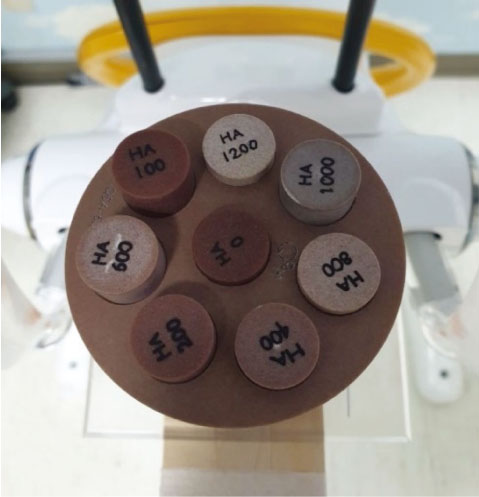
Fig. 2 The phantoms available for use in this study. Of these, HA0, HA400, HA800, HA1000, and HA1200 are used.
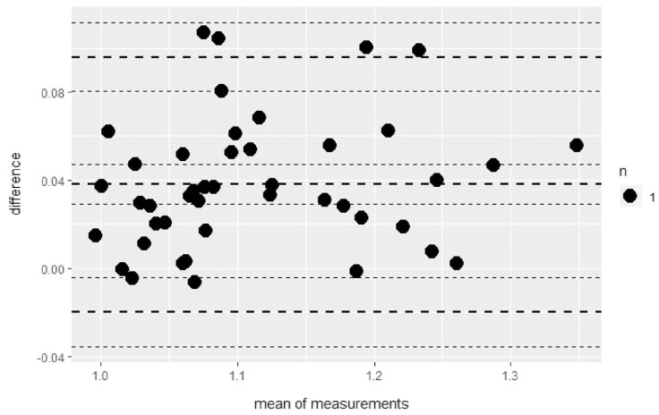
Fig. 3 Bland-Altman analysis of bone mineral density (g/cm3) as measured with dual-energy cone-beam computed tomography (DE-CBCT) and multislice computed tomography (MSCT). The upper and lower limits of agreement are 0.10 and −0.02, respectively.
Reference
-
1. Campos MJ, de Souza TS, Mota Júnior SL, Fraga MR, Vitral RW. Bone mineral density in cone beam computed tomography: only a few shades of gray. World J Radiol. 2014; 6:607–612.
Article2. Cassetta M, Stefanelli LV, Pacifici A, Pacifici L, Barbato E. How accurate is CBCT in measuring bone density? A comparative CBCT-CT in vitro study. Clin Implant Dent Relat Res. 2014; 16:471–478.
Article3. Pauwels R, Jacobs R, Singer SR, Mupparapu M. CBCT-based bone quality assessment: are Hounsfield units applicable? Dentomaxillofac Radiol. 2015; 44:20140238.
Article4. Razi T, Niknami M, Alavi Ghazani F. Relationship between Hounsfield unit in CT scan and gray scale in CBCT. J Dent Res Dent Clin Dent Prospects. 2014; 8:107–110.5. Kim DG. Can dental cone beam computed tomography assess bone mineral density? J Bone Metab. 2014; 21:117–126.
Article6. Miracle AC, Mukherji SK. Conebeam CT of the head and neck, part 1: physical principles. AJNR Am J Neuroradiol. 2009; 30:1088–1095.
Article7. Aranyarachkul P, Caruso J, Gantes B, Schulz E, Riggs M, Dus I, et al. Bone density assessments of dental implant sites: 2. Quantitative cone-beam computerized tomography. Int J Oral Maxillofac Implants. 2005; 20:416–424.8. Nackaerts O, Maes F, Yan H, Couto Souza P, Pauwels R, Jacobs R. Analysis of intensity variability in multislice and cone beam computed tomography. Clin Oral Implants Res. 2011; 22:873–879.
Article9. Silva IM, Freitas DQ, Ambrosano GM, Bóscolo FN, Almeida SM. Bone density: comparative evaluation of Hounsfield units in multislice and cone-beam computed tomography. Braz Oral Res. 2012; 26:550–556.
Article10. Turkyilmaz I, Tözüm TF, Tumer C. Bone density assessments of oral implant sites using computerized tomography. J Oral Rehabil. 2007; 34:267–272.
Article11. Shapurian T, Damoulis PD, Reiser GM, Griffin TJ, Rand WM. Quantitative evaluation of bone density using the Hounsfield index. Int J Oral Maxillofac Implants. 2006; 21:290–297.12. Fuh LJ, Huang HL, Chen CS, Fu KL, Shen YW, Tu MG, et al. Variations in bone density at dental implant sites in different regions of the jawbone. J Oral Rehabil. 2010; 37:346–351.
Article13. Aksoy U, Eratalay K, Tözüm TF. The possible association among bone density values, resonance frequency measurements, tactile sense, and histomorphometric evaluations of dental implant osteotomy sites: a preliminary study. Implant Dent. 2009; 18:316–325.
Article14. Jain S, Choudhary K, Nagi R, Shukla S, Kaur N, Grover D. New evolution of cone-beam computed tomography in dentistry: combining digital technologies. Imaging Sci Dent. 2019; 49:179–190.
Article15. American Academy of Oral and Maxillofacial Radiology. Clinical recommendations regarding use of cone beam computed tomography in orthodontics. [corrected]. Position statement by the American Academy of Oral and Maxillofacial Radiology. Oral Surg Oral Med Oral Pathol Oral Radiol. 2013; 116:238–257.
- Full Text Links
-
- Actions
-
Cited
- CITED
-
- Close
- Share
-
- Similar articles
-
- Can Dental Cone Beam Computed Tomography Assess Bone Mineral Density?
- Utility of the computed tomography indices on cone beam computed tomography images in the diagnosis of osteoporosis in women
- Management of root canal perforation by using cone-beam computed tomography
- Analysis of Beam Hardening of Modulation Layers for Dual Energy Cone-beam CT
- Three-dimensional imaging modalities in endodontics
