Can Dental Cone Beam Computed Tomography Assess Bone Mineral Density?
- Affiliations
-
- 1Division of Orthodontics, Ohio State University College of Dentistry, Columbus, OH, USA. kim.2508@osu.edu
- KMID: 2241532
- DOI: http://doi.org/10.11005/jbm.2014.21.2.117
Abstract
- Mineral density distribution of bone tissue is altered by active bone modeling and remodeling due to bone complications including bone disease and implantation surgery. Clinical cone beam computed tomography (CBCT) has been examined whether it can assess oral bone mineral density (BMD) in patient. It has been indicated that CBCT has disadvantages of higher noise and lower contrast than conventional medical computed tomography (CT) systems. On the other hand, it has advantages of a relatively lower cost and radiation dose but higher spatial resolution. However, the reliability of CBCT based mineral density measurement has not yet been fully validated. Thus, the objectives of this review are to discuss 1) why assessment of BMD distribution is important and 2) whether the clinical CBCT can be used as a potential tool to measure the BMD. Brief descriptions of image artefacts associated with assessment of gray value, which has been used to account for mineral density, in CBCT images are provided. Techniques to correct local and conversion errors in obtaining the gray values in CBCT images are also introduced. This review can be used as a quick reference for users who may encounter these errors during analysis of CBCT images.
MeSH Terms
Figure
-

Fig. 1 (A) Detailed tissue mineral density (TMD) distribution in vertebral trabecular bone. A darker color represents less TMD. (B) A typical TMD histogram of a micro-computed tomography image (voxel size 16×16×16 µm3). The TMD distribution was different between the control sham surgery (black) and ovariectomized (OVX) (gray) groups. [Reprinted from "Increased variability of bone tissue mineral density resulting from estrogen deficiency influences creep behavior in a rat vertebral body", by Kim DG, Navalgund AR, Tee BC, Noble GJ, Hart RT, Lee HR, 2012, Bone, 51(5), pp. 868-75. Copyright 2012 by the Elsevier. Reprinted with permission].

Fig. 2 (A) Micro-computed tomography (CT) image (27×27×27 µm3 voxel size) and (B) cone beam CT image (200×200×200 µm3 voxel size) of the same human condyle.

Fig. 3 X-ray beam projection scheme comparing acquisition geometry of conventional or "fan" beam (right) and "cone" beam (left) imaging geometry and resultant image production. The amount of scatter generated (sinusoidal lines) and recorded by cone-beam image acquisition is substantially higher, reducing image contrast and increasing image noise. [Reprinted from "What is cone-beam CT and how does it work?", by Scarfe WC, Farman AG, 2008, Dent Clin North Am, 52(4), pp.707-30. Copyright 2008 by the Elsevier. Reprinted with permission].
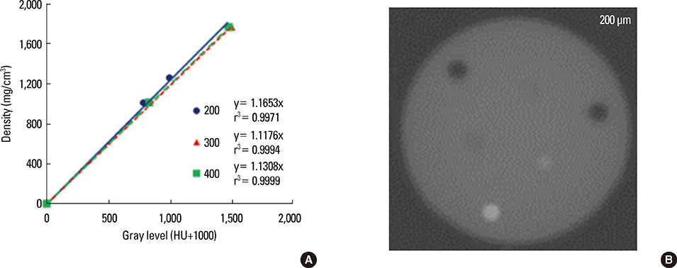
Fig. 4 (A) Strong positive correlations in the calibration curves of gray values for (B) phantoms of bone materials (hydroxyapatite) with 3 different densities (1,000, 1,250, and 1,750 mg/cm3) scanned using 3 different resolutions (200, 300, and 400 µm) of cone beam computed tomography. HU, Hounsfield units.
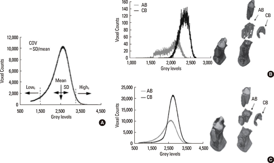
Fig. 5 (A) Degree of bone mineralization parameters determined using a grey level histogram, (B) comparison of grey level histograms between alveolar bone (AB, grey line) and basal cortical bone (CB, black line) regions using a three-dimensional (3D) cone beam computed tomography (CT) image (200×200×200 µm3 voxel size), and (C) using 3D micro-CT image (20×20×20 µm3 voxel size) for the same specimen. COV, coefficient of variation; Highs, grey level at the 95th percentile; LOWs, grey level at the 5th percentile; SD, standard deviation; AB, alveolar bone; CB, control bone. [Modified from "Comparison of micro-CT and cone beam CT-based assessments for relative difference of grey level distribution in a human mandible" by Taylor TT, Gans SI, Jones EM, Firestone AR, Johnston WM, Kim DG, 2013, Dentomaxillofac Radiol, 42(3), pp. 25117764. Copyright 2013 by British Institute of Radiology. Reprinted with permission].
Cited by 2 articles
-
Effect of field-of-view size on gray values derived from cone-beam computed tomography compared with the Hounsfield unit values from multidetector computed tomography scans
Abbas Shokri, Leila Ramezani, Mohsen Bidgoli, Mahdi Akbarzadeh, Karim Ghazikhanlu-Sani, Hamed Fallahi-Sichani
Imaging Sci Dent. 2018;48(1):31-39. doi: 10.5624/isd.2018.48.1.31.A clinical pilot study of jawbone mineral density measured by the newly developed dual-energy cone-beam computed tomography method compared to calibrated multislice computed tomography
Hyun Jeong Kim, Ji Eun Kim, Jiyeon Choo, Jeonghee Min, Sungho Chang, Sang Chul Lee, Woong Beom Pyun, Kwang-Suk Seo, Myong-Hwan Karm, Ki-Tae Koo, In-Chul Rhyu, Hoon Myoung, Min-Suk Heo
Imaging Sci Dent. 2019;49(4):295-299. doi: 10.5624/isd.2019.49.4.295.
Reference
-
1. Martins RB, Burr DB, Sharkey NA. Skeletal tissue mechanics. New York, NY: Springer;1998.2. Carter DR, Beaupré GS. Skeletal function and form: mechanobiology of skeletal development, aging, and regeneration. New York, NY: Cambridge University Press;2007.3. Parfitt AM. The physiologic and clinical significance of bone histomorphometric data. In : Recker R, editor. Bone histomorphometry: techniques and interpretations. Boca Raton, FL: CRC Press;1983. p. 143–223.4. Allen MR, Turek JJ, Phipps RJ, et al. Greater magnitude of turnover suppression occurs earlier after treatment initiation with risedronate than alendronate. Bone. 2011; 49:128–132.
Article5. Kim DG, Shertok D, Ching Tee B, et al. Variability of tissue mineral density can determine physiological creep of human vertebral cancellous bone. J Biomech. 2011; 44:1660–1665.
Article6. Turner RT, Riggs BL, Spelsberg TC. Skeletal effects of estrogen. Endocr Rev. 1994; 15:275–300.
Article7. Lerner UH. Bone remodeling in post-menopausal osteoporosis. J Dent Res. 2006; 85:584–595.
Article8. Keaveny TM, Hayes WC. A 20-year perspective on the mechanical properties of trabecular bone. J Biomech Eng. 1993; 115:534–542.
Article9. Keaveny TM. Biomechanical computed tomography-noninvasive bone strength analysis using clinical computed tomography scans. Ann N Y Acad Sci. 2010; 1192:57–65.
Article10. Genant HK, Engelke K, Fuerst T, et al. Noninvasive assessment of bone mineral and structure: state of the art. J Bone Miner Res. 1996; 11:707–730.
Article11. Genant HK, Jiang Y. Advanced imaging assessment of bone quality. Ann N Y Acad Sci. 2006; 1068:410–428.
Article12. Boonrungsiman S, Gentleman E, Carzaniga R, et al. The role of intracellular calcium phosphate in osteoblast-mediated bone apatite formation. Proc Natl Acad Sci U S A. 2012; 109:14170–14175.
Article13. Roschger P, Paschalis EP, Fratzl P, et al. Bone mineralization density distribution in health and disease. Bone. 2008; 42:456–466.
Article14. Boivin G, Meunier PJ. Methodological considerations in measurement of bone mineral content. Osteoporos Int. 2003; 14:Suppl 5. S22–S28.
Article15. Lee BC, Yeo IS, Kim DJ, et al. Bone formation around zirconia implants combined with rhBMP-2 gel in the canine mandible. Clin Oral Implants Res. 2013; 24:1332–1338.
Article16. Kim DG, Huja SS, Tee BC, et al. Bone ingrowth and initial stability of titanium and porous tantalum dental implants: a pilot canine study. Implant Dent. 2013; 22:399–405.
Article17. Ames MS, Hong S, Lee HR, et al. Estrogen deficiency increases variability of tissue mineral density of alveolar bone surrounding teeth. Arch Oral Biol. 2010; 55:599–605.
Article18. Kim DG, Navalgund AR, Tee BC, et al. Increased variability of bone tissue mineral density resulting from estrogen deficiency influences creep behavior in a rat vertebral body. Bone. 2012; 51:868–875.
Article19. Renders GA, Mulder L, van Ruijven LJ, et al. Degree and distribution of mineralization in the human mandibular condyle. Calcif Tissue Int. 2006; 79:190–196.
Article20. McCreadie BR, Goldstein SA. Biomechanics of fracture: is bone mineral density sufficient to assess risk? J Bone Miner Res. 2000; 15:2305–2308.
Article21. Genant HK, Cooper C, Poor G, et al. Interim report and recommendations of the World Health Organization Task-Force for Osteoporosis. Osteoporos Int. 1999; 10:259–264.
Article22. Heaney RP. Is the paradigm shifting? Bone. 2003; 33:457–465.
Article23. Hernandez CJ, Keaveny TM. A biomechanical perspective on bone quality. Bone. 2006; 39:1173–1181.
Article24. Donnelly E. Methods for assessing bone quality: a review. Clin Orthop Relat Res. 2011; 469:2128–2138.
Article25. Kim DG, Christopherson GT, Dong XN, et al. The effect of microcomputed tomography scanning and reconstruction voxel size on the accuracy of stereological measurements in human cancellous bone. Bone. 2004; 35:1375–1382.
Article26. Hou FJ, Lang SM, Hoshaw SJ, et al. Human vertebral body apparent and hard tissue stiffness. J Biomech. 1998; 31:1009–1015.
Article27. Morgan EF, Keaveny TM. Dependence of yield strain of human trabecular bone on anatomic site. J Biomech. 2001; 34:569–577.
Article28. Kim DG, Huja SS, Lee HR, et al. Relationships of viscosity with contact hardness and modulus of bone matrix measured by nanoindentation. J Biomech Eng. 2010; 132:024502.
Article29. Kim DG, Huja SS, Navalgund A, et al. Effect of estrogen deficiency on regional variation of a viscoelastic tissue property of bone. J Biomech. 2013; 46:110–115.
Article30. Isaksson H, Nagao S, Małkiewicz M, et al. Precision of nanoindentation protocols for measurement of viscoelasticity in cortical and trabecular bone. J Biomech. 2010; 43:2410–2417.
Article31. Isaksson H, Malkiewicz M, Nowak R, et al. Rabbit cortical bone tissue increases its elastic stiffness but becomes less viscoelastic with age. Bone. 2010; 47:1030–1038.
Article32. Guermazi A, Mohr A, Grigorian M, et al. Identification of vertebral fractures in osteoporosis. Semin Musculoskelet Radiol. 2002; 6:241–252.
Article33. Uchiyama T, Tanizawa T, Muramatsu H, et al. A morphometric comparison of trabecular structure of human ilium between microcomputed tomography and conventional histomorphometry. Calcif Tissue Int. 1997; 61:493–498.
Article34. Ibrahim N, Parsa A, Hassan B, et al. Diagnostic imaging of trabecular bone microstructure for oral implants: a literature review. Dentomaxillofac Radiol. 2013; 42:20120075.
Article35. Scarfe WC, Farman AG. What is cone-beam CT and how does it work? Dent Clin North Am. 2008; 52:707–730. v
Article36. Scarfe WC, Farman AG, Sukovic P. Clinical applications of cone-beam computed tomography in dental practice. J Can Dent Assoc. 2006; 72:75–80.37. Molteni R. Prospects and challenges of rendering tissue density in Hounsfield units for cone beam computed tomography. Oral Surg Oral Med Oral Pathol Oral Radiol. 2013; 116:105–119.
Article38. Katsumata A, Hirukawa A, Okumura S, et al. Relationship between density variability and imaging volume size in cone-beam computerized tomographic scanning of the maxillofacial region: an in vitro study. Oral Surg Oral Med Oral Pathol Oral Radiol Endod. 2009; 107:420–425.
Article39. Schulze R, Heil U, Gross D, et al. Artefacts in CBCT: a review. Dentomaxillofac Radiol. 2011; 40:265–273.
Article40. Mozzo P, Procacci C, Tacconi A, et al. A new volumetric CT machine for dental imaging based on the cone-beam technique: preliminary results. Eur Radiol. 1998; 8:1558–1564.
Article41. Ludlow JB, Davies-Ludlow LE, Brooks SL, et al. Dosimetry of 3 CBCT devices for oral and maxillofacial radiology: CB Mercuray, NewTom 3G and i-CAT. Dentomaxillofac Radiol. 2006; 35:219–226.
Article42. Ketcham RA, Carlson WD. Acquisition, optimization and interpretation of X-ray computed tomographic imagery: applications to the geosciences. Comput Geosci. 2001; 27:381–400.
Article43. Barrett JF, Keat N. Artifacts in CT: recognition and avoidance. Radiographics. 2004; 24:1679–1691.
Article44. Feldkamp LA, Davis LC, Kress JW. Practical cone-beam algorithm. J Opt Soc Am A. 1984; 1:612–619.
Article45. Siltanen S, Kolehmainen V, Järvenpää S, et al. Statistical inversion for medical x-ray tomography with few radiographs: I. General theory. Phys Med Biol. 2003; 48:1437–1463.
Article46. Bryant JA, Drage NA, Richmond S. Study of the scan uniformity from an i-CAT cone beam computed tomography dental imaging system. Dentomaxillofac Radiol. 2008; 37:365–374.
Article47. Mah P, Reeves TE, McDavid WD. Deriving Hounsfield units using grey levels in cone beam computed tomography. Dentomaxillofac Radiol. 2010; 39:323–335.
Article48. Reeves TE, Mah P, McDavid WD. Deriving Hounsfield units using grey levels in cone beam CT: a clinical application. Dentomaxillofac Radiol. 2012; 41:500–508.
Article49. Rührnschopf EP, Klingenbeck K. A general framework and review of scatter correction methods in x-ray cone-beam computerized tomography. Part 1: Scatter compensation approaches. Med Phys. 2011; 38:4296–4311.
Article50. Dong X, Petrongolo M, Niu T, et al. Low-dose and scatter-free cone-beam CT imaging using a stationary beam blocker in a single scan: phantom studies. Comput Math Methods Med. 2013; 2013:637614.
Article51. Li J, Yao W, Xiao Y, et al. Feasibility of improving cone-beam CT number consistency using a scatter correction algorithm. J Appl Clin Med Phys. 2013; 14:4346.
Article52. Staub D, Murphy MJ. A digitally reconstructed radiograph algorithm calculated from first principles. Med Phys. 2013; 40:011902.
Article53. Hunter AK, McDavid WD. Characterization and correction of cupping effect artefacts in cone beam CT. Dentomaxillofac Radiol. 2012; 41:217–223.
Article54. Nomura Y, Watanabe H, Honda E, et al. Reliability of voxel values from cone-beam computed tomography for dental use in evaluating bone mineral density. Clin Oral Implants Res. 2010; 21:558–562.
Article55. Naitoh M, Hirukawa A, Katsumata A, et al. Evaluation of voxel values in mandibular cancellous bone: relationship between cone-beam computed tomography and multislice helical computed tomography. Clin Oral Implants Res. 2009; 20:503–506.
Article56. Parsa A, Ibrahim N, Hassan B, et al. Reliability of voxel gray values in cone beam computed tomography for preoperative implant planning assessment. Int J Oral Maxillofac Implants. 2012; 27:1438–1442.57. Parsa A, Ibrahim N, Hassan B, et al. Bone quality evaluation at dental implant site using multislice CT, micro-CT, and cone beam CT. Clin Oral Implants Res. 2013.
Article58. Pittman JW, Navalgund A, Byun SH, et al. Primary migration of a mini-implant under a functional orthodontic loading. Clin Oral Investig. 2014; 18:721–728.
Article59. Cha JY, Kil JK, Yoon TM, et al. Miniscrew stability evaluated with computerized tomography scanning. Am J Orthod Dentofacial Orthop. 2010; 137:73–79.
Article60. Huang H, Richards M, Bedair T, et al. Effects of orthodontic treatment on human alveolar bone density distribution. Clin Oral Investig. 2013; 17:2033–2040.
Article61. Taylor TT, Gans SI, Jones EM, et al. Comparison of micro-CT and cone beam CT-based assessments for relative difference of grey level distribution in a human mandible. Dentomaxillofac Radiol. 2013; 42:25117764.
Article62. Agbaje JO, Jacobs R, Maes F, et al. Volumetric analysis of extraction sockets using cone beam computed tomography: a pilot study on ex vivo jaw bone. J Clin Periodontol. 2007; 34:985–990.
Article63. Huja SS, Beck FM. Bone remodeling in maxilla, mandible, and femur of young dogs. Anat Rec (Hoboken). 2008; 291:1–5.
Article64. Kennedy KS, Jones EM, Kim DG, et al. A prospective clinical study to evaluate early success of short implants. Int J Oral Maxillofac Implants. 2013; 28:170–177.
Article65. Leblebicioglu B, Salas M, Ort Y, et al. Determinants of alveolar ridge preservation differ by anatomic location. J Clin Periodontol. 2013; 40:387–395.
Article66. Benavides E, Rios HF, Ganz SD, et al. Use of cone beam computed tomography in implant dentistry: the International Congress of Oral Implantologists consensus report. Implant Dent. 2012; 21:78–86.67. Santiago RC, de Paula FO, Fraga MR, et al. Correlation between miniscrew stability and bone mineral density in orthodontic patients. Am J Orthod Dentofacial Orthop. 2009; 136:243–250.
Article68. Song YD, Jun SH, Kwon JJ. Correlation between bone quality evaluated by cone-beam computerized tomography and implant primary stability. Int J Oral Maxillofac Implants. 2009; 24:59–64.69. Aranyarachkul P, Caruso J, Gantes B, et al. Bone density assessments of dental implant sites: 2. Quantitative cone-beam computerized tomography. Int J Oral Maxillofac Implants. 2005; 20:416–424.70. Guerrero ME, Jacobs R, Loubele M, et al. State-of-the-art on cone beam CT imaging for preoperative planning of implant placement. Clin Oral Investig. 2006; 10:1–7.
Article71. Hsu JT, Chen YJ, Tsai MT, et al. Predicting cortical bone strength from DXA and dental cone-beam CT. PLoS One. 2012; 7:e50008.
Article72. Hsu JT, Chang HW, Huang HL, et al. Bone density changes around teeth during orthodontic treatment. Clin Oral Investig. 2011; 15:511–519.
Article73. dos Anjos Pontual ML, Freire JS, Barbosa JM, et al. Evaluation of bone changes in the temporomandibular joint using cone beam CT. Dentomaxillofac Radiol. 2012; 41:24–29.
Article74. Hohlweg-Majert B, Metzger MC, Kummer T, et al. Morphometric analysis - Cone beam computed tomography to predict bone quality and quantity. J Craniomaxillofac Surg. 2011; 39:330–334.
Article75. Loubele M, Maes F, Schutyser F, et al. Assessment of bone segmentation quality of cone-beam CT versus multislice spiral CT: a pilot study. Oral Surg Oral Med Oral Pathol Oral Radiol Endod. 2006; 102:225–234.
Article76. Nackaerts O, Depypere M, Zhang G, et al. Segmentation of Trabecular Jaw Bone on Cone Beam CT Datasets. Clin Implant Dent Relat Res. 2014.
Article77. Hangartner TN. Thresholding technique for accurate analysis of density and geometry in QCT, pQCT and microCT images. J Musculoskelet Neuronal Interact. 2007; 7:9–16.78. Klintstrom E, Smedby O, Moreno R, et al. Trabecular bone structure parameters from 3D image processing of clinical multi-slice and cone-beam computed tomography data. Skeletal Radiol. 2014; 43:197–204.
Article79. Wang L, Chen KC, Gao Y, et al. Automated bone segmentation from dental CBCT images using patch-based sparse representation and convex optimization. Med Phys. 2014; 41:043503.
Article80. Burghardt AJ, Link TM, Majumdar S. High-resolution computed tomography for clinical imaging of bone microarchitecture. Clin Orthop Relat Res. 2011; 469:2179–2193.
Article81. Loubele M, Jacobs R, Maes F, et al. Image quality vs radiation dose of four cone beam computed tomography scanners. Dentomaxillofac Radiol. 2008; 37:309–318.82. Link TM. Osteoporosis imaging: state of the art and advanced imaging. Radiology. 2012; 263:3–17.
Article
- Full Text Links
-
- Actions
-
Cited
- CITED
-
- Close
- Share
-
- Similar articles
-
- Utility of the computed tomography indices on cone beam computed tomography images in the diagnosis of osteoporosis in women
- Evaluation of imaging reformation with cone beam computed tomography for the assessment of bone density and shape in mandible
- Management of root canal perforation by using cone-beam computed tomography
- ASSESSMENT OF BONE DENSITY ON MAXILLA AFTER IMPLANTATION WITH CONE BEAM COMPUTED TOMOGRAPHY
- Three-dimensional imaging modalities in endodontics
