The Impact of Iterative Reconstruction in Low-Dose Computed Tomography on the Evaluation of Diffuse Interstitial Lung Disease
- Affiliations
-
- 1Department of Radiology and Center for Imaging Science, Samsung Medical Center, Sungkyunkwan University School of Medicine, Seoul 06351, Korea. mj1.chung@samsung.com
- KMID: 2466292
- DOI: http://doi.org/10.3348/kjr.2016.17.6.950
Abstract
OBJECTIVE
To evaluate the impact of iterative reconstruction (IR) on the assessment of diffuse interstitial lung disease (DILD) using CT.
MATERIALS AND METHODS
An American College of Radiology (ACR) phantom (module 4 to assess spatial resolution) was scanned with 10-100 effective mAs at 120 kVp. The images were reconstructed using filtered back projection (FBP), adaptive statistical iterative reconstruction (ASIR), with blending ratios of 0%, 30%, 70% and 100%, and model-based iterative reconstruction (MBIR), and their spatial resolution was objectively assessed by the line pair structure method. The patient study was based on retrospective interpretation of prospectively acquired data, and it was approved by the institutional review board. Chest CT scans of 23 patients (mean age 64 years) were performed at 120 kVp using 1) standard dose protocol applying 142-275 mA with dose modulation (high-resolution computed tomography [HRCT]) and 2) low-dose protocol applying 20 mA (low dose CT, LDCT). HRCT images were reconstructed with FBP, and LDCT images were reconstructed using FBP, ASIR, and MBIR. Matching images were randomized and independently reviewed by chest radiologists. Subjective assessment of disease presence and radiological diagnosis was made on a 10-point scale. In addition, semi-quantitative results were compared for the extent of abnormalities estimated to the nearest 5% of parenchymal involvement.
RESULTS
In the phantom study, ASIR was comparable to FBP in terms of spatial resolution. However, for MBIR, the spatial resolution was greatly decreased under 10 mA. In the patient study, the detection of the presence of disease was not significantly different. The values for area under the curve for detection of DILD by HRCT, FBP, ASIR, and MBIR were as follows: 0.978, 0.979, 0.972, and 0.963. LDCT images reconstructed with FBP, ASIR, and MBIR tended to underestimate reticular or honeycombing opacities (-2.8%, -4.1%, and -5.3%, respectively) and overestimate ground glass opacities (+4.6%, +8.9%, and +8.5%, respectively) compared to the HRCT images. However, the reconstruction methods did not differ with respect to radiologic diagnosis.
CONCLUSION
The diagnostic performance of LDCT with MBIR was similar to that of HRCT in typical DILD cases. However, caution should be exercised when comparing disease extent, especially in follow-up studies with IR.
Keyword
MeSH Terms
Figure
-

Fig. 1 Example results of phantom study with model-based iterative reconstruction (MBIR) are shown (A, scanned under 120 kVp, 10 mA); B (120 kVp, 50 mA).(A) shows significant decrease in difference between maximal and minimum values (NB[i]–NA[i]) in higher spatial resolution phantom images (7 lp/cm) at lower dose (120 kVp, 10 mA) compared with (B) (120 kVp, 50 mA). (C) shows changes in numerator value when using same radiation dose and reconstruction method. MBIR images obtained at 10 mA demonstrated significant decrease in numerator value for bar resolution pattern with 7 lp/cm. ASIR = adaptive statistical iterative reconstruction, FBP = filtered back projection, HU = Hounsfield unit
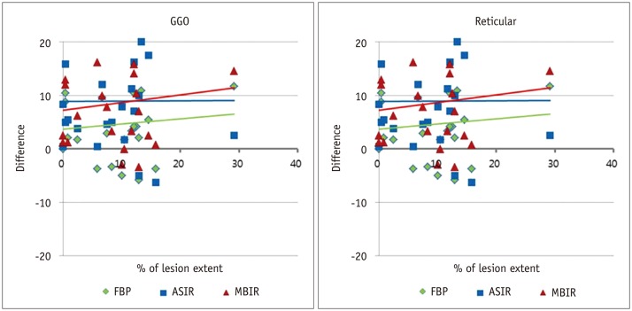
Fig. 2 Bland-Altman plots for each of four observers illustrating differences in measurement of disease extent based on FBP, ASIR, and MBIR images.With MBIR images, observers overestimated extent of GGO and underestimated extent of reticular opacity. However, there were no significant differences in evaluation of extent of consolidation (data not shown). ASIR = adaptive statistical iterative reconstruction, FBP = filtered back projection, GGO = ground glass opacity, MBIR = model-based iterative reconstruction
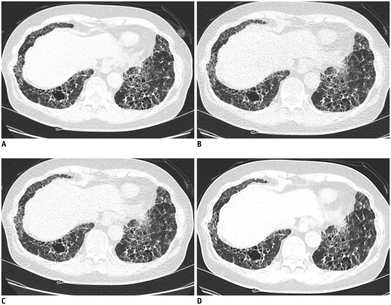
Fig. 3 Computed tomography (CT) images from a 69-year-old man with biopsy-proven usual interstitial pneumonia.A. Standard-dose HRCT image with FBP reconstruction. B-D. Exact same level images from raw data of low-dose CT reconstructed with (B) FBP, (C) ASIR, and (D) MBIR. Blurring phenomenon due to effect of MBIR, not due to respiratory artifact is shown in (D). ASIR = adaptive statistical iterative reconstruction, FBP = filtered back projection, HRCT = high resolution computed tomography, MBIR = model-based iterative reconstruction
Cited by 4 articles
-
Effect of Hybrid Kernel and Iterative Reconstruction on Objective and Subjective Analysis of Lung Nodule Calcification in Low-Dose Chest CT
Seul Gi Hong, Eun-Ju Kang, Jae Hyung Park, Won Jin Choi, Ki-Nam Lee, Hee Jin Kwon, Dong-Ho Ha, Dong Won Kim, Sang Hyeon Kim, Jeong-Hyun Jo, Jongmin Lee
Korean J Radiol. 2018;19(5):888-896. doi: 10.3348/kjr.2018.19.5.888.Quantitative Image Quality and Histogram-Based Evaluations of an Iterative Reconstruction Algorithm at Low-to-Ultralow Radiation Dose Levels: A Phantom Study in Chest CT
Ki Baek Lee, Hyun Woo Goo
Korean J Radiol. 2018;19(1):119-129. doi: 10.3348/kjr.2018.19.1.119.Comparison of Filtered Back Projection, Hybrid Iterative Reconstruction, Model-Based Iterative Reconstruction, and Virtual Monoenergetic Reconstruction Images at Both Low- and Standard-Dose Settings in Measurement of Emphysema Volume and Airway Wall Thickness: A CT Phantom Study
Cherry Kim, Ki Yeol Lee, Chol Shin, Eun-Young Kang, Yu-Whan Oh, Moin Ha, Chang Sub Ko, Jaehyung Cha
Korean J Radiol. 2018;19(4):809-817. doi: 10.3348/kjr.2018.19.4.809.Contrast-Enhanced CT with Knowledge-Based Iterative Model Reconstruction for the Evaluation of Parotid Gland Tumors: A Feasibility Study
Chae Jung Park, Ki Wook Kim, Ho-Joon Lee, Myeong-Jin Kim, Jinna Kim
Korean J Radiol. 2018;19(5):957-964. doi: 10.3348/kjr.2018.19.5.957.
Reference
-
1. Kubo T, Lin PJ, Stiller W, Takahashi M, Kauczor HU, Ohno Y, et al. Radiation dose reduction in chest CT: a review. AJR Am J Roentgenol. 2008; 190:335–343. PMID: 18212218.
Article2. Mayo JR. CT evaluation of diffuse infiltrative lung disease: dose considerations and optimal technique. J Thorac Imaging. 2009; 24:252–259. PMID: 19935222.3. Tsapaki V, Aldrich JE, Sharma R, Staniszewska MA, Krisanachinda A, Rehani M, et al. Dose reduction in CT while maintaining diagnostic confidence: diagnostic reference levels at routine head, chest, and abdominal CT--IAEA-coordinated research project. Radiology. 2006; 240:828–834. PMID: 16837668.
Article4. Kalra MK, Maher MM, Toth TL, Hamberg LM, Blake MA, Shepard JA, et al. Strategies for CT radiation dose optimization. Radiology. 2004; 230:619–628. PMID: 14739312.
Article5. Heyer CM, Mohr PS, Lemburg SP, Peters SA, Nicolas V. Image quality and radiation exposure at pulmonary CT angiography with 100- or 120-kVp protocol: prospective randomized study. Radiology. 2007; 245:577–583. PMID: 17940308.
Article6. Diel J, Perlmutter S, Venkataramanan N, Mueller R, Lane MJ, Katz DS. Unenhanced helical CT using increased pitch for suspected renal colic: an effective technique for radiation dose reduction? J Comput Assist Tomogr. 2000; 24:795–801. PMID: 11045705.
Article7. Kalra MK, Maher MM, Sahani DV, Blake MA, Hahn PF, Avinash GB, et al. Low-dose CT of the abdomen: evaluation of image improvement with use of noise reduction filters pilot study. Radiology. 2003; 228:251–256. PMID: 12832586.8. Thibault JB, Sauer KD, Bouman CA, Hsieh J. A three-dimensional statistical approach to improved image quality for multislice helical CT. Med Phys. 2007; 34:4526–4544. PMID: 18072519.
Article9. Machida H, Tanaka I, Fukui R, Kita K, Shen Y, Ueno E, et al. Improved delineation of the anterior spinal artery with model-based iterative reconstruction in CT angiography: a clinical pilot study. AJR Am J Roentgenol. 2013; 200:442–446. PMID: 23345369.
Article10. Shuman WP, Green DE, Busey JM, Kolokythas O, Mitsumori LM, Koprowicz KM, et al. Model-based iterative reconstruction versus adaptive statistical iterative reconstruction and filtered back projection in liver 64-MDCT: focal lesion detection, lesion conspicuity, and image noise. AJR Am J Roentgenol. 2013; 200:1071–1076. PMID: 23617492.
Article11. Goldman LW. Principles of CT: radiation dose and image quality. J Nucl Med Technol. 2007; 35:213–225. quiz 226-228. PMID: 18006597.
Article12. Goldman LW. Principles of CT and CT technology. J Nucl Med Technol. 2007; 35:115–128. quiz 129-130. PMID: 17823453.
Article13. Ichikawa Y, Kitagawa K, Nagasawa N, Murashima S, Sakuma H. CT of the chest with model-based, fully iterative reconstruction: comparison with adaptive statistical iterative reconstruction. BMC Med Imaging. 2013; 13:27. PMID: 23927627.
Article14. Pontana F, Billard AS, Duhamel A, Schmidt B, Faivre JB, Hachulla E, et al. Effect of iterative reconstruction on the detection of systemic sclerosis-related interstitial lung disease: clinical experience in 55 patients. Radiology. 2016; 279:297–305. PMID: 26583761.
Article15. AAPM. The measurement, reporting, and management of radiation dose in CT. Alexandria, VA: AAPM;2008.16. Yoon HJ, Chung MJ, Hwang HS, Moon JW, Lee KS. Adaptive statistical iterative reconstruction-applied ultra-low-dose CT with radiography-comparable radiation dose: usefulness for lung nodule detection. Korean J Radiol. 2015; 16:1132–1141. PMID: 26357505.
Article17. Staude A, Goebbels J. Determining the spatial resolution in computed tomography-comparison of MTF and line-pair structures. In : International symposium on digital industrial radiology and computed tomography-Tu.4.1; 2011 June 20-22; Berlin, Germany.18. Bland JM, Altman DG. Statistical methods for assessing agreement between two methods of clinical measurement. Lancet. 1986; 1:307–310. PMID: 2868172.
Article19. Dorfman DD, Berbaum KS, Metz CE. Receiver operating characteristic rating analysis. Generalization to the population of readers and patients with the jackknife method. Invest Radiol. 1992; 27:723–731. PMID: 1399456.20. Obuchowski NA. New methodological tools for multiple-reader ROC studies. Radiology. 2007; 243:10–12. PMID: 17392244.
Article21. Richeldi L, du Bois RM, Raghu G, Azuma A, Brown KK, Costabel U, et al. Efficacy and safety of nintedanib in idiopathic pulmonary fibrosis. N Engl J Med. 2014; 370:2071–2082. PMID: 24836310.
Article22. King TE Jr, Bradford WZ, Castro-Bernardini S, Fagan EA, Glaspole I, Glassberg MK, et al. A phase 3 trial of pirfenidone in patients with idiopathic pulmonary fibrosis. N Engl J Med. 2014; 370:2083–2092. PMID: 24836312.
Article23. Sverzellati N, Zompatori M, De Luca G, Chetta A, Bnà C, Ormitti F, et al. Evaluation of quantitative CT indexes in idiopathic interstitial pneumonitis using a low-dose technique. Eur J Radiol. 2005; 56:370–375. PMID: 15978764.
Article24. Yabuuchi H, Matsuo Y, Tsukamoto H, Horiuchi T, Sunami S, Kamitani T, et al. Evaluation of the extent of ground-glass opacity on high-resolution CT in patients with interstitial pneumonia associated with systemic sclerosis: comparison between quantitative and qualitative analysis. Clin Radiol. 2014; 69:758–764. PMID: 24824977.
Article25. Bendaoud S, Remy-Jardin M, Wallaert B, Deken V, Duhamel A, Faivre JB, et al. Sequential versus volumetric computed tomography in the follow-up of chronic bronchopulmonary diseases: comparison of diagnostic information and radiation dose in 63 adults. J Thorac Imaging. 2011; 26:190–195. PMID: 21785287.26. Xu J, Mahesh M, Tsui BM. Is iterative reconstruction ready for MDCT? J Am Coll Radiol. 2009; 6:274–276. PMID: 19327661.
Article27. Silva AC, Lawder HJ, Hara A, Kujak J, Pavlicek W. Innovations in CT dose reduction strategy: application of the adaptive statistical iterative reconstruction algorithm. AJR Am J Roentgenol. 2010; 194:191–199. PMID: 20028923.
Article28. Jiao de C, Li TF, Han XW, Wu G, Ma J, Fu MT, et al. Clinical applications of the C-arm cone-beam CT-based 3D needle guidance system in performing percutaneous transthoracic needle biopsy of pulmonary lesions. Diagn Interv Radiol. 2014; 20:470–474. PMID: 25323838.
Article29. Smith EA, Dillman JR, Goodsitt MM, Christodoulou EG, Keshavarzi N, Strouse PJ. Model-based iterative reconstruction: effect on patient radiation dose and image quality in pediatric body CT. Radiology. 2014; 270:526–534. PMID: 24091359.
Article30. Ghetti C, Ortenzia O, Serreli G. CT iterative reconstruction in image space: a phantom study. Phys Med. 2012; 28:161–165. PMID: 21497530.
Article31. Yamada Y, Jinzaki M, Tanami Y, Shiomi E, Sugiura H, Abe T, et al. Model-based iterative reconstruction technique for ultralow-dose computed tomography of the lung: a pilot study. Invest Radiol. 2012; 47:482–489. PMID: 22766910.32. Katsura M, Matsuda I, Akahane M, Sato J, Akai H, Yasaka K, et al. Model-based iterative reconstruction technique for radiation dose reduction in chest CT: comparison with the adaptive statistical iterative reconstruction technique. Eur Radiol. 2012; 22:1613–1623. PMID: 22538629.
Article33. Yu Z, Thibault JB, Bouman CA, Sauer KD, Hsieh J. Fast model-based X-ray CT reconstruction using spatially nonhomogeneous ICD optimization. IEEE Trans Image Process. 2011; 20:161–175. PMID: 20643609.
Article
- Full Text Links
-
- Actions
-
Cited
- CITED
-
- Close
- Share
-
- Similar articles
-
- Dosimetric Effects of Low Dose 4D CT Using a Commercial Iterative Reconstruction on Dose Calculation in Radiation Treatment Planning: A Phantom Study
- Update in Diagnosis of Idiopathic Pulmonary Fibrosis and Interstitial Lung Abnormality
- Performance of Half-dose Chest Computed Tomography in Lung Malignancy Using an Iterative Reconstruction Technique
- Contrast-Enhanced CT with Knowledge-Based Iterative Model Reconstruction for the Evaluation of Parotid Gland Tumors: A Feasibility Study
- Quantitative Image Quality and Histogram-Based Evaluations of an Iterative Reconstruction Algorithm at Low-to-Ultralow Radiation Dose Levels: A Phantom Study in Chest CT
