Kosin Med J.
2017 Jun;32(1):47-57. 10.7180/kmj.2017.32.1.47.
Performance of Half-dose Chest Computed Tomography in Lung Malignancy Using an Iterative Reconstruction Technique
- Affiliations
-
- 1Department of Radiology, College of Medicine, Kosin University, Busan, Korea. cibertim@naver.com
- 2Department of Radiology, Dong-A University Medical Center, Busan, Korea.
- KMID: 2384833
- DOI: http://doi.org/10.7180/kmj.2017.32.1.47
Abstract
OBJECTIVES
The purpose of this study was to evaluate the performance of half-dose chest CT using an iterative reconstruction technique in patients with lung malignancies.
METHODS
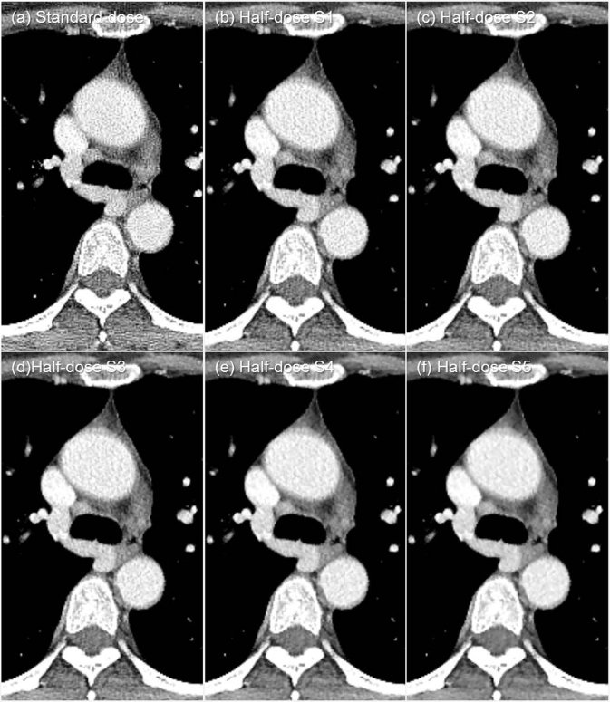
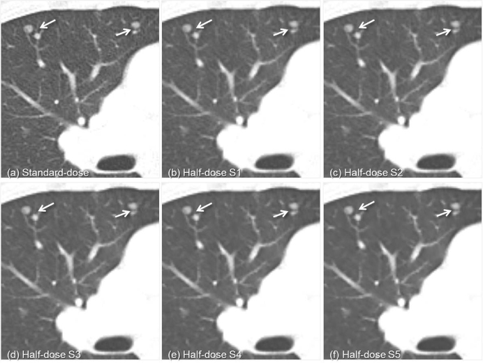
The Dual-source CT scans were obtained and half-dose datasets were reconstructed with 5 different strengths in 38 adults with lung malignancies. Two radiologists graded subjective image quality; noise, contrast and sharpness at the central/peripheral lung, mediastinum and chest wall of the reconstructed half-dose images, compared with those of standard-dose images, using a three-point scale. A lesion assessment; lesion conspicuity and diagnostic confidence, was also performed. The quantitative image noises; contrast-to-noise ratio (CNR) and signal-to-noise ratio (SNR) were measured and compared with those of standard-dose images.
RESULTS
The subjective image noise in the half-dose images was less than that of the standard-dose images. The contrast in strengths 2 to 5 was superior, the sharpness of the lung parenchyma in strengths 3 to 5 was inferior, and the CNR/SNR in all strengths were higher than those of standard-dose images (P < 0.05). The improvement of subjective image noise and contrast, the decrease in sharpness, were correlated with strength level (P < 0.05). The lesion conspicuity in half-dose images of strengths 4 and 5 was decreased. The diagnostic confidence of the half-dose images of all strengths was comparable to that of the standard-dose images (P < 0.05).
CONCLUSIONS
Half-dose chest CT images using an iterative reconstruction technique show decreased image noise, increased contrast, and diagnostic confidence comparable to standard-dose images. Images reconstructed with strength 2 and 3 appear to be the optimal choice in clinical practice.
Keyword
MeSH Terms
Figure
Reference
-
1. Park MY, Jung SE. CT radiation dose and radiation reduction strategies. J Korean Med Assoc. 2011; 54:1262–1268.
Article2. Crawley MT, Booth A, Wainwright A. A practical approach to the first iteration in the optimization of radiation dose and image quality in CT: estimates of the collective dose savings achieved. Br J Radiol. 2001; 74:607–614.
Article3. Greffier J, Fernandez A, Macri F, Freitag C, Metge L, Beregi JP. Which dose for what image? Iterative reconstruction for CT scan. Diagn Interv Imaging. 2013; 94:1117–1121.
Article4. Hara AK, Paden RG, Silva AC, Kujak JL, Lawder HJ, Pavlicek W. Iterative reconstruction technique for reducing body radiation dose at CT: feasibility study. AJR Am J Roentgenol. 2009; 193:764–771.
Article5. Liu YJ, Zhu PP, Chen B, Wang JY, Yuan QX, Huang WX, et al. A new iterative algorithm to reconstruct the refractive index. Phys Med Biol. 2007; 52:L5–L13.
Article6. Moscariello A, Takx RA, Schoepf UJ, Renker M, Zwerner PL, O'Brien TX, et al. Coronary CT angiography: image quality, diagnostic accuracy, and potential for radiation dose reduction using a novel iterative image reconstruction technique-comparison with traditional filtered back projection. Eur Radiol. 2011; 21:2130–2138.
Article7. Winklehner A, Karlo C, Puippe G, Schmidt B, Flohr T, Goetti R, et al. Raw data-based iterative reconstruction in body CTA: evaluation of radiation dose saving potential. Eur Radiol. 2011; 21:2521–2526.
Article8. Wang R, Schoepf UJ, Wu R, Gibbs KP, Yu W, Li M, et al. CT coronary angiography: image quality with sinogram-affirmed iterative reconstruction compared with filtered back-projection. Clin Radiol. 2013; 68:272–278.
Article9. Kalra MK, Wittram C, Maher MM, Sharma A, Avinash GB, Karau K, et al. Can noise reduction filters improve low-radiation-dose chest CT images? Pilot study. Radiology. 2003; 228:257–264.
Article10. Kalra MK, Woisetschläger M, Dahlström N, Singh S, Digumarthy S, Do S, et al. Sinogram-Affirmed Iterative Reconstruction of Low-Dose Chest CT: Effect on Image Quality and Radiation Dose. AJR Am J Roentgenol. 2013; 201:W235–W244.
Article11. McNitt-Gray MF. AAPM/RSNA Physics Tutorial for Residents: Topics in CT. Radiation dose in CT. Radiographics. 2002; 22:1541–1553.
Article12. Lee TY, Chhem RK. Impact of new technologies on dose reduction in CT. Eur J Radiol. 2010; 76:28–35.
Article13. Prasad SR, Wittram C, Shepard JA, McLoud T, Rhea J. Standard-dose and 50%-reduced-dose chest CT: comparing the effect on image quality. AJR Am J Roentgenol. 2002; 179:461–465.
Article14. Kalra MK, Woisetschläger M, Dahlström N, Singh S, Lindblom M, Choy G, et al. Radiation dose reduction with Sinogram Affirmed Iterative Reconstruction technique for abdominal computed tomography. J Comput Assist Tomogr. 2012; 36:339–346.
Article15. Wang R, Schoepf UJ, Wu R, Reddy RP, Zhang C, Yu W, et al. Image quality and radiation dose of low dose coronary CT angiography in obese patients: sinogram affirmed iterative reconstruction versus filtered back projection. Eur J Radiol. 2012; 81:3141–3145.
Article16. Baumueller S, Winklehner A, Karlo C, Goetti R, Flohr T, Russi EW, et al. Low-dose CT of the lung: potential value of iterative reconstructions. Eur Radiol. 2012; 22:2597–2606.
Article17. Hwang HJ, Seo JB, Lee HJ, Lee SM, Kim EY, Oh SY, et al. Low-dose chest computed tomography with sinogram-affirmed iterative reconstruction, iterative reconstruction in image space, and filtered back projection: studies on image quality. J Comput Assist Tomogr. 2013; 37:610–617.
Article18. Wang H, Tan B, Zhao B, Liang C, Xu Z. Raw-data-based iterative reconstruction versus filtered back projection: image quality of low-dose chest computed tomography examinations in 87 patients. Clin Imaging. 2013; 37:1024–1032.
Article19. Yang WJ, Yan FH, Liu B, Pang LF, Hou L, Zhang H, et al. Can sinogram-affirmed iterative (SAFIRE) reconstruction improve imaging quality on low-dose lung CT screening compared with traditional filtered back projection (FBP) reconstruction? J Comput Assist Tomogr. 2013; 37:301–305.
Article20. Singh S, Kalra MK, Gilman MD, Hsieh J, Pien HH, Digumarthy SR, et al. Adaptive statistical iterative reconstruction technique for radiation dose reduction in chest CT: a pilot study. Radiology. 2011; 259:565–573.
Article21. Prakash P, Kalra MK, Ackman JB, Digumarthy SR, Hsieh J, Do S, et al. Diffuse lung disease: CT of the chest with adaptive statistical iterative reconstruction technique. Radiology. 2010; 256:261–269.
Article22. Leipsic J, Nguyen G, Brown J, Sin D, Mayo JR. A prospective evaluation of dose reduction and image quality in chest CT using adaptive statistical iterative reconstruction. AJR Am J Roentgenol. 2010; 195:1095–1099.
Article23. Singh S, Kalra MK, Hsieh J, Licato PE, Do S, Pien HH, et al. Abdominal CT: comparison of adaptive statistical iterative and filtered back projection reconstruction techniques. Radiology. 2010; 257:373–383.
Article24. Marin D, Nelson RC, Schindera ST, Richard S, Youngblood RS, Yoshizumi TT, et al. Low-tube-voltage, high-tube-current multi-detector abdominal CT: improved image quality and decreased radiation dose with adaptive statistical iterative reconstruction algorithm--initial clinical experience. Radiology. 2010; 254:145–153.
Article25. Yoshimura N, Sabir A, Kubo T, Lin PJ, Clouse ME, Hatabu H. Correlation between image noise and body weight in coronary CTA with 16-row MDCT. Acad Radiol. 2006; 13:324–328.
Article
- Full Text Links
- Actions
-
Cited
- CITED
-
- Close
- Share
- Similar articles
-
- Dosimetric Effects of Low Dose 4D CT Using a Commercial Iterative Reconstruction on Dose Calculation in Radiation Treatment Planning: A Phantom Study
- Radiation Dose Reduction of Chest CT with Iterative Reconstruction in Image Space - Part II: Assessment of Radiologists' Preferences Using Dual Source CT
- Radiation Dose Reduction of Chest CT with Iterative Reconstruction in Image Space - Part I: Studies on Image Quality Using Dual Source CT
- Effects of Iterative Reconstruction Algorithm, Automatic Exposure Control on Image Quality, and Radiation Dose: Phantom Experiments with Coronary CT Angiography Protocols
- Deep Learning-Based Reconstruction Algorithm With Lung Enhancement Filter for Chest CT: Effect on Image Quality and Ground Glass Nodule Sharpness


