Eosinophilic Enteritis Presenting as Massive Ascites after Influenza A Virus Infection in a Young Female
- Affiliations
-
- 1Division of Gastroenterology, Department of Internal Medicine, CHA University School of Medicine, CHA Gumi Medical Center, Gumi, Korea. zenus1@hanmail.net
- KMID: 2458697
- DOI: http://doi.org/10.4166/kjg.2019.74.3.163
Abstract
- Eosinophilic gastrointestinal disorder (EGID) is an uncommon disease that is accompanied by intestinal eosinophil infiltration without a secondary cause of eosinophilia. Eosinophilic enteritis is a secondary portion of EGID that can present a range of gastrointestinal symptoms according to the affected depth of the intestinal layer. The subserosal type of eosinophilic enteritis presenting as ascites is relatively rarer than the mucosal type. In general, eosinophilic enteritis occurs in patients with food allergies, but its mechanism is unclear. The authors experienced a 29-year-old female patient with a large amount of ascites with diarrhea and abdominal pain. The patient was diagnosed with an influenza A infection one week earlier. Peripheral eosinophilia (absolute eosinophil count: 6,351 cells/mm³) and eosinophilic ascites (97% of white blood cells in the ascites are eosinophil) were present. Abdominal CT revealed a large amount of ascites and edematous changes in the ileum and ascending colon wall. A diagnosis of eosinophilic enteritis was confirmed as eosinophilic ascites by paracentesis, with eosinophil infiltration of the bowel wall by an endoscopic biopsy. The patient's symptoms improved rapidly after using steroids. To the best of the author's knowledge, this is the first report of eosinophilic enteritis with massive ascites after an influenza A virus infection in a Korean adult.
MeSH Terms
Figure
-
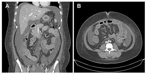
Fig. 1 Abdominal computed tomography showing a massive amount of ascites and diffuse edematous wall thickening at the ileum and ascending colon. (A) Coronal view. (B) Axial view.
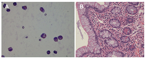
Fig. 2 Histologic images. (A) Most leukocytes in the ascitic fluid were observed as eosinophils (Wright stain, ×1,000). (B) Microscopic findings in a colonoscopic biopsy showing that numerous eosinophils are infiltrated in lamina propria (H&E, ×200).
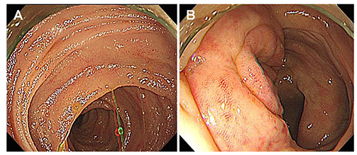
Fig. 3 Colonoscopy showing edematous and focally hyperemic mucosa at the (A) terminal ileum and (B) ileocecal valve.

Fig. 4 Dramatic decrease in peripheral blood eosinophil count is seen from the next day of steroid treatment. HD, hospital day.

Fig. 5 Abdominal computed tomography at the end of the treatment showing disappearance of ascites and improvement of edematous change of bowel wall. (A) Coronal view. (B) Axial view.
Reference
-
1. Rothenberg ME. Eosinophilic gastrointestinal disorders (EGID). J Allergy Clin Immunol. 2004; 113:11–28.
Article2. Pineton de Chambrun G, Dufour G, Tassy B, et al. Diagnosis, natural history and treatment of eosinophilic enteritis: a review. Curr Gastroenterol Rep. 2018; 20:37.
Article3. Lee J, Dierkhising R, Wu TT, Alexander J, Weiler C. Eosinophilic gastrointestinal disorders (EGID) with peripheral eosinophilia: a retrospective review at Mayo Clinic. Dig Dis Sci. 2011; 56:3254–3261.
Article4. Mansoor E, Saleh MA, Cooper GS. Prevalence of eosinophilic gastroenteritis and colitis in a population-based study, from 2012 to 2017. Clin Gastroenterol Hepatol. 2017; 15:1733–1741.
Article5. Klein NC, Hargrove RL, Sleisenger MH, Jeffries GH. Eosinophilic gastroenteritis. Medicine (Baltimore). 1970; 49:299–319.
Article6. Kwon JY, Huh JS, Je BK, Hong KD, Lee JH. Eosinophilic gastrointestinal disorder presenting as intractable vomiting and ascites in a young girl. Pediatr Gastroenterol Hepatol Nutr. 2017; 20:198–203.
Article7. Sheikh RA, Prindiville TP, Pecha RE, Ruebner BH. Unusual presentations of eosinophilic gastroenteritis: case series and review of literature. World J Gastroenterol. 2009; 15:2156–2161.
Article8. Minodier L, Charrel RN, Ceccaldi PE, et al. Prevalence of gastrointestinal symptoms in patients with influenza, clinical significance, and pathophysiology of human influenza viruses in faecal samples: what do we know? Virol J. 2015; 12:215.
Article9. Zhang M, Li Y. Eosinophilic gastroenteritis: a state-of-the-art review. J Gastroenterol Hepatol. 2017; 32:64–72.
Article10. Takeyama J, Abukawa D, Miura K. Eosinophilic gastroenteritis with cytomegalovirus infection in an immunocompetent child. World J Gastroenterol. 2007; 13:4653–4654.
Article11. Koga M, Fujiwara M, Hotta N, Matsubara T, Suzuki E, Furukawa S. Eosinophilic gastroenteritis associated with Epstein-Barr virus infection in a young boy. J Pediatr Gastroenterol Nutr. 2001; 33:610–612.
Article12. Terai M, Honda T, Yamamoto S, et al. Early induction of interleukin-5 and peripheral eosinophilia in acute pneumonia in Japanese children infected by pandemic 2009 influenza A in the Tokyo area. Microbiol Immunol. 2011; 55:341–346.
Article13. Sládková T, Kostolanský F. The role of cytokines in the immune response to influenza A virus infection. Acta Virol. 2006; 50:151–162.14. Zhu FL, Ling AS, Wei Q, Ma J, Lu G. Tumor markers in serum and ascites in the diagnosis of benign and malignant ascites. Asian Pac J Cancer Prev. 2015; 16:719–722.
Article15. Hallal H, Más Mercader P, Alberca de las Parras F, et al. Eosinophilic gastroenteritis and CA-125 elevation. Gastroenterol Hepatol. 2004; 27:292–293.
- Full Text Links
-
- Actions
-
Cited
- CITED
-
- Close
- Share
-
- Similar articles
-
- Eosinophilic Gastrointestinal Disorder Presenting as Intractable Vomiting and Ascites in a Young Girl
- A Case of Anisakiasis Diagnosed after Partial Resection of Ileum Due to Eosinophilic Ascites and Ileal Abscess
- Eosinophilic Enteritis Diagnosed by Laparoscopic Biopsy
- Eosinophilic Enteritis with Eosinophilic Ascites without Eosinophilia
- A Case of Eosinophilic Gastroenteritis Diagnosed by Repeated Abdominal Pain with Eosinophilic Ascites and Cystitis
