Synchronous Primary Leiomyosarcoma in the Thoracic Vertebra and the Liver
- Affiliations
-
- 1Department of Internal Medicine, Seoul Paik Hospital, Inje University College of Medicine, Seoul, Korea. rshdrryu@hanmail.net
- 2Department of Hemato-oncology, Kyung Hee University Hospital at Gangdong, School of Medicine, Kyung Hee University, Seoul, Korea.
- 3Department of Radiology, Seoul Paik Hospital, Inje University College of Medicine, Seoul, Korea.
- 4Department of Pathology, Seoul Paik Hospital, Inje University College of Medicine, Seoul, Korea.
- 5Piedmont Transplant Institute of Atlanta, Atlanta, GA, USA.
- KMID: 2453260
- DOI: http://doi.org/10.4166/kjg.2019.74.1.57
Abstract
- This is a case report of simultaneous primary leiomyosarcomas in the spine and liver. A 64-year-old woman presented to the Seoul Paik Hospital with epigastric discomfort and constipation that she had experienced for two months. A physical examination revealed severe tenderness around the thoraco-lumbar junction. Esophagogastroduodenoscopy showed an ulceroinfiltrative lesion on the gastric angle. An abdominopelvic CT scan revealed two low attenuated lesions in the S4 and S8 regions of the liver, as well as a soft tissue mass at the T10 vertebra. Percutaneous ultrasonography-guided needle biopsy of the hepatic nodules revealed a leiomyosarcoma. The tumor at the T10 vertebra was removed to avoid spinal cord compression. The histology of this tumor was compatible with that of leiomyosarcoma. The potential primary sites for leiomyosarcoma, including the lung, thyroid, breast, kidney, genitourinary organs, and gastrointestinal tract, were subsequently investigated. No detectable abnormal findings that would suggest the origin of the tumor were found. Synchronous primary leiomyosarcomas in the spine and liver are quite rare and have a poor prognosis.
MeSH Terms
Figure
-
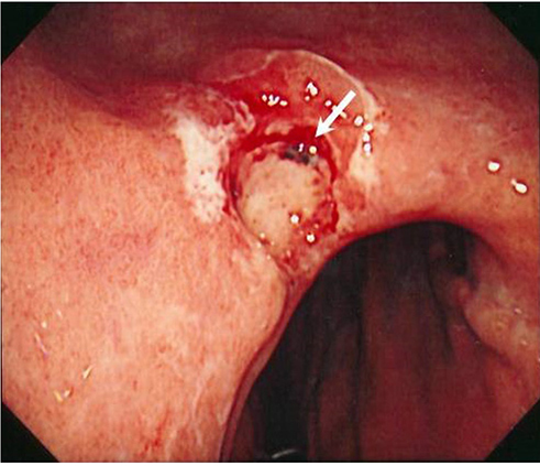
Fig. 1 Esophagogastroduodenoscopy shows an ulceroinfiltrative lesion (arrow) on the gastric angle.
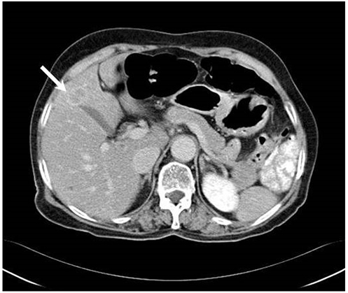
Fig. 2 Abdominopelvic computed tomography scan shows an oval low attenuation nodule with rim enhancement (arrow) at S4 in the liver.
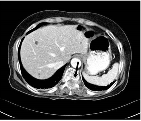
Fig. 3 Computed tomography scan shows a lobulated heterogeneous low attenuation mass (arrow) involving the left pedicle and both laminae extending into the spinal canal.
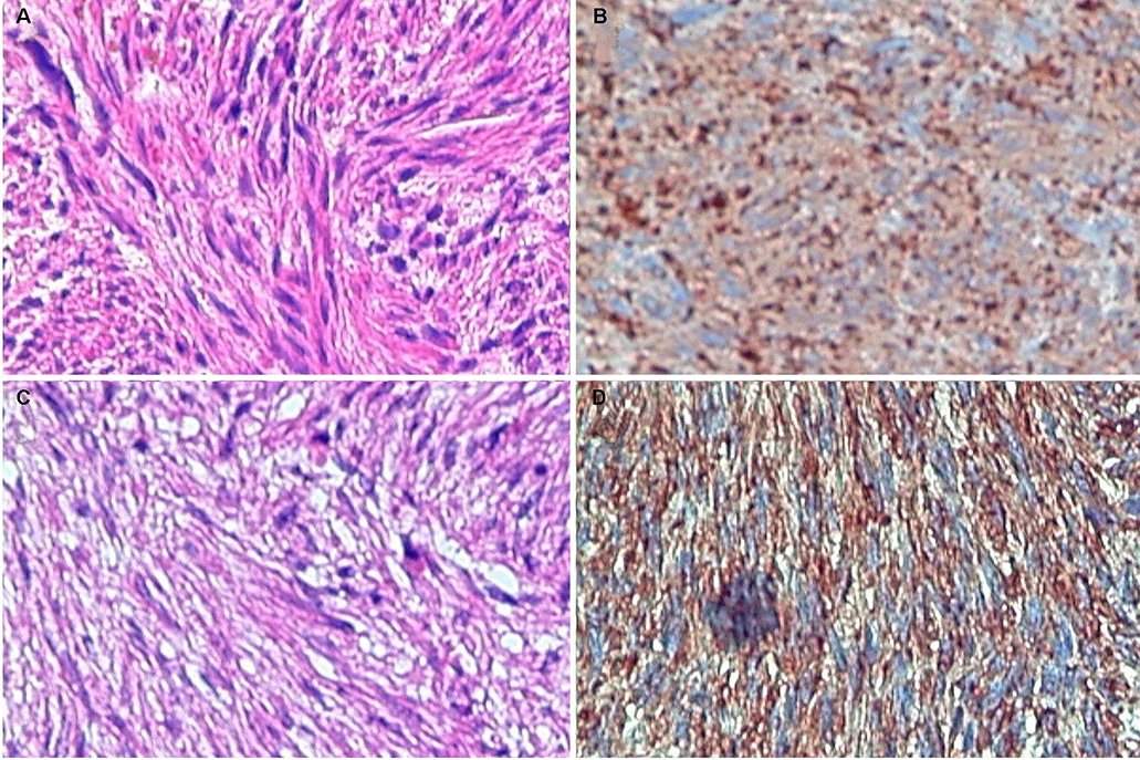
Fig. 4 (A) Microscopic view of the hepatic lesion shows an interlacing bundle of atypical spindle cells (H&E, ×200). (B) Immunohistochemical staining of the hepatic lesion shows a positive reaction for desmin (×200). (C) Microscopic view of the spinal lesion showing an interlacing bundle of atypical spindle cells (H&E, ×400). (D) Immunohistochemical staining of the spinal lesion showing a positive reaction for smooth muscle actin (×200).
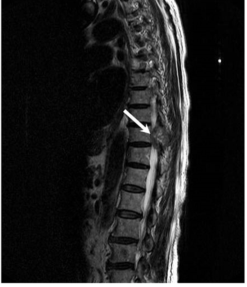
Fig. 5 Magnetic resonance imaging shows a heterogeneous low-signal intensity mass compressing the dural sac at the T10 level (arrow) on a sagittal T2-weighted image.
Reference
-
1. Russell WO, Cohen J, Enzinger F, et al. A clinical and pathological staging system for soft tissue sarcomas. Cancer. 1977; 40:1562–1570.
Article2. Antonescu CR, Erlandson RA, Huvos AG. Primary leiomyosarcoma of bone: a clinicopathologic, immunohistochemical, and ultrastructural study of 33 patients and a literature review. Am J Surg Pathol. 1997; 21:1281–1294.
Article3. Potsi M, Stavrinou P, Patsinakidis N, et al. Primary osseous leiomyosarcoma of the spine: a rare entity--case report and review of the literature. J Neurol Surg A Cent Eur Neurosurg. 2012; 73:238–242.
Article4. Mirra JM. Bone tumors: clinical, radiologic, and pathologic correlations. illustrated ed. Philadelphia: Lea & Febiger;1989.5. von Hochstetter AR, Eberle H, Rüttner JR. Primary leiomyosarcoma of extragnathic bones. Case report and review of literature. Cancer. 1984; 53:2194–2200.
Article6. Sasaguri T, Tanimoto A, Kimura S, et al. Primary leiomyosarcoma of the vertebra: case report and review of the literature. Pathol Int. 2004; 54:73–76.
Article7. Ganau S, Tomás X, Mallofré C, Macho JM, Pomés J, Combalia A. Leiomyosarcoma of sacrum: imaging and histopathologic findings. Eur Radiol. 2002; 12:Suppl 3. S35–S39.
Article8. Khoddami M, Bedard YC, Bell RS, Kandel RA. Primary leiomyosarcoma of bone: report of seven cases and review of the literature. Arch Pathol Lab Med. 1996; 120:671–675.9. Lopez-Barea F, Rodriguez-Peralto JL, Sanchez-Herrera S, Gonzalez-Lopez J, Burgos-Lizaldez E. Primary epithelioid leiomyosarcoma of bone. Case report and literature review. Virchows Arch. 1999; 434:367–371.10. Watanabe K, Kusakabe T, Hoshi N, Saito A, Suzuki T. h-Caldesmon in leiomyosarcoma and tumors with smooth muscle cell-like differentiation: its specific expression in the smooth muscle cell tumor. Hum Pathol. 1999; 30:392–396.
Article11. Young MP, Freemont AJ. Primary leiomyosarcoma of bone. Histopathology. 1991; 19:257–262.
Article12. Shen SH, Steinbach LS, Wang SF, Chen WY, Chen WM, Chang CY. Primary leiomyosarcoma of bone. Skeletal Radiol. 2001; 30:600–603.
Article13. Fornasier VL, Paley D. Leiomyosarcoma in bone: primary or secondary? A case report and review of the literature. Skeletal Radiol. 1983; 10:147–153.14. Soyer P, Poccard M, Boudiaf M, et al. Detection of hypovascular hepatic metastases at triple-phase helical CT: sensitivity of phases and comparison with surgical and histopathologic findings. Radiology. 2004; 231:413–420.
Article15. Meltzer CC, Fishman EK, Scott WW Jr. Computed tomography appearance of bone metastases of leiomyosarcoma. Skeletal Radiol. 1992; 21:445–447.
Article16. Sundaresan N, DiGiacinto GV, Krol G, Hughes JE. Spondylectomy for malignant tumors of the spine. J Clin Oncol. 1989; 7:1485–1491.
Article17. Krepler P, Windhager R, Bretschneider W, Toma CD, Kotz R. Total vertebrectomy for primary malignant tumours of the spine. J Bone Joint Surg Br. 2002; 84:712–715.
Article18. Kepka L, DeLaney TF, Suit HD, Goldberg SI. Results of radiation therapy for unresected soft-tissue sarcomas. Int J Radiat Oncol Biol Phys. 2005; 63:852–859.
Article19. Patel SR, Vadhan-Raj S, Burgess MA, et al. Results of two consecutive trials of dose-intensive chemotherapy with doxorubicin and ifosfamide in patients with sarcomas. Am J Clin Oncol. 1998; 21:317–321.
Article20. Maki RG, Wathen JK, Patel SR, et al. Randomized phase II study of gemcitabine and docetaxel compared with gemcitabine alone in patients with metastatic soft tissue sarcomas: results of sarcoma alliance for research through collaboration study 002 [corrected]. J Clin Oncol. 2007; 25:2755–2763.
