Pathological Classification of Focal Cortical Dysplasia (FCD) : Personal Comments for Well Understanding FCD Classification
- Affiliations
-
- 1Department of Pathology, Severance Hospital, Yonsei University College of Medicine, Seoul, Korea. paxco@yuhs.ac
- 2Yonsei Institute of Pharmaceutical Sciences, College of Pharmacy, Yonsei University, Incheon, Korea.
- KMID: 2453233
- DOI: http://doi.org/10.3340/jkns.2019.0025
Abstract
- In 2011, the International League against Epilepsy (ILAE) proposed a first international consensus of the classification of focal cortical dysplasia (FCD). This FCD classification had been widely used in worldwide. In this review paper, the authors would like to give helpful comments for better understanding of the current FCD classification. Especially, the basic concepts of FCD type I, such as "radial", "tangential" and "microcolumn" will be discussed with figures. In addition, the limitations, genetic progress and prospect of FCD will be suggested.
MeSH Terms
Figure
-
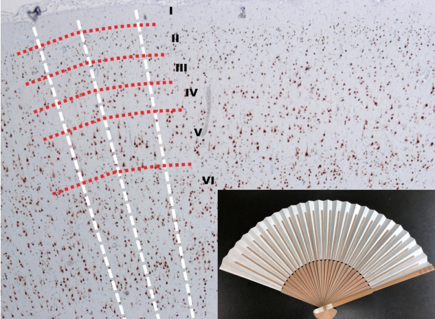
Fig. 1. The low power view of normal neocortex (NeuN, ×40) and a Korean folding pan (inlet). The imaginary white lines show the direction of radial migrations of neural cells. The lines look like the bones of a folding pan (inlet). The cortical six-layers were seen with red imaginary lines. The red imaginary lines show the tangential composition of neocortex. In the normal neocortex, it is unusual that the neuronal cells exactly align along the white imaginary lines.
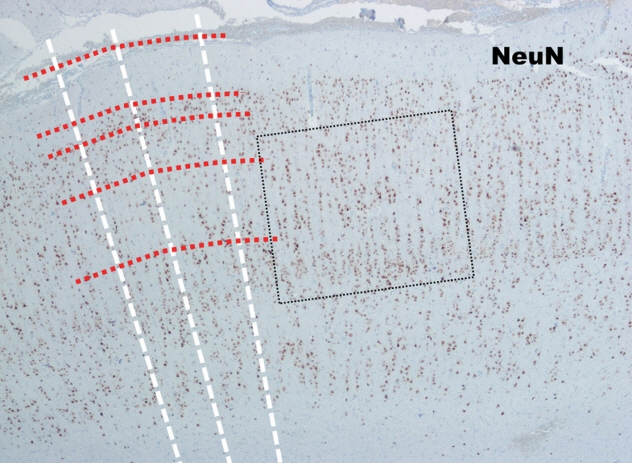
Fig. 2. The low power view of the neocortex in FCD type Ia (NeuN, ×40). In contrast to the normal neocortex, many neuronal cells in FCD type Ia show linear alignment as shown in the dot-line box. FCD : focal cortical dysplasia.
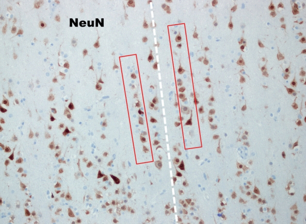
Fig. 3. In high power view of the box (NeuN, ×200), the so-called many microcolumns, which were defined more than eight neurons aligned in a vertical direction, are noted (red boxes).
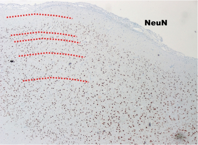
Fig. 4. The low power view of FCD type Ib (NeuN, ×40). The cortical sixlayers are deranged (too chaotic) compared to those in normal neocortex (Fig. 1). FCD : focal cortical dysplasia.
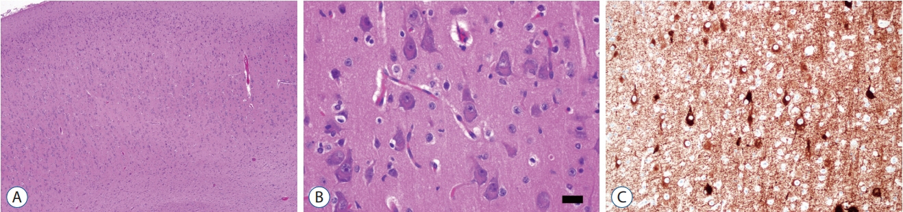
Fig. 5. The histopathological findings of FCD type IIa. The low power view (A; H-E, ×40) shows cortical dyslamination which means derangement of normal neocortical six-layers. High power view shows many large dysmorphic neurons with coarse Nissl substances. The size of dysmorphic neuron cytoplasm is more than 20 µm (B; H-E, ×400; black bar, 20 µm). Dysmorphic neurons are highlighted by immunostaining with nonphosphorylated neurofilament (C; SMI132, ×200). FCD : focal cortical dysplasia.
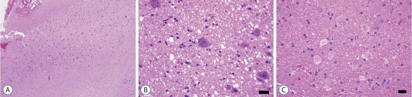
Fig. 6. The histopathological findings of FCD type IIb. The low power view (A; H-E, ×40) shows cortical dyslamination as FCD type IIa. High power view shows many large dysmorphic neurons with coarse Nissl substances (B; H-E, ×400; black bar, 20 µm) and balloon cells showing eosinophilic homogenous cytoplasm (C; H-E, ×400; black bar, 20 µm). FCD : focal cortical dysplasia.
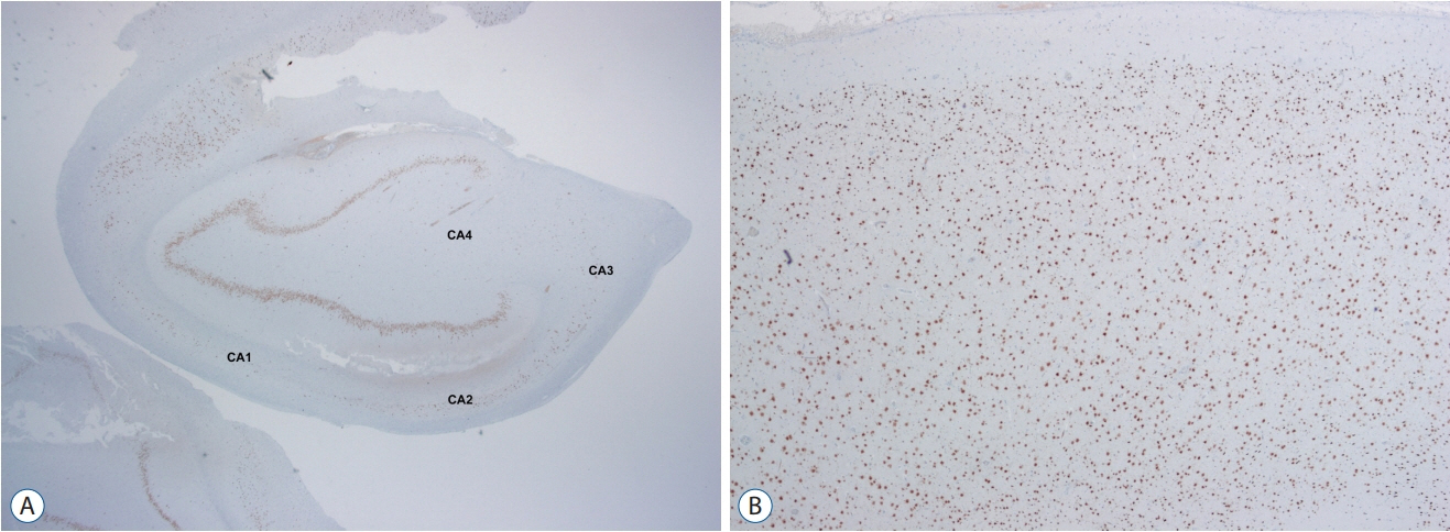
Fig. 7. FCD type IIIa is a hippocampal sclerosis (ILAE type Ia) showing neuronal loss in CA4, 3, 2 and 1 (A; NeuN, ×12) with associating FCD type Ib which shows a breakdown of cortical tangential composition in overlying cortex (B; NeuN, ×40). FCD : focal cortical dysplasia.
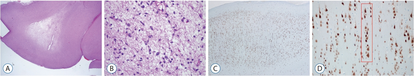
Fig. 8. FCD type IIIb is combined with a low grade-epilepsy associated tumor (LEAT). Diagnosis of “the associated tumor” in this case is dysembryoplastic neuroepithelial tumor (DNT) (A; H-E, ×12 and B; H-E, ×200). Overlying cortex shows FCD type Ia (C; NeuN, ×40) characterized by microcolumns (D; red box; NeuN, ×200). FCD : focal cortical dysplasia.
Reference
-
References
1. Aronica E, Becker AJ, Spreafico R. Malformations of cortical development. Brain Pathol. 22:380–401. 2012.
Article2. Barkovich AJ, Guerrini R, Kuzniecky RI, Jackson GD, Dobyns WB. A developmental and genetic classification for malformations of cortical development: update 2012. Brain. 135:1348–1369. 2012.
Article3. Blümcke I, Aronica E, Miyata H, Sarnat HB, Thom M, Roessler K, et al. International recommendation for a comprehensive neuropathologic workup of epilepsy surgery brain tissue: a consensus task force report from the ILAE commission on diagnostic methods. Epilepsia. 57:348–358. 2016.
Article4. Blümcke I, Spreafico R, Haaker G, Coras R, Kobow K, Bien CG, et al. Histopathological findings in brain tissue obtained during epilepsy surgery. N Engl J Med. 377:1648–1656. 2017.
Article5. Blümcke I, Thom M, Aronica E, Armstrong DD, Vinters HV, Palmini A, et al. The clinicopathologic spectrum of focal cortical dysplasias: a consensus classification proposed by an ad hoc task force of the ILAE diagnostic methods commission. Epilepsia. 52:158–174. 2011.
Article6. Cepeda C, André VM, Levine MS, Salamon N, Miyata H, Vinters HV, et al. Epileptogenesis in pediatric cortical dysplasia: the dysmature cerebral developmental hypothesis. Epilepsy Behav. 9:219–235. 2006.
Article7. Crome L. Infantile cerebral gliosis with giant nerve cells. J Neurol Neurosurg Psychiatry. 20:117–124. 1957.
Article8. Hildebrandt M, Pieper T, Winkler P, Kolodziejczyk D, Holthausen H, Blümcke I. Neuropathological spectrum of cortical dysplasia in children with severe focal epilepsies. Acta Neuropathol. 110:1–11. 2005.
Article9. Meencke HJ, Veith G. Migration disturbances in epilepsy. Epilepsy Res Suppl. 9:31–39. discussion 39-40. 1992.10. Najm IM, Sarnat HB, Blümcke I. Review: the international consensus classification of focal cortical dysplasia - a critical update 2018. Neuropathol Appl Neurobiol. 44:18–31. 2018.
Article11. Palmini A, Najm I, Avanzini G, Babb T, Guerrini R, Foldvary-Schaefer N, et al. Terminology and classification of the cortical dysplasias. Neurology. 62:S2–S8. 2004.
Article12. Prayson RA. Pathology of epilepsy. In : Perry A, Brat DJ, editors. Parctical surgical neuropathology : A diagnostic approach. ed 2. Philadelphia: Elsevier;2018. p. 617–632.13. Taylor DC, Falconer MA, Bruton CJ, Corsellis JA. Focal dysplasia of the cerebral cortex in epilepsy. J Neurol Neurosurg Psychiatry. 34:369–387. 1971.
Article14. The Free Dictionary. Dysplasia. Available at : https://medical-dictionary.thefreedictionary.com/dysplasia.15. Thom M, Eriksson S, Martinian L, Caboclo LO, McEvoy AW, Duncan JS, et al. Temporal lobe sclerosis associated with hippocampal sclerosis in temporal lobe epilepsy : neuropathological features. J Neuropathol Exp Neurol. 68:928–938. 2009.
Article16. Wikipedia. Dysplasia. Available at : https://en.wikipedia.org/wiki/Dysplasia.
- Full Text Links
-
- Actions
-
Cited
- CITED
-
- Close
- Share
-
- Similar articles
-
- Focal Cortical Dysplasia in Pediatric Epilepsy
- Discontinuing Antiepileptic Drugs after Pediatric Epilepsy Surgery for Focal Cortical Dysplasia
- Lesion Detection Through MRI Postprocessing in Pathology-Proven Focal Cortical Dysplasia: Experience at a Single Institution in the Republic of Korea
- The Surgical and Cognitive Outcomes of Focal Cortical Dysplasia
- Surgically Treated Intractable Child Epilepsy with Focal Cortical Dysplasia : Clinical and Electroencephalographic Findings
