Imaging Spectrum and Pitfalls of 11C-Methionine Positron Emission Tomography in a Series of Patients with Intracranial Lesions
- Affiliations
-
- 1Department of Radiology, Tokyo Metropolitan Geriatric Hospital and Institute of Gerontology, Tokyo 173-0015, Japan. itoukimiteru@yahoo.co.jp
- 2Integrative Brain Imaging Center, National Center of Neurology and Psychiatry, Tokyo 187-8551, Japan.
- 3Division of Nuclear Medicine, National Center for Global Health and Medicine, Tokyo 162-8655, Japan.
- KMID: 2451416
- DOI: http://doi.org/10.3348/kjr.2016.17.3.424
Abstract
- 11C-methionine (Met) positron emission tomography (PET) is one of the most commonly used PET tracers for evaluating brain tumors. However, few reports have described tips and pitfalls of 11C-Met PET for general practitioners. Physiological 11C-Met uptake, anatomical variations, vascular disorders, non-tumorous lesions such as inflammation or dysplasia, benign brain tumors and patient condition during 11C-Met PET examination can potentially affect the image interpretation and cause false positives and negatives. These pitfalls in the interpretation of 11C-Met PET images are important for not only nuclear medicine physicians but also general radiologists. Familiarity with the spectrum and pitfalls of 11C-Met images could help prevent unfavorable clinical results caused by misdiagnoses.
MeSH Terms
-
Adolescent
Adult
Aged
Aged, 80 and over
Brain Neoplasms/*diagnostic imaging/pathology
Carbon Radioisotopes/chemistry
Child
Child, Preschool
Female
Glioma/diagnostic imaging/pathology
Humans
Male
Methionine/*chemistry
Middle Aged
Neoplasm Recurrence, Local
*Positron-Emission Tomography
Vascular Diseases/*diagnostic imaging/pathology
Young Adult
Carbon Radioisotopes
Methionine
Figure
-

Fig. 1 52-year-old woman with epilepsy. A. Axial 11C-methionine (Met) positron emission tomography/computed tomography (CT) image showing high 11C-Met uptake in bilateral thickened frontal bone (arrows). B. Axial CT bone window showing hyperostosis (arrows). Hyperostosis occasionally shows high 11C-Met uptake.

Fig. 2 75-year-old man with amygdala enlargement and Rathke's cleft cyst. Sagittal 11C-methionine (Met) positron emission tomography/CT image showing no 11C-Met uptake in Rathke's cleft cyst, whereas physiological 11C-Met uptake is observed in pituitary gland in anterior part.
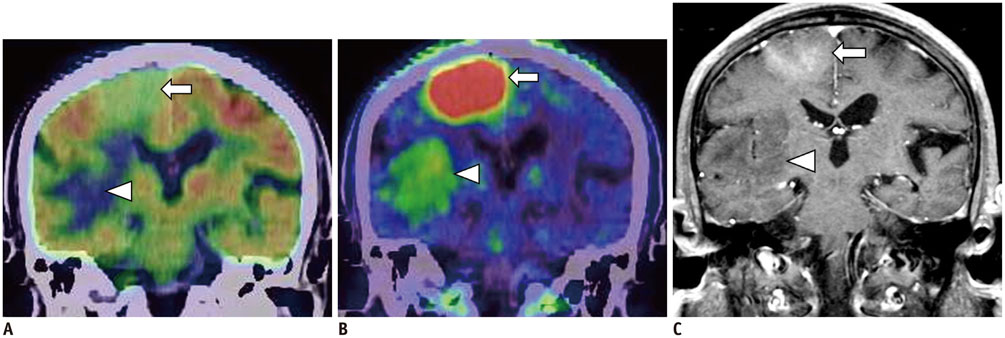
Fig. 3 76-year-old man with brain tumor. A. Coronal 18F-fluorodeoxyglucose (FDG) positron emission tomography (PET)/CT image showing lower uptake of convex lesion than of cortex (arrow). Lesion in white matter at right temporal lobes (arrowhead) shows markedly low uptake. B. Coronal 11C-methionine (Met) PET/CT image showing higher uptake at convex lesion (arrow), with tumor-to-normal (T/N) ratio of 7.2. However, lesion at right temporal lobes and opaque part showing lower 11C-Met uptake (arrowhead), with T/N ratio of 2.3. C. Axial contrast-enhanced MR T1-weighted imaging showing contrast-enhanced lesions in convex (arrow). Lesion at right temporal lobes (arrowhead) shows no contrast-enhancing. In biopsy specimens, high- and low-11C-Met-uptake areas corresponded to high- and low-grade astrocytomas.
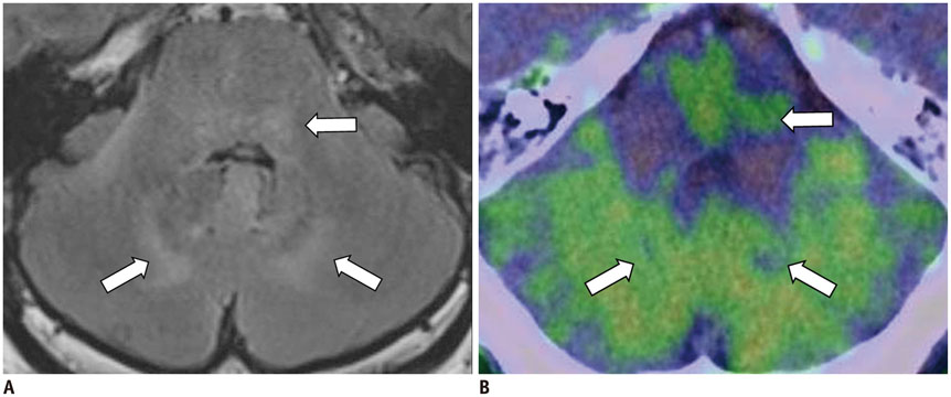
Fig. 4 48-year-old man with gliomatosis cerebri. A. Axial fluid-attenuated inversion recovery image of magnetic resonance imaging showing high intensity with unclear boundaries in tegmentum of pons and bilateral internal portion of cerebellum (arrows). Lesion shows no contrast enhancement. B. Axial 11C-methionine (Met) positron emission tomography/CT showing unclear uptake of lesion, which is almost indistinguishable from background uptake in cerebellum (arrows), with tumor-to-normal ratio of 1.2. 11C-Met uptake appears vague or patchy in lesions.

Fig. 5 26-year-old woman with anaplastic oligoastrocytoma and coarse calcifications. Axial 11C-methionine positron emission tomography (PET)/CT image showing high uptake in lesion with coarse calcifications (arrows) at upper right frontal lobe, with tumor-to-normal ratio of 3.9. CT imaging during PET/CT is helpful for diagnosis of brain lesions.
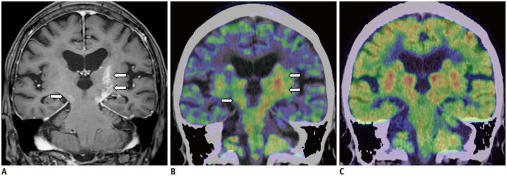
Fig. 6 84-year-old woman with primary central nervous system lymphoma. A. Contrast-enhanced T1-weighted imaging (T1WI) of MRI showing contrast-enhanced lesions in left basal ganglia and bilateral cerebral peduncles (arrows). B. Axial 11C-methionine (Met) positron emission tomography (PET)/CT images clearly showing high uptakes in left basal ganglia and bilateral cerebral peduncles (arrows). Area of 11C-Met uptake is more widely spread out than that of contrast-enhanced T1WI. C. Lesion is poorly defined on coronal 18F-fluorodeoxyglucose PET/CT because of physiological uptake.
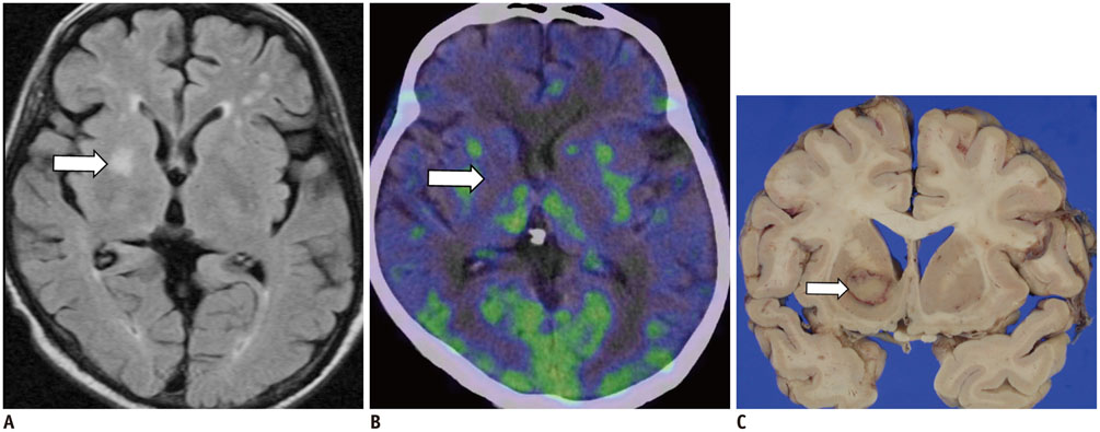
Fig. 7 56-year-old man with recurrent leukemia after chemotherapy. A. Axial fluid-attenuated inversion recovery image of MRI showing high intensity lesion at right basal ganglia (arrow) without contrast enhancement. B. Axial 11C-methionine positron emission tomography/CT images showing low uptake corresponding to lesion (arrow), with tumor-to-normal ratio of 1.5. C. Postmortem brain specimen showing ring-like lesion, which was confirmed as infiltration of leukemia cells (arrow).
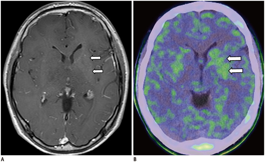
Fig. 8 15-year-old man with germinoma. A. Contrast-enhanced T1-weighted image showing slightly enhanced lesions at left basal ganglia (arrows). B. Axial 11C-methionine positron emission tomography/CT image showing mild uptake in lesion (arrows).
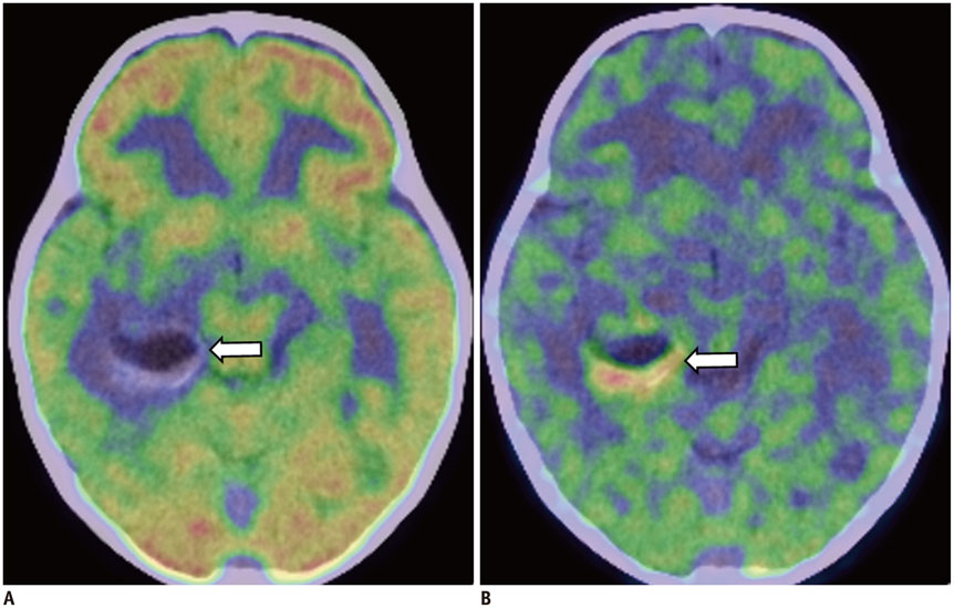
Fig. 9 2-year-old girl with ganglioglioma. A. Axial 18F-fluorodeoxyglucose positron emission tomography (PET)/CT image showing lower uptake at cystic lesion surrounding solid portion with vague calcification at right temporal lobe (arrow). B. Axial 11C-methionine PET/CT image showing high uptake at solid component (arrow).

Fig. 10 5-year-old boy with dysembryoplastic neuroepithelial tumors. A. T2-weighted image (T2WI) showing high-intensity mass with relatively distinct border at right temporal lobe (arrows). B. Axial 11C-methionine positron emission tomography/CT image showing low uptake corresponding to lesion with T2WI high-intensity area (arrows).

Fig. 11 22-year-old woman with central neurocytoma. A. Axial CT image showing tumor with coarse calcifications in lateral ventricle (arrows). B. Axial 11C-methionine positron emission tomography/CT image showing high uptake at tumor (arrows), with tumor-to-normal ratio of 4.9.
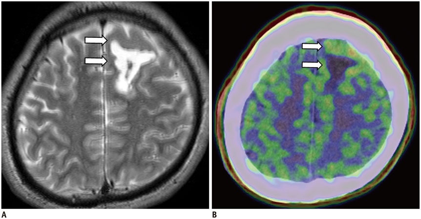
Fig. 12 26-year-old man with hematoma. A. Axial T2-weighted image showing high-intensity mass at left temporal lobes (arrows). B. Axial 11C-methionine positron emission tomography/CT image showing low uptake at subcortical area surrounding slightly high uptake at cortex in left frontal lobe (arrows).
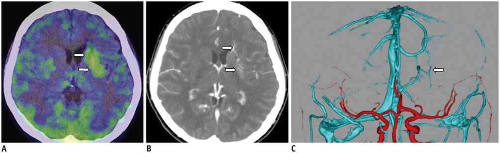
Fig. 13 25-year-old woman having venous malformation with plugged venous flow. A. Axial 11C-methionine positron emission tomography/CT image showing high uptake at left basal ganglia (arrows), with tumor-to-normal ratio of 2.6. B. Axial contrast-enhanced CT image showing vascular anomalies in left basal ganglia (arrows). C. Volume-rendered CT angiographic image showing venous malformation at left basal ganglia (arrow).

Fig. 14 64-year-old man with tumefactive demyelination in post-treatment state. A. Axial contrast-enhanced T1-weighted image showing lesion with non-enhancement of deep white matter in left posterior temporal lobe (arrow). B. Axial 11C-methionine positron emission tomography/CT image taken at almost same time as in (A) showing high uptake corresponding to lesion (arrow).
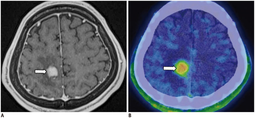
Fig. 15 28-year-old man with toxoplasmosis caused by acquired immunodeficiency syndrome. A. Axial contrast-enhanced T1-weighted image showing clearly enhanced lesion with edema (arrow) in right parietal lobe. B. Coronal 11C-methionine positron emission tomography/CT image showing high uptake (arrow) corresponding to lesion, with tumor-to-normal ratio of 4.0.

Fig. 16 25-year-old woman with focal cortical dysplasia. A. Axial T2-weighted image showing slight thickening in cortex with unclear boundary between white matter and cortex in right occipital lobe (arrow). B. Axial 11C-methionine positron emission tomography/CT image showing slightly higher uptake of lesion than background (arrow).

Fig. 17 8-year-old boy with hamartoma. A. Axial fluid-attenuated inversion recovery image showing normal intensity of lesion in left cerebellar peduncle (arrow). B. Axial 11C-methionine positron emission tomography/CT image showing low uptake in lesion similar to background (arrow).
Reference
-
1. Zhao C, Zhang Y, Wang J. A meta-analysis on the diagnostic performance of (18)F-FDG and (11)C-methionine PET for differentiating brain tumors. AJNR Am J Neuroradiol. 2014; 35:1058–1065.2. Singhal T, Narayanan TK, Jain V, Mukherjee J, Mantil J. 11C-L-methionine positron emission tomography in the clinical management of cerebral gliomas. Mol Imaging Biol. 2008; 10:1–18.3. Glaudemans AW, Enting RH, Heesters MA, Dierckx RA, van Rheenen RW, Walenkamp AM, et al. Value of 11C-methionine PET in imaging brain tumours and metastases. Eur J Nucl Med Mol Imaging. 2013; 40:615–635.4. Nagata T, Tsuyuguchi N, Uda T, Terakawa Y, Takami T, Ohata K. Examination of 11C-methionine metabolism by the standardized uptake value in the normal brain of children. J Nucl Med. 2011; 52:201–205.5. Lindholm P, Leskinen-Kallio S, Kirvelä O, Någren K, Lehikoinen P, Pulkki K, et al. Head and neck cancer: effect of food ingestion on uptake of C-11 methionine. Radiology. 1994; 190:863–867.6. Torii K, Tsuyuguchi N, Kawabe J, Sunada I, Hara M, Shiomi S. Correlation of amino-acid uptake using methionine PET and histological classifications in various gliomas. Ann Nucl Med. 2005; 19:677–683.7. Tomura N, Ito Y, Matsuoka H, Saginoya T, Numazawa SI, Mizuno Y, et al. PET findings of intramedullary tumors of the spinal cord using [18F] FDG and [11C] methionine. AJNR Am J Neuroradiol. 2013; 34:1278–1283.8. Kawase Y, Yamamoto Y, Kameyama R, Kawai N, Kudomi N, Nishiyama Y. Comparison of 11C-methionine PET and 18F-FDG PET in patients with primary central nervous system lymphoma. Mol Imaging Biol. 2011; 13:1284–1289.9. Okochi Y, Nihashi T, Fujii M, Kato K, Okada Y, Ando Y, et al. Clinical use of (11)C-methionine and (18)F-FDG-PET for germinoma in central nervous system. Ann Nucl Med. 2014; 28:94–102.10. Phi JH, Paeng JC, Lee HS, Wang KC, Cho BK, Lee JY, et al. Evaluation of focal cortical dysplasia and mixed neuronal and glial tumors in pediatric epilepsy patients using 18F-FDG and 11C-methionine pet. J Nucl Med. 2010; 51:728–734.11. Arita H, Kinoshita M, Okita Y, Hirayama R, Watabe T, Ishohashi K, et al. Clinical characteristics of meningiomas assessed by 11C-methionine and 18F-fluorodeoxyglucose positron-emission tomography. J Neurooncol. 2012; 107:379–386.12. Takao H, Momose T, Ohtomo K. Methionine and glucose metabolism of central neurocytoma: a PET study. Clin Nucl Med. 2004; 29:838–839.13. Nakagawa M, Kuwabara Y, Sasaki M, Koga H, Chen T, Kaneko O, et al. 11C-methionine uptake in cerebrovascular disease: a comparison with 18F-fDG PET and 99mTc-HMPAO SPECT. Ann Nucl Med. 2002; 16:207–211.14. Harada Y, Hirata K, Kobayashi H, Usui R, Shiga T, Terae S, et al. A pitfall of C-11 methionine PET: cerebral venous infarction mimicked a glioma. Clin Nucl Med. 2012; 37:110–111.15. O'Doherty MJ, Barrington SF, Campbell M, Lowe J, Bradbeer CS. PET scanning and the human immunodeficiency virus-positive patient. J Nucl Med. 1997; 38:1575–1583.16. Sasaki M, Kuwabara Y, Yoshida T, Fukumura T, Morioka T, Nishio S, et al. Carbon-11-methionine PET in focal cortical dysplasia: a comparison with fluorine-18-FDG PET and technetium-99m-ECD SPECT. J Nucl Med. 1998; 39:974–977.
- Full Text Links
-
- Actions
-
Cited
- CITED
-
- Close
- Share
-
- Similar articles
-
- A Case of Basal Ganglia Germinoma Presenting Only with Cerebral Hemiatrophy Diagnosed by Using 11C-Methionine Positron Emission Tomography
- Combination of Magnetic Resonance Spectroscopy and ¹¹C-Methionine Positron Emission Tomography for the Accurate Diagnosis of Non-Enhancing Supratentorial Glioma
- Positron Emission Tomography: Application in Pediatric Epilepsy
- Assessment of Therapeutic Effect of Sunitinib by 11C-Acetate PET Compared with FDG PET Imaging in a Patient with Metastatic Renal Cell Carcinoma
- Current Radiopharmaceuticals for Positron Emission Tomography of Brain Tumors
