Contrast-Enhanced Ultrasound and Shear Wave Elastography Evaluation of Crohn's Disease Activity in Three Adolescent Patients
- Affiliations
-
- 1The Johns Hopkins University School of Medicine, Baltimore, MD, USA.
- 2Division of Pediatric Gastroenterology, Johns Hopkins Hospital, Baltimore, MD, USA.
- 3Division of Pediatric Surgery, Johns Hopkins Hospital, Baltimore, MD, USA.
- 4Department of Radiology, Children's Hospital of Philadelphia, Perelman School of Medicine, University of Pennsylvania, Philadelphia, PA, USA. Hwangm@email.chop.edu
- KMID: 2444854
- DOI: http://doi.org/10.5223/pghn.2019.22.3.282
Abstract
- Characterizing inflammation and fibrosis in Crohn's disease (CD) is necessary to guide clinical management, but distinguishing the two remains challenging. Novel ultrasound (US) techniques: contrast-enhanced US (CEUS) and shear wave elastography (SWE) offer great potential in evaluating disease activity in pediatric patients. Three patients ages 16 to 20 with known CD underwent CEUS and SWE to characterize bowel wall inflammation and fibrosis. Magnetic resonance enterography, endoscopy, or surgical pathology findings are also described when available. The patients' disease activity included acute inflammation, chronic inflammation with stricture formation, and a fibrotic surgical anastomosis without inflammation. CEUS was useful in determining the degree of inflammation, and SWE identified bowel wall fibrosis. Used together these techniques allow for better characterization of the degree of fibrosis and inflammation in bowel strictures. With further validation CEUS and SWE may allow for improved characterization of bowel strictures and disease flares in pediatric patients suffering from CD.
Keyword
MeSH Terms
Figure
-
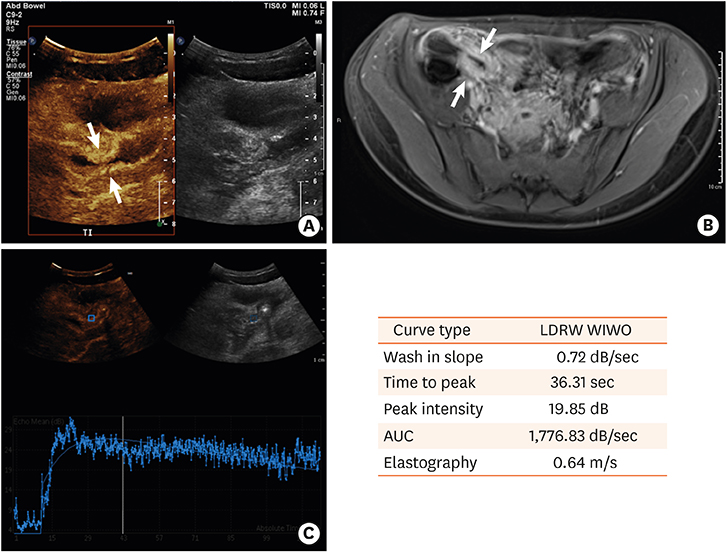
Fig. 1 A case of acute inflammation in a child with Crohn's disease. (A) Contrast-enhanced ultrasound demonstrates avid contrast enhancement in the narrowed terminal ileum. (B) Transverse contrast magnetic resonance imaging of the abdomen demonstrating bowel wall thickening and inflammation in the terminal ileum. (C) Contrast enhancement curve and quantitative parameters generated from the time-intensity curves based on the drawn region of interest in the terminal ileum. LDRW: local density random walk, WIWO: wash-in, wash-out, AUC: area under the curve.
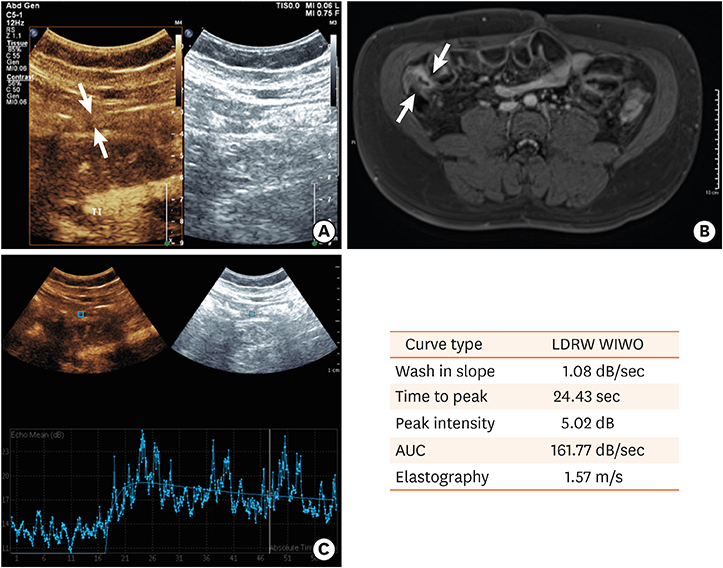
Fig. 2 A case of a fibrotic stricture in a child with Crohn's disease. (A) Contrast-enhanced ultrasound demonstrates minimal contrast enhancement in the surgical anastomosis. (B) Transverse magnetic resonance imaging of the abdomen demonstrates mild hyperenhancement of the small bowel loops without definite wall thickening or mesenteric inflammation. (C) Contrast enhancement curve and quantitative parameters generated from the time-intensity curves based on the drawn region of interest in the surgical anastomosis. LDRW: local density random walk, WIWO: wash-in, wash-out, AUC: area under the curve.

Fig. 3 A case of chronic inflammation and stricture formation in a child with Crohn's disease. (A) Intraoperative picture from ileocecectomy demonstrating the stretched-out stricture. The surgical instrument on the top indicates the area of the worst disease activity. (B) Contrast enhancement curve and quantitative parameters generated from the time-intensity curves based on the drawn region of interest in the terminal ileum. LDRW: local density random walk, WIWO: wash-in, wash-out, AUC: area under the curve.
Reference
-
1. Kappelman MD, Rifas-Shiman SL, Kleinman K, Ollendorf D, Bousvaros A, Grand RJ, et al. The prevalence and geographic distribution of Crohn's disease and ulcerative colitis in the United States. Clin Gastroenterol Hepatol. 2007; 5:1424–1429.
Article2. Rieder F, Zimmermann EM, Remzi FH, Sandborn WJ. Crohn's disease complicated by strictures: a systematic review. Gut. 2013; 62:1072–1084.
Article3. Malgras B, Pautrat K, Dray X, Pasquier P, Valleur P, Pocard M, et al. Multidisciplinary management of gastrointestinal fibrotic stenosis in Crohn's disease. Dig Dis Sci. 2015; 60:1152–1168.
Article4. Conti CB, Giunta M, Gridavilla D, Conte D, Fraquelli M. Role of bowel ultrasound in the diagnosis and follow-up of patients with Crohn's disease. Ultrasound Med Biol. 2017; 43:725–734.
Article5. Yusuf GT, Sellars ME, Deganello A, Cosgrove DO, Sidhu PS. Retrospective analysis of the safety and cost implications of pediatric contrast-enhanced ultrasound at a single center. AJR Am J Roentgenol. 2017; 208:446–452.
Article6. Migaleddu V, Scanu AM, Quaia E, Rocca PC, Dore MP, Scanu D, et al. Contrast-enhanced ultrasonographic evaluation of inflammatory activity in Crohn's disease. Gastroenterology. 2009; 137:43–52.
Article7. Romanini L, Passamonti M, Navarria M, Lanzarotto F, Villanacci V, Grazioli L, et al. Quantitative analysis of contrast-enhanced ultrasonography of the bowel wall can predict disease activity in inflammatory bowel disease. Eur J Radiol. 2014; 83:1317–1323.
Article8. Wong DD, Forbes GM, Zelesco M, Mason R, Pawlik J, Mendelson RM. Crohn's disease activity: quantitative contrast-enhanced ultrasound assessment. Abdom Imaging. 2012; 37:369–376.
Article9. Shiina T. JSUM ultrasound elastography practice guidelines: basics and terminology. J Med Ultrason (2001). 2013; 40:309–323.
Article10. Bailey C, Huisman TA, de Jong RM, Hwang M. Contrast-enhanced ultrasound and elastography imaging of the neonatal brain: a review. J Neuroimaging. 2017; 27:437–441.
Article11. Baumgart DC, Müller HP, Grittner U, Metzke D, Fischer A, Guckelberger O, et al. US-based real-time elastography for the detection of fibrotic gut tissue in patients with stricturing Crohn disease. Radiology. 2015; 275:889–899.
Article12. Dillman JR, Stidham RW, Higgins PD, Moons DS, Johnson LA, Rubin JM. US elastography-derived shear wave velocity helps distinguish acutely inflamed from fibrotic bowel in a Crohn disease animal model. Radiology. 2013; 267:757–766.
Article13. Torkzad MR, Ullberg U, Nyström N, Blomqvist L, Hellström P, Fagerberg UL. Manifestations of small bowel disease in pediatric Crohn's disease on magnetic resonance enterography. Inflamm Bowel Dis. 2012; 18:520–528.
Article14. Park SK, Yang SK, Park SH, Park SH, Kim JW, Yang DH, et al. Long-term prognosis of the jejunal involvement of Crohn's disease. J Clin Gastroenterol. 2013; 47:400–408.
Article15. Pallotta N, Civitelli F, Di Nardo G, Vincoli G, Aloi M, Viola F, et al. Small intestine contrast ultrasonography in pediatric Crohn's disease. J Pediatr. 2013; 163:778–784.e1.
Article16. Kljucevsek D, Vidmar D, Urlep D, Dezman R. Dynamic contrast-enhanced ultrasound of the bowel wall with quantitative assessment of Crohn's disease activity in childhood. Radiol Oncol. 2016; 50:347–354.
Article17. Fufezan O, Asavoaie C, Tamas A, Farcau D, Serban D. Bowel elastography - a pilot study for developing an elastographic scoring system to evaluate disease activity in pediatric Crohn's disease. Med Ultrason. 2015; 17:422–430.
Article18. Anupindi SA, Podberesky DJ, Towbin AJ, Courtier J, Gee MS, Darge K, et al. Pediatric inflammatory bowel disease: imaging issues with targeted solutions. Abdom Imaging. 2015; 40:975–992.
Article19. Coelho R, Ribeiro H, Maconi G. Bowel thickening in Crohn's disease: fibrosis or inflammation? Diagnostic ultrasound imaging tools. Inflamm Bowel Dis. 2017; 23:23–34.20. Ripollés T, Rausell N, Paredes JM, Grau E, Martínez MJ, Vizuete J. Effectiveness of contrast-enhanced ultrasound for characterisation of intestinal inflammation in Crohn's disease: a comparison with surgical histopathology analysis. J Crohns Colitis. 2013; 7:120–128.
Article21. Kucharzik T, Kannengiesser K, Petersen F. The use of ultrasound in inflammatory bowel disease. Ann Gastroenterol. 2017; 30:135–144.
Article22. Stidham RW, Xu J, Johnson LA, Kim K, Moons DS, McKenna BJ, et al. Ultrasound elasticity imaging for detecting intestinal fibrosis and inflammation in rats and humans with Crohn's disease. Gastroenterology. 2011; 141:819–826.e1.
Article23. Dillman JR, Rubin JM, Johnson LA, Moons DS, Higgins PD. Can contrast-enhanced sonography detect bowel wall fibrosis in mixed inflammatory and fibrotic Crohn disease lesions in an animal model? J Ultrasound Med. 2017; 36:523–530.
Article24. Quaia E, Gennari AG, van Beek EJ. Differentiation of inflammatory from fibrotic ileal strictures among patients with Crohn's disease through analysis of time-intensity curves obtained after microbubble contrast agent injection. Ultrasound Med Biol. 2017; 43:1171–1178.
Article25. Quaia E, Gennari AG, Cova MA, van Beek EJ. Differentiation of inflammatory from fibrotic ileal strictures among patients with Crohn's disease based on visual analysis: feasibility study combining conventional B-mode ultrasound, contrast-enhanced ultrasound and strain elastography. Ultrasound Med Biol. 2018; 44:762–770.
Article
- Full Text Links
-
- Actions
-
Cited
- CITED
-
- Close
- Share
-
- Similar articles
-
- Diagnostic Performance of Quantitative Shear Wave Ultrasound Elastography for Thyroid Cancer
- Ultrasound Elastography for Liver Disease with Focus on Hepatic Fibrosis
- Elastography of the Pancreas, Current View
- Ultrasound elastography of the thyroid: principles and current status
- Future of breast elastography
