Dement Neurocogn Disord.
2016 Mar;15(1):15-19. 10.12779/dnd.2016.15.1.15.
Phenotypic Features of Cerebral Autosomal-Dominant Arteriopathy with Subcortical Infarcts and Leukoencephalopathy Subjects with R544C Mutation
- Affiliations
-
- 1Department of Neurology, Jeju National University Hospital, Jeju, Korea. nrlee71@naver.com
- 2Department of Psychiatry, Jeju National University Hospital, Jeju, Korea.
- 3Department of Radiology, Jeju National University Hospital, Jeju, Korea.
- KMID: 2442866
- DOI: http://doi.org/10.12779/dnd.2016.15.1.15
Abstract
- BACKGROUND AND PURPOSE
Cerebral autosomal-dominant arteriopathy with subcortical infarcts and leukoencephalopathy (CADASIL) is the most-common single gene disorder of cerebral small vessel disease. There is no definite evidence of genotype-phenotype correlation in CADASIL. However, recent studies have shown the unique phenotypic feature of NOTCH3 R544C mutation.
METHODS
We investigated the phenotypic spectrum of NOTCH3 R544C mutation in 73 CADASIL patients in Jeju between April 2012 and January 2014.
RESULTS
Of the 73 subjects from 60 unrelated families included in this study, 40 (55%) were men. The mean age of the subjects was 62.2±12.2 (range 34-86 years). Cerebral infarction was the most frequent manifestation (37%), followed by cognitive impairment (32%), headache (17%), psychiatric symptom (16%), intracerebral hemorrhage (12%), transient ischemic attack (7%), and seizure (1%). The mean age of the subjects with ischemic or hemorrhagic episodes was 64.9±10.9 (range 41-86 years). A diagnosis of dementia was made in 12 subjects (16%). The mean age of the subjects with dementia was 75.6±6.5 (range 62-86 years). About 3% of subjects were unable to walk without assistance at assessment. Only one subject had developed chronic headache before the 40s.
CONCLUSIONS
Our data support the hypothesis that CADASIL patients with R544C mutation in Jeju have relatively late onset disease.
Keyword
MeSH Terms
Figure
-
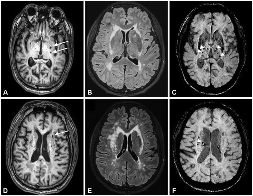
Fig. 1 Axial MR images in the subjects with R544C mutation. A and D: 3D-T1 weighted image showing multiple lacunes in the basal ganglia and corona radiate. B and E: Fluid-attenuated inversion recovery image showing confluent hyperintensity of external capsule. C and F: Susceptibility Weighted Image showing multiple cerebral microbleeds in the thalamus and corona radiate. Arrow indicates lacunes. Arrow-head points to cerebral microbleeds.
Reference
-
1. Joutel A, Corpechot C, Ducros A, Vahedi K, Chabriat H, Mouton P, et al. Notch3 mutations in CADASIL, a hereditary adult-onset condition causing stroke and dementia. Nature. 1996; 383:707–710.
Article2. Ghosh M, Balbi M, Hellal F, Dichgans M, Lindauer U, Plesnila N. Pericytes are involved in the pathogenesis of cerebral autosomal dominant arteriopathy with subcortical infarcts and leukoencephalopathy. Ann Neurol. 2015; 78:887–900.
Article3. Chabriat H, Vahedi K, Iba-Zizen MT, Joutel A, Nibbio A, Nagy TG, et al. Clinical spectrum of CADASIL: a study of 7 families. Cerebral autosomal dominant arteriopathy with subcortical infarcts and leukoencephalopathy. Lancet. 1995; 346:934–939.
Article4. Lee JS, Kang CH, Park SQ, Choi HA, Sim KB. Clinical significance of cerebral microbleeds locations in CADASIL with R544C NOTCH3 mutation. PLoS One. 2015; 10:e0118163.
Article5. Liao YC, Hsiao CT, Fuh JL, Chern CM, Lee WJ, Guo YC, et al. Characterization of CADASIL among the Han Chinese in Taiwan: distinct genotypic and phenotypic profiles. PLoS One. 2015; 10:e0136501.
Article6. Chabriat H, Joutel A, Dichgans M, Tournier-Lasserve E, Bousser MG. Cadasil. Lancet Neurol. 2009; 8:643–653.
Article7. Adib-Samii P, Brice G, Martin RJ, Markus HS. Clinical spectrum of CADASIL and the effect of cardiovascular risk factors on phenotype: study in 200 consecutively recruited individuals. Stroke. 2010; 41:630–634.
Article8. Lee JH, Lee KU, Lee DY, Kim KW, Jhoo JH, Kim JH, et al. Development of the Korean version of the Consortium to Establish a Registry for Alzheimer’s Disease Assessment Packet (CERAD-K): clinical and neuropsychological assessment batteries. J Gerontol B Psychol Sci Soc Sci. 2002; 57:P47–P53.
Article9. Oberstein SA, Ferrari MD, Bakker E, van Gestel J, Kneppers AL, Frants RR, et al. Diagnostic Notch3 sequence analysis in CADASIL: three new mutations in Dutch patients. Dutch CADASIL Research Group. Neurology. 1999; 52:1913–1915.
Article10. Opherk C, Peters N, Herzog J, Luedtke R, Dichgans M. Long-term prognosis and causes of death in CADASIL: a retrospective study in 411 patients. Brain. 2004; 127(Pt 11):2533–2539.
Article11. Mykkänen K, Savontaus ML, Juvonen V, Sistonen P, Tuisku S, Tuominen S, et al. Detection of the founder effect in Finnish CADASIL families. Eur J Hum Genet. 2004; 12:813–819.
Article12. Bianchi S, Zicari E, Carluccio A, Di Donato I, Pescini F, Nannucci S, et al. CADASIL in central Italy: a retrospective clinical and genetic study in 229 patients. J Neurol. 2015; 262:134–141.
Article13. Liu X, Zuo Y, Sun W, Zhang W, Lv H, Huang Y, et al. The genetic spectrum and the evaluation of CADASIL screening scale in Chinese patients with NOTCH3 mutations. J Neurol Sci. 2015; 354:63–69.
Article14. Ueda A, Ueda M, Nagatoshi A, Hirano T, Ito T, Arai N, et al. Genotypic and phenotypic spectrum of CADASIL in Japan: the experience at a referral center in Kumamoto University from 1997 to 2014. J Neurol. 2015; 262:1828–1836.
Article15. Kim YE, Yoon CW, Seo SW, Ki CS, Kim YB, Kim JW, et al. Spectrum of NOTCH3 mutations in Korean patients with clinically suspicious cerebral autosomal dominant arteriopathy with subcortical infarcts and leukoencephalopathy. Neurobiol Aging. 2014; 35:726.e1–726.e6.
Article16. Vahedi K, Chabriat H, Levy C, Joutel A, Tournier-Lasserve E, Bousser MG. Migraine with aura and brain magnetic resonance imaging abnormalities in patients with CADASIL. Arch Neurol. 2004; 61:1237–1240.
Article17. Chabriat H, Tournier-Lasserve E, Vahedi K, Leys D, Joutel A, Nibbio A, et al. Autosomal dominant migraine with MRI white-matter abnormalities mapping to the CADASIL locus. Neurology. 1995; 45:1086–1091.
Article18. Choi JC, Song SK, Lee JS, Kang SY, Kang JH. Headache among CADASIL patients with R544C mutation: prevalence, characteristics, and associations. Cephalalgia. 2014; 34:22–28.
Article19. Dichgans M, Mayer M, Uttner I, Brüning R, Müller-Höcker J, Rungger G, et al. The phenotypic spectrum of CADASIL: clinical findings in 102 cases. Ann Neurol. 1998; 44:731–739.20. Lee JS, Chio JC, Kang SY, Na HR, Kang JH. Vascular risk factors in CADASIL patients with Notch R544C mutation. Dement Neurocogn Dis. 2009; 8:98–103.
- Full Text Links
-
- Actions
-
Cited
- CITED
-
- Close
- Share
-
- Similar articles
-
- Clinical Characteristics of Cerebral Autosomal-Dominant Arteriopathy with Subcortical Infarcts and Leukoencephalopathy Patients with R544C Mutation Aged 90 or Older
- Parkinsonism in Cerebral Autosomal Dominant Arteriopathy With Subcortical Infarcts and Leukoencephalopathy: Clinical Features and Biomarkers
- Possible Role of a Missense Mutation of p.P167S on NOTCH3 Gene Associated with Cerebral Autosomal Dominant Arteriopathy with Subcortical Infarcts and Leukoencephalopathy
- A Case of CADASIL (Cerebral Autosomal Dominant Arteriopathy with Subcortical Infarcts and Leukoencephalopathy) Diagnosed by Skin Biopsy
- Cerebral Autosomal Dominant Arteriopathy with Subcortical Infarcts and Leukoencephalopathy: A Genetic Cause of Cerebral Small Vessel Disease
