Korean J Leg Med.
2019 Feb;43(1):23-27. 10.7580/kjlm.2019.43.1.23.
Traumatic Rupture of the Middle Cerebral Artery Followed by Acute Basal Subarachnoid Hemorrhage: Tailored Approach in Forensic Pathology by Aid of Post-mortem Angiographic Findings
- Affiliations
-
- 1Medical Examiner's Office, National Forensic Service, Wonju, Korea. zzeva@korea.kr
- 2Forensic Examination Division, Seoul Institute, National Forensic Service, Seoul, Korea.
- 3Department of Radiology, Soonchunhyang University Hospital, Bucheon, Korea.
- KMID: 2440395
- DOI: http://doi.org/10.7580/kjlm.2019.43.1.23
Abstract
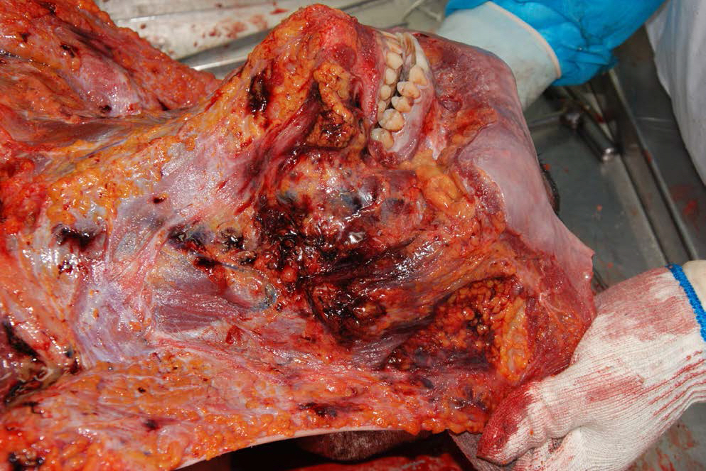
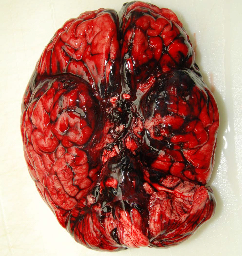
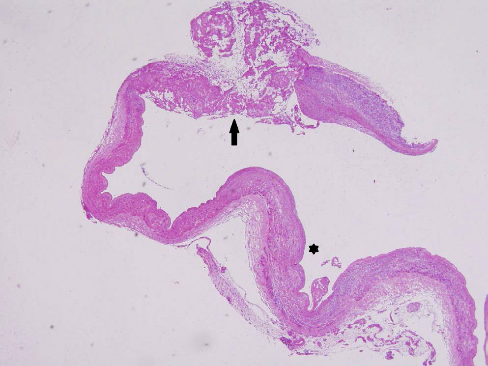
- We present the case of a 23-year-old man who suddenly collapsed during a physical altercation with his friends while in a drunken state. The post-mortem computed tomography (CT) with angiography revealed acute basal subarachnoid hemorrhage with rupture of the left middle cerebral artery. On autopsy, the head, face, mandible and neck showed multifocal hemorrhages with fracture of the hyoid bone, and the pathologic findings of the brain was consistent with CT findings. However, the vascular rupture site was not observed macroscopically. On histologic examination, a microscopic focal rupture was identified at the proximal portion of the middle cerebral artery, and possibility of arteriopathy was considered. This case illustrates that other parts of intracerebral arteries (other than the vertebral arteries) can be the culprit of rupture in the case of traumatic basal subarachnoid hemorrhage, and the post-mortem angiographic findings can be helpful in targeting the site of vascular injury. Furthermore, meticulous sampling of intracranial vessels could help find the vascular rupture site and identify any histologic findings suspicious of arteriopathy. Therefore, we suggest that post-mortem angiography can be an effective and adjunctive tool for a tailored approach in finding the vascular injury, and that histologic examination of both the intracranial and extracranial arteries be important to medicolegally ensure the death of traumatic basal subarachnoid hemorrhage and to examine presence of arteriopathy as a predisposing factor.
Keyword
MeSH Terms
Figure
Reference
-
1. Contostavlos DL. Massive subarachnoid hemorrhage due to laceration of the vertebral artery associated with fracture of the transverse process of the atlas. J Forensic Sci. 1971; 16:40–56.2. Itabashi HH, Andrews JM, Tomiyasu U, et al. Foresensic neuropathology. Burlington: Elsevier Inc.;2007.3. Ellison D, Love S, Chimelli LM, et al. Neuropathology: a reference text of CNS pathology. 3rd ed.Edinburgh: Mosby & Elsevier;2013.4. Wong B, Ong BB, Milne N. The source of haemorrhage in traumatic basal subarachnoid haemorrhage. J Forensic Leg Med. 2015; 29:18–23.
Article5. Djokic V, Savic S, Atanasijevic T. Medicolegal diagnostic value and clinical significance of traumatic incomplete tears of the basilar artery. Am J Forensic Med Pathol. 2003; 24:208–13.
Article6. Contostavlos DL. Isolated basilar traumatic subarachnoid hemorrhage: an observer's 25 year re-evaluation of the pathogenetic possibilities. Forensic Sci Int. 1995; 73:61–74.
Article7. Gray JT, Puetz SM, Jackson SL, et al. Traumatic subarachnoid haemorrhage: a 10-year case study and review. Forensic Sci Int. 1999; 105:13–23.
Article8. Kim TH, Lee SH, Kim DH, et al. Non-aneurysmal and non-traumatic subarachnoid hemorrhage after attempted suicide by incomplete hanging. Clin Exp Emerg Med. 2017; 4:56–9.
Article9. Kim GN, Lee SH, Chae JM, et al. Analysis of traumatic subarachnoid hemorrhage due to blunt force trauma on facial region. Korean J Leg Med. 2006; 30:1–13.10. Salvatori M, Kodikara S, Pollanen M. Fatal subarachnoid hemorrhage following traumatic rupture of the internal carotid artery. Leg Med (Tokyo). 2012; 14:328–30.
Article11. An TL. Fatal dissecting aneurysm of the internal carotid artery with delayed symptoms following facial impact. J Forensic Sci. 1992; 37:646–51.
Article12. Blanco Pampin J, Morte Tamayo N, Hinojal Fonseca R, et al. Delayed presentation of carotid dissection, cerebral ischemia, and infarction following blunt trauma: two cases. J Clin Forensic Med. 2002; 9:136–40.
Article13. Kindelberger D, Gilmore K, Catanese CA, et al. Hyperextension and rotation of head causing internal carotid artery laceration with basilar subarachnoid hematoma. J Forensic Sci. 2003; 48:1366–8.
Article14. Moar JJ. Traumatic rupture of the cervical carotid arteries: an autopsy and histopathological study of 200 cases. Forensic Sci Int. 1987; 34:227–44.
Article15. Pickup MJ, Pollanen MS. Traumatic subarachnoid hemorrhage and the COL3A1 gene: emergence of a potential causal link. Forensic Sci Med Pathol. 2011; 7:192–7.
- Full Text Links
- Actions
-
Cited
- CITED
-
- Close
- Share
- Similar articles
-
- Inconspicuous longitudinal tears of the intracranial vertebral artery in traumatic basal subarachnoid hemorrhage
- Terson Syndrome after Subarachnoid Hemorrhage Occurred by Thrombolysis and Mechanical Thrombectomy to Treat Acute Ischemic Stroke: A Case Report
- Subarachnoid Hemorrhage Due to a Ruptured Middle Cerebral Artery Bifurcation Aneurysm Superimposed by an Idiopathic Intracerebral Hematoma
- The Forensic Pathology in Scotland and the Analysis of Post-mortem Examinations at Glasgow
- Traumatic Intracranial Aneurysm Presenting with Delayed Subarachnoid Hemorrhage


