Accuracy comparison of buccal bite scans by five intra-oral scanners
- Affiliations
-
- 1Department of Prosthodontics, College of Dentistry, Yonsei University, Seoul, Republic of Korea.
- 2Department of Prosthodontics and Dental Research Institute, School of Dentistry, Seoul National University, Seoul, Republic of Korea. 0504heo@hanmail.net
- KMID: 2439252
- DOI: http://doi.org/10.14368/jdras.2018.34.1.17
Abstract
- PURPOSE
The purpose of this study was to investigate the accuracy of the interocclusal relationship between upper and lower teeth according to the buccal interocclusal record scan using various intraoral scanner systems.
MATERIALS AND METHODS
The upper and lower full arch Models with normal occlusion were scanned with 5 intraoral scanners (Cerec Omnicam, CS3500, iTero, Trios, True Definition). Buccal interocclusal record scan was taken only at the left side while occlusion was intentionally raised by 1 mm, 2 mm, 3 mm, and 4 mm with metal cylinder core embedded within polyvinylsiloxane bite registration material at the right molar region. The superimposition analysis was done to evaluate overall three-dimensional deviation and cross-section analysis was done to evaluate the degree and the direction of deviation of interocclusal relationship.
RESULTS
From the superimposition study, Cerec Omnicam showed the least deviation (165.5 µm) and CS3500 (369.0 µm) showed the largest (P < 0.01). And the deviation was greater in 3, 4, 2 mm group than 1 mm (P < 0.01). From the cross-section study, Cerec Omnicam showed the farthest deviation (−242.8 µm) and CS3500 showed the closest deviation (312.5 µm) and a significantly high value was shown in 3 mm group.
CONCLUSION
Every intraoral scanner has different accuracy in reproducing interocclusal relationship.
MeSH Terms
Figure
-

Fig. 1 Unilateral 4 jigs were made with 1 mm, 2 mm, 3 mm and 4 mm metal cylinder core embedded by bite registration material.

Fig. 2 Intraoral scan data were aligned with reference points of maxillary arch and refined with best fit algorithm before mandibular teeth deviations were measured.
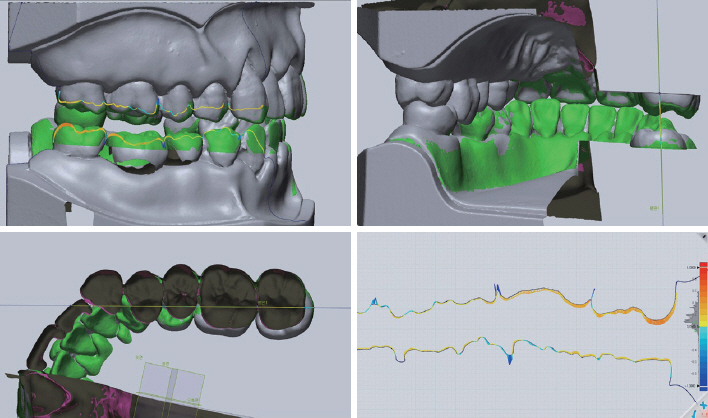
Fig. 3 Reference plane that passes the palatal cusp of maxillary first molar and the central fossa of mandibular first molar was set. Cross-section images of the reference plane were obtained and deviations of occlusal surface from reference scanner were calculated.
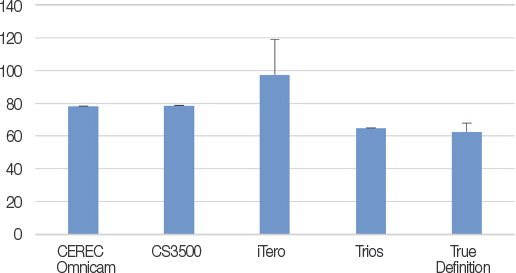
Fig. 4 Mean deviations of maxillary arch. Phantom was scanned once and duplicated for each buccal vestibular interocclusal record registration in CEREC Omnicam, CS3500, Trios, whereas in iTero and True Definition, it was scanned repeatedly with interocclusal record registration (unit: μm).
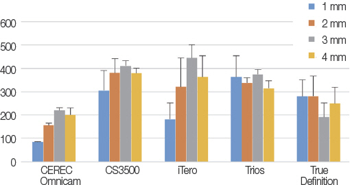
Fig. 5 Mean deviations of intraoral scanner in superimposition study (unit: μm).
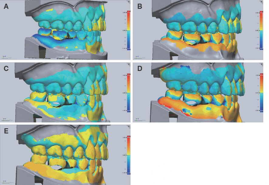
Fig. 6 The color maps of deviations shown with 1 mm open condition. (A) CEREC Omnicam, (B) CS3500, (C) iTero, (D) Trios, (E) True Definition.
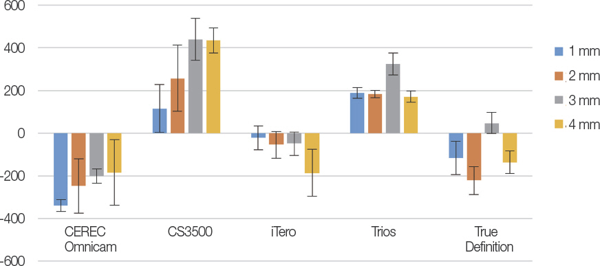
Fig. 7 Two-dimensional deviations of intraoral scanner data in a reference plane. The positive value means that upper and lower teeth got closer and the negative value means they got farther from each other (unit: μm).
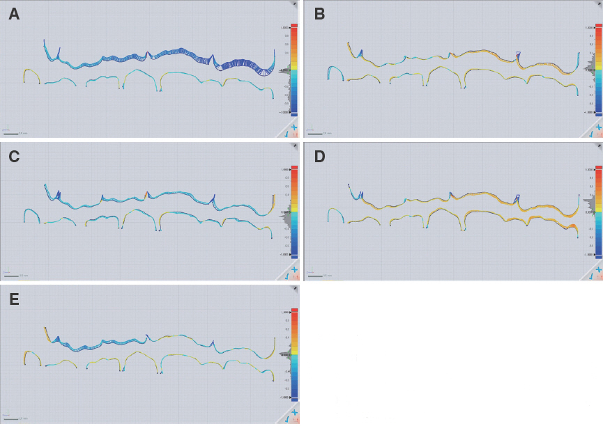
Fig. 8 The color maps of two-dimensional deviations in cross-section. Blue means negative, red means positive. (A) CEREC Omnicam, (B) CS3500, (C) iTero, (D) Trios, (E) True Definition.
Reference
-
References
1. Duret F, Blouin JL, Duret B. CAD-CAM in dentistry. J Am Dent Assoc. 1988; 117:715–20. DOI: 10.14219/jada.archive.1988.0096. PMID: 3058771.2. Mörmann WH, Brandestini M, Lutz F, Barbakow F. Chairside computer-aided direct ceramic inlays. Quintessence Int. 1989; 20:329–39. PMID: 2756089.3. Mörmann WH, Brandestini M, Lutz F. The Cerec system: computer-assisted preparation of direct ceramic inlays in 1 setting. Quintessenz. 1987; 38:45770.4. Miyazaki T, Hotta Y, Kunii J, Kuriyama S, Tamaki Y. A review of dental CAD/CAM: current status and future perspectives from 20 years of experience. Dent Mater J. 2009; 28:44–56. DOI: 10.4012/dmj.28.44. PMID: 19280967.5. van Noort R. The future of dental devices is digital. Dent Mater. 2012; 28:3–12. DOI: 10.1016/j.dental.2011.10.014. PMID: 22119539.6. Patzelt SB, Lamprinos C, Stampf S, Att W. The time efficiency of intraoral scanners: an in vitro comparative study. J Am Dent Assoc. 2014; 145:54251. DOI: 10.14219/jada.2014.23. PMID: 24878708.7. Yuzbasioglu E, Kurt H, Turunc R, Bilir H. Comparison of digital and conventional impression techniques: evaluation of patients'perception, treatment comfort, effectiveness and clinical outcomes. BMC Oral Health. 2014; 14:10. DOI: 10.1186/1472-6831-14-10. PMID: 24479892. PMCID: PMC3913616.8. Lee SJ, Gallucci GO. Digital vs. conventional implant impressions: efficiency outcomes. Clin Oral Implants Res. 2013; 24:111–5. DOI: 10.1111/j.1600-0501.2012.02430.x. PMID: 22353208.9. Ghazal M, Kern M. Mounting casts on an articulator using interocclusal records. J Prosthet Dent. 2008; 100:408–9. DOI: 10.1016/S0022-3913(08)60247-4.10. Michalakis KX, Pissiotis A, Anastasiadou V, Kapari D. An experimental study on particular physical properties of several interocclusal recording media. Part I: consistency prior to setting. J Prosthodont. 2004; 13:42–6. DOI: 10.1111/j.1532-849X.2004.04005.x. PMID: 15032895.11. Michalakis KX, Pissiotis A, Anastasiadou V, Kapari D. An experimental study on particular physical properties of several interocclusal recording media. Part II: linear dimensional change and accompanying weight change. J Prosthodont. 2004; 13:150–9. DOI: 10.1111/j.1532-849X.2004.04024.x. PMID: 15345014.12. Lassila V. Comparison of five interocclusal recording materials. J Prosthet Dent. 1986; 55:215–8. DOI: 10.1016/0022-3913(86)90347-1.13. Müller J, Götz G, Hörz W, Kraft E. An experimental study on the influence of the derived casts on the accuracy of different recording materials. Part II: polyether, acrylic resin, and corrected wax wafer. J Prosthet Dent. 1990; 63:389–95. DOI: 10.1016/0022-3913(90)90225-2.14. Müller J, Götz G, Hörz W, Kraft E. An experimental study on the influence of the derived casts on the accuracy of different recording materials. Part I: plaster, impression compound, and wax. J Prosthet Dent. 1990; 63:263–9. DOI: 10.1016/0022-3913(90)90225-2.15. Ghazal M, Albashaireh ZS, Kern M. The ability of different materials to reproduce accurate records of interocclusal relationships in the vertical dimension. J Oral Rehabil. 2008; 35:816–20. DOI: 10.1111/j.1365-2842.2008.01870.x. PMID: 18482345.16. Skurnik H. Accurate interocclusal records. J Prosthet Dent. 1969; 21:154–65. DOI: 10.1016/0022-3913(69)90088-2.17. Balthazar-Hart Y, Sandrik JL, Malone WF, Mazur B, Hart T. Accuracy and dimensional stability of four interocclusal recording materials. J Prosthet Dent. 1981; 45:586–91. DOI: 10.1016/0022-3913(81)90416-9.18. Fattore L, Malone WF, Sandrik JL, Mazur B, Hart T. Clinical evaluation of the accuracy of interocclusal recording materials. J Prosthet Dent. 1984; 51:152–7. DOI: 10.1016/0022-3913(84)90251-8.19. Balthazar YM, Ziebert GJ, Donegan SJ. Effect of interocclusal records on transverse axis position. J Prosthet Dent. 1984; 52:804–9. DOI: 10.1016/S0022-3913(84)80008-6.20. Breeding LC, Dixon DL, Kinderknecht KE. Accuracy of three interocclusal recording materials used to mount a working cast. J Prosthet Dent. 1994; 71:265–70. DOI: 10.1016/0022-3913(94)90465-0.21. Vergos VK, Tripodakis AP. Evaluation of vertical accuracy of interocclusal records. Int J Prosthodont. 2003; 16:365–8. PMID: 12956489.22. Frank E, Frank S. Bite registration in Cerec and in lab. Int J Comput Dent. 2012; 15:149–158. PMID: 22891418.23. Freilich MA, Altieri JV, Wahle JJ. Principles for selecting interocclusal records for articulation of dentate and partially dentate casts. J Prosthet Dent. 1992; 68:361–7. DOI: 10.1016/0022-3913(92)90346-C.24. Ruge S, Quooss A, Kordass B. Variability of closing movements, dynamic occlusion, and occlusal contact patterns during mastication. Int J Comput Dent. 2011; 14:119–27. PMID: 21877378.25. Ruge S, Kordass B. 3D-VAS-initial results from computerized visualization of dynamic occlusion. Int J Comput Dent. 2008; 11:9–16. PMID: 18780558.26. Mehl A, Ender A, Mörmann W, Attin T. Accuracy testing of a new intraoral 3D camera. Int J Comput Dent. 2009; 12:11–28. PMID: 19213357.27. Syrek A, Reich G, Ranftl D, Klein C, Cerny B, Brodesser J. Clinical evaluation of all-ceramic crowns fabricated from intraoral digital impressions based on the principle of active wavefront sampling. J Dent. 2010; 38:553–9. DOI: 10.1016/j.jdent.2010.03.015. PMID: 20381576.28. Luthardt RG, Loos R, Quaas S. Accuracy of intraoral data acquisition in comparison to the conventional impression. Int J Comput Dent. 2005; 8:28394.29. Ender A, Mehl A. Influence of scanning strategies on the accuracy of digital intraoral scanning systems. Int J Comput Dent. 2013; 16:11–21. PMID: 23641661.30. Ender A, Mehl A. Full arch scans: conventional versus digital impressions-an in-vitro study. Int J Comput Dent. 2011; 14:11–21. PMID: 21657122.31. Ender A, Mehl A. Accuracy of complete-arch dental impressions: a new method of measuring trueness and precision. J Prosthet Dent. 2013; 109:1218. DOI: 10.1016/S0022-3913(13)60028-1.32. Flügge TV, Schlager S, Nelson K, Nahles S, Metzger MC. Precision of intraoral digital dental impressions with iTero and extraoral digitization with the iTero and a model scanner. Am J Orthod Dentofacial Orthop. 2013; 144:471–8. DOI: 10.1016/j.ajodo.2013.04.017. PMID: 23992820.33. Sorensen JA. Accuracy of Full-Arch Scanning With Intra-Oral Scanners. Available from: https://iadr.abstractarchives.com/abstract/43am-185064/accuracy-of-full-arch-scanning-with-intra-oral-scanners. (updated 2018 Mar 16).34. Patzelt SB, Emmanouilidi A, Stampf S, Strub JR, Att W. Accuracy of full-arch scans using intraoral scanners. Clin Oral Investig. 2014; 18:1687–94. DOI: 10.1007/s00784-013-1132-y. PMID: 24240949.35. Ender A, Mehl A. Accuracy in dental medicine, a new way to measure trueness and precision. J Vis Exp. 2014; Apr. 29. 86.36. Keeling AJ, Brunton PA, Holt RJ. Optical inter-oc-clusal records are repeatable and accurate. Available from: https://iadr.abstractarchives.com/abstract/14iags-188378/optical-interocclusal-records-arerepeatable-and-accurate. (updated 2018 Mar 16).37. Solaberrieta E, Arias A, Brizuela A, Garikano X, Pradies G. Determining the requirements, section quantity, and dimension of the virtual occlusal record. J Prosthet Dent. 2016; 115:52–6. DOI: 10.1016/j.prosdent.2015.06.013. PMID: 26386482.38. Solaberrieta E, Garmendia A, Brizuela A, Otegi JR, Pradies G, Szentpétery A. Intraoral digital impressions for virtual occlusal records: section quantity and dimensions. Biomed Res Int. 2016; 2016:7173824. DOI: 10.1155/2016/7173824. PMID: 26881226. PMCID: PMC4736956.
- Full Text Links
-
- Actions
-
Cited
- CITED
-
- Close
- Share
-
- Similar articles
-
- Effects of inter-implant distance on the accuracy of intraoral scanner: An in vitro study
- A novel method for testing accuracy of bite registration using intraoral scanners
- Evaluation of the accuracy of three different intraoral scanners for endocrown digital impression: An in vitro study
- Accuracy of bite registration according to the buccal bite scan range of intra-oral scanner
- Full-arch accuracy of five intraoral scanners:In vivo analysis of trueness and precision
