Full-arch accuracy of five intraoral scanners:In vivo analysis of trueness and precision
- Affiliations
-
- 1Department of Orthodontics, Korea University Graduate School of Clinical Dentistry, Seoul, Korea
- 2Department of Mechanical Engineering, Korea University School of Engineering, Seoul, Korea
- 3Department of Orthodontics, Asan Medical Center, University of Ulsan College of Medicine, Seoul, Korea
- KMID: 2513578
- DOI: http://doi.org/10.4041/kjod.2021.51.2.95
Abstract
Objective
To evaluate the trueness and precision of full-arch scans acquired using five intraoral scanners and investigate the factors associated with the dimensional accuracy of the intraoral scan data.
Methods
Nine adult participants (mean age, 34.3 ± 8.3 years) were recruited. Four zirconium spheres (Ø 6 mm) were bonded to the canines and the molars. Following acquisition of reference scans using an industrial-grade scanner, five intraoral scanners, namely i500, CS3600, Trios 3, iTero, and CEREC Omnicam, were used to scan the arches. Linear distances between the four reference spheres were automatically calculated, and linear mixed model analysis was performed to compare the trueness and precision of the intraoral scan data among the different scanners.
Results
The absolute mean trueness and precision values for all intraoral scanners were 76.6 ± 79.3 and 56.6 ± 52.4 µm, respectively. The type of scanner and the measured linear distances had significant effects on the accuracy of the intraoral scan data. With regard to trueness, errors in the intermolar dimension and the distance from the canine to the contralateral molar were greater with Omnicam than with the other scanners. With regard to precision, the error in the linear distance from the canine to the molar in the same quadrant was greater with Omnicam and CS3600 than with the other scanners.
Conclusions
The dimensional accuracy of intraoral scan data may differ significantly according to the type of scanner, with the amount of error in terms of trueness being clinically significant.
Keyword
Figure
-

Figure 1 Placement of reference spheres for the in vivo measurement of linear distances. Four reference spheres have been attached to the lingual or palatal surface of the right and left canines and the occlusal surface of the right and left first molars. Intraoral scan data are acquired for accuracy analysis.
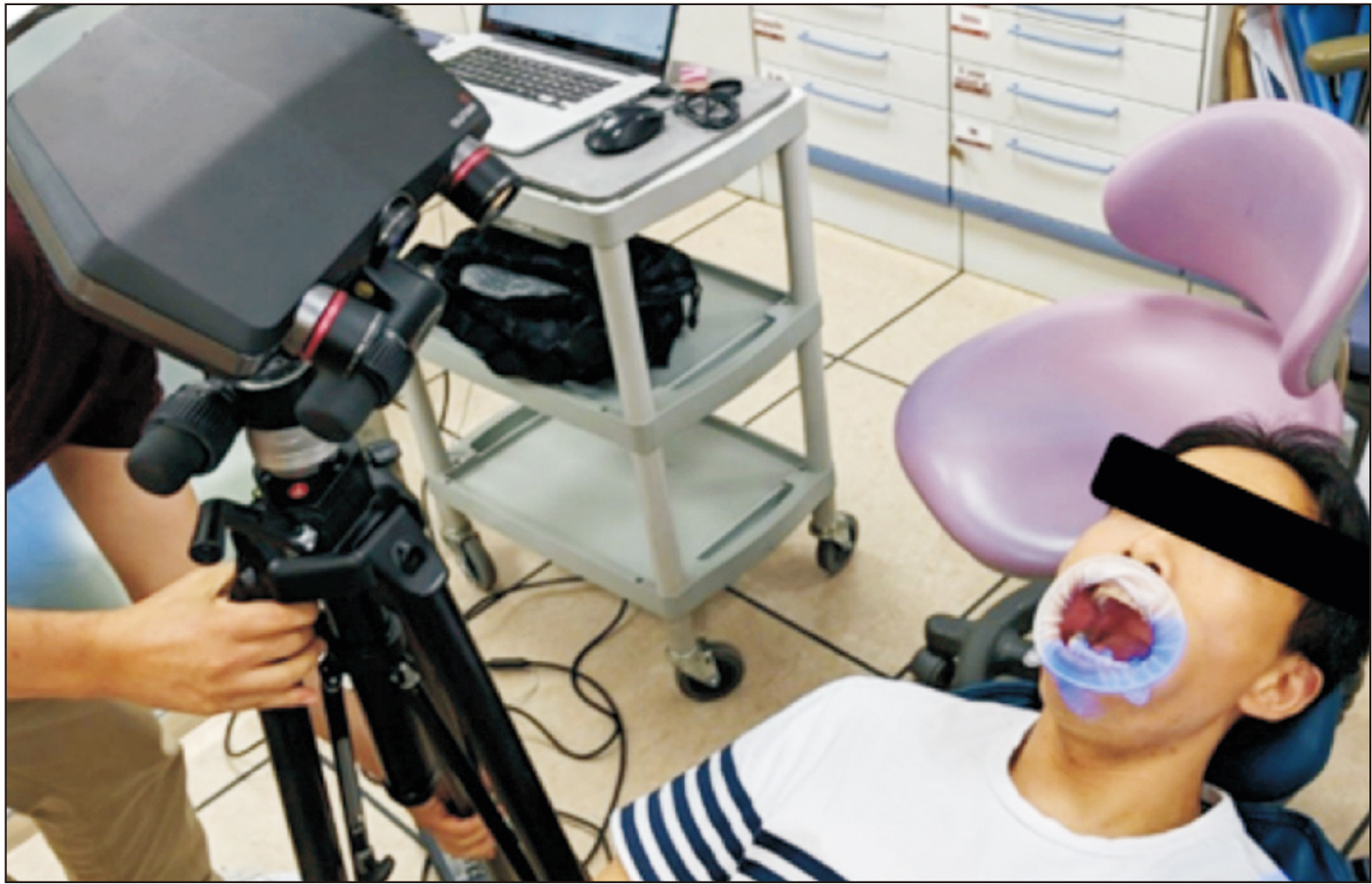
Figure 2 Reference scans for in vivo analysis of trueness. Reference scans of the four spheres are acquired using an industrial-grade scanner (Solutionix C500; Medit Corp., Seoul, Korea) for in vivo analysis of trueness.
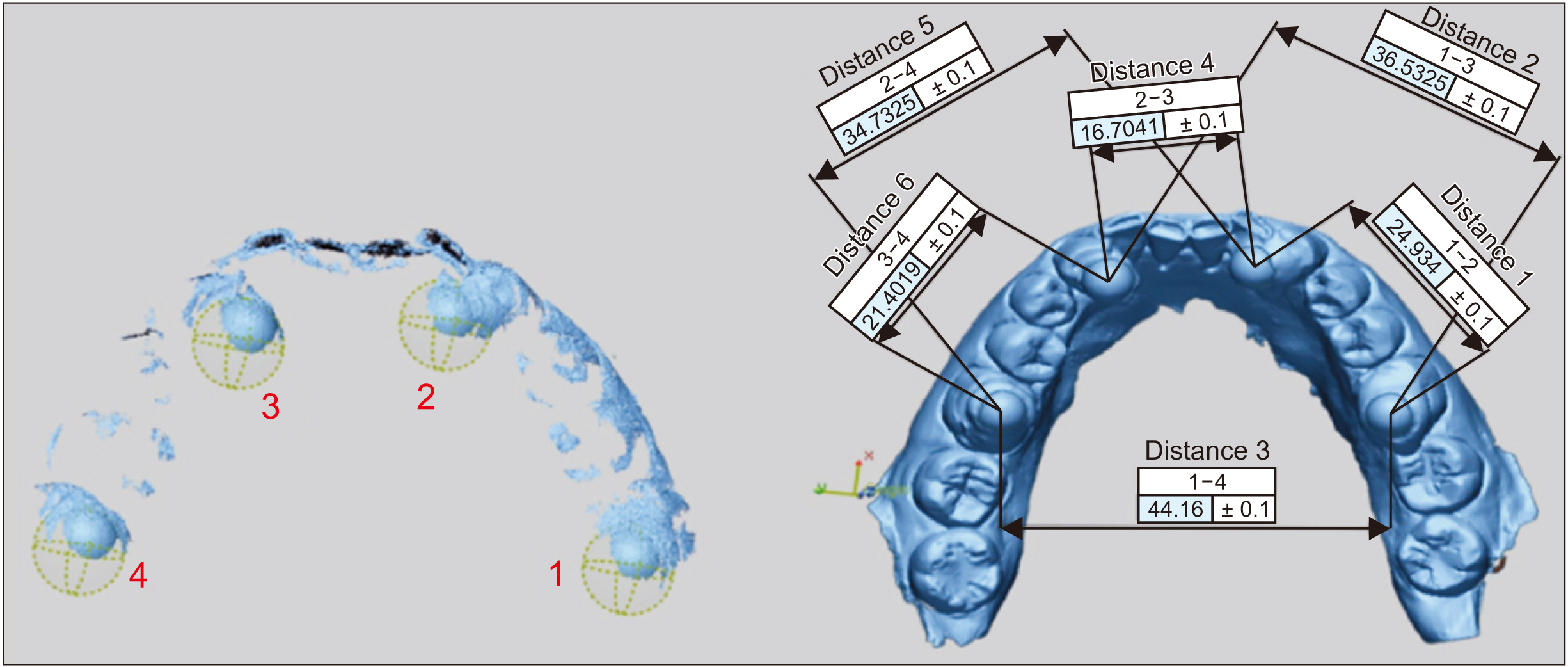
Figure 3 Measurement of linear distances. Linear distances between spheres are automatically calculated by matching with pre-imputed specification data. Distance 1, between reference spheres 1 and 2; Distance 2, between reference spheres 1 and 3; Distance 3, between reference spheres 1 and 4; Distance 4, between reference spheres 2 and 3; Distance 5, between reference spheres 2 and 4; Distance 6, between reference spheres 3 and 4.
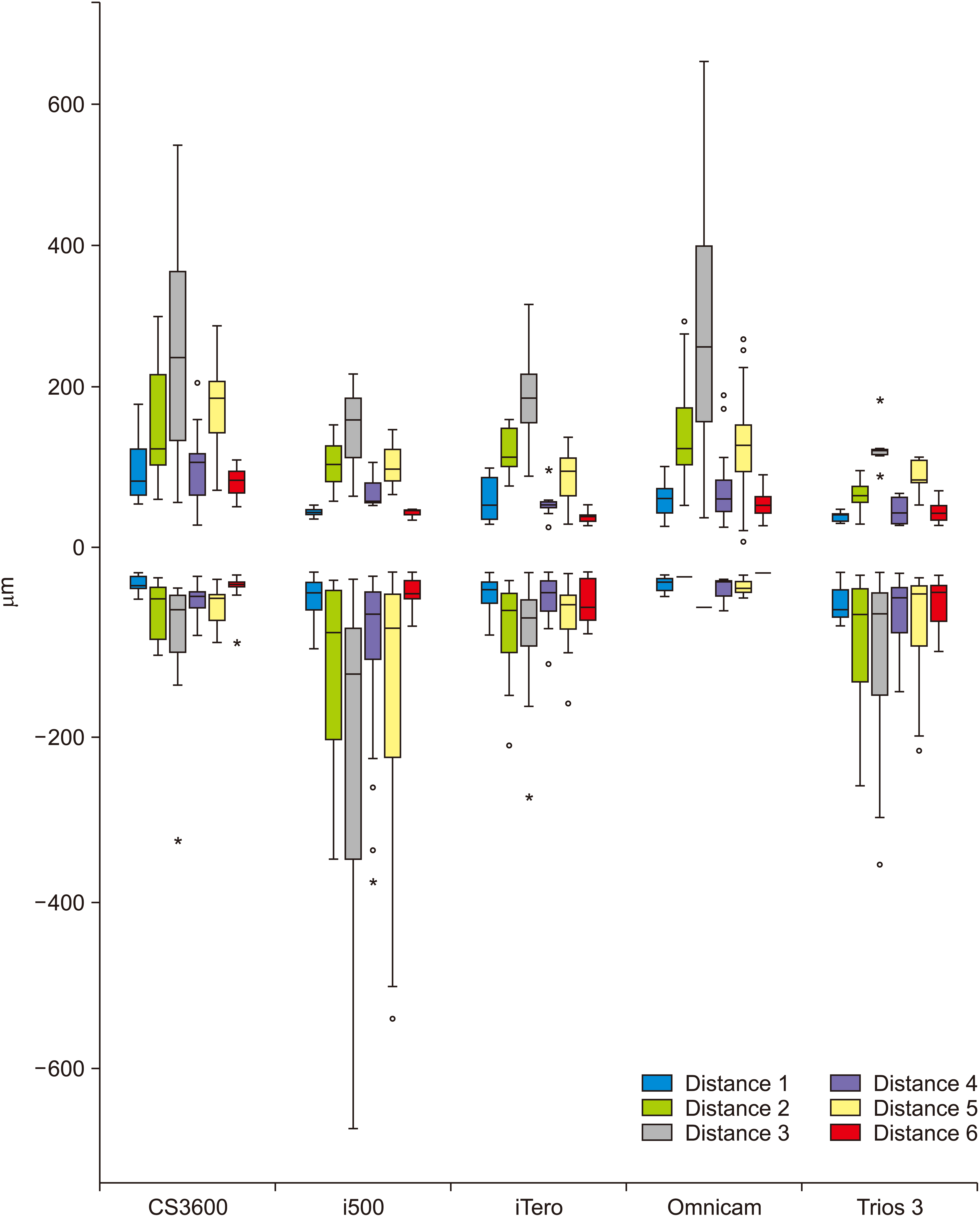
Figure 4 Mean positive and negative errors in terms of trueness according to the scanner type (µm). Distance 1, between reference spheres 1 and 2; Distance 2, between reference spheres 1 and 3; Distance 3, between reference spheres 1 and 4; Distance 4, between reference spheres 2 and 3; Distance 5, between reference spheres 2 and 4; Distance 6, between reference spheres 3 and 4.
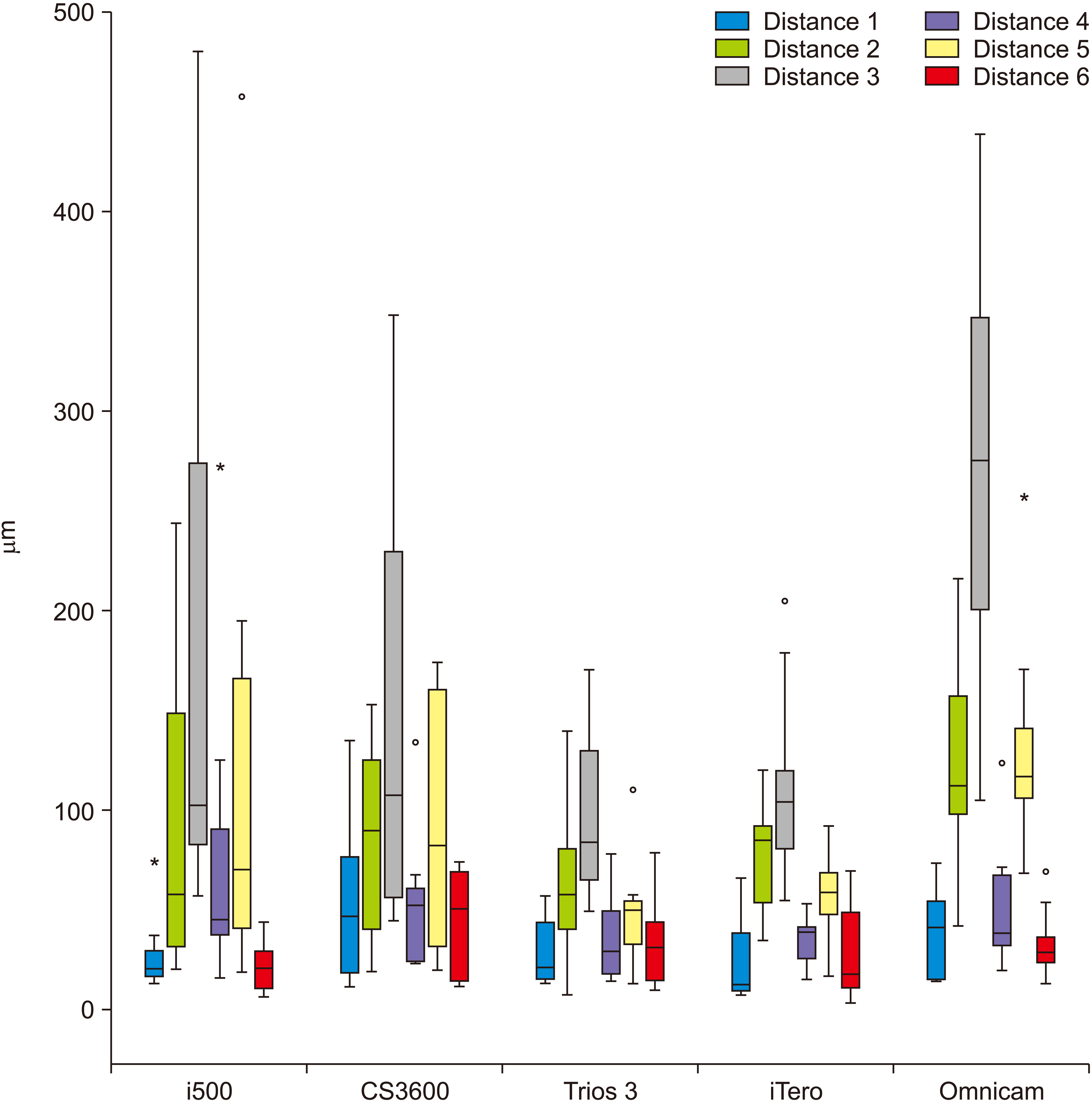
Figure 5 Mean absolute error in trueness according to the scanner type (µm). Distance 1, between reference spheres 1 and 2; Distance 2, between reference spheres 1 and 3; Distance 3, between reference spheres 1 and 4; Distance 4, between reference spheres 2 and 3; Distance 5, between reference spheres 2 and 4; Distance 6, between reference spheres 3 and 4.
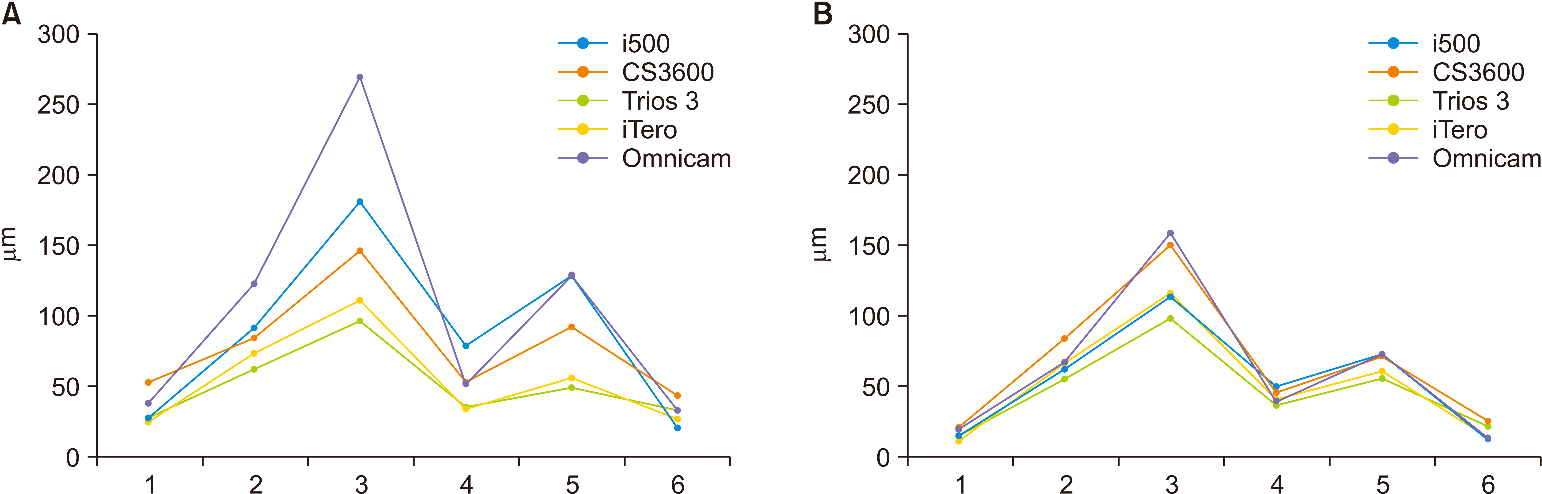
Figure 6 Mean absolute errors in terms of the trueness (A) and precision (B) of intraoral scanners according to the measured linear distances (µm). 1, distance between reference spheres 1 and 2; 2, distance between reference spheres 1 and 3; 3 distance between reference spheres 1 and 4; 4, distance between reference spheres 2 and 3; 5, distance between reference spheres 2 and 4; 6, distance between reference spheres 3 and 4.
Reference
-
1. Güth JF, Keul C, Stimmelmayr M, Beuer F, Edelhoff D. 2013; Accuracy of digital models obtained by direct and indirect data capturing. Clin Oral Investig. 17:1201–8. DOI: 10.1007/s00784-012-0795-0. PMID: 22847854.
Article2. Boeddinghaus M, Breloer ES, Rehmann P, Wöstmann B. 2015; Accuracy of single-tooth restorations based on intraoral digital and conventional impressions in patients. Clin Oral Investig. 19:2027–34. DOI: 10.1007/s00784-015-1430-7. PMID: 25693497.
Article3. Berrendero S, Salido MP, Valverde A, Ferreiroa A, Pradíes G. 2016; Influence of conventional and digital intraoral impressions on the fit of CAD/CAM-fabricated all-ceramic crowns. Clin Oral Investig. 20:2403–10. DOI: 10.1007/s00784-016-1714-6. PMID: 26800669.
Article4. Atieh MA, Ritter AV, Ko CC, Duqum I. 2017; Accuracy evaluation of intraoral optical impressions: a clinical study using a reference appliance. J Prosthet Dent. 118:400–5. DOI: 10.1016/j.prosdent.2016.10.022. PMID: 28222869. PMCID: PMC5812952.
Article5. Ender A, Mehl A. 2013; Influence of scanning strategies on the accuracy of digital intraoral scanning systems. Int J Comput Dent. 16:11–21. PMID: 23641661.6. Kuhr F, Schmidt A, Rehmann P, Wöstmann B. 2016; A new method for assessing the accuracy of full arch impressions in patients. J Dent. 55:68–74. DOI: 10.1016/j.jdent.2016.10.002. PMID: 27717754.
Article7. Rhee YK, Huh YH, Cho LR, Park CJ. 2015; Comparison of intraoral scanning and conventional impression techniques using 3-dimensional superimposition. J Adv Prosthodont. 7:460–7. DOI: 10.4047/jap.2015.7.6.460. PMID: 26816576. PMCID: PMC4722150.
Article8. Sousa MV, Vasconcelos EC, Janson G, Garib D, Pinzan A. 2012; Accuracy and reproducibility of 3-dimensional digital model measurements. Am J Orthod Dentofacial Orthop. 142:269–73. DOI: 10.1016/j.ajodo.2011.12.028. PMID: 22858338.
Article9. Lee KM. 2018; Comparison of two intraoral scanners based on three-dimensional surface analysis. Prog Orthod. 19:6. DOI: 10.1186/s40510-018-0205-5. PMID: 29430612. PMCID: PMC6890885.
Article10. Li H, Lyu P, Wang Y, Sun Y. 2017; Influence of object translucency on the scanning accuracy of a powder-free intraoral scanner: a laboratory study. J Prosthet Dent. 117:93–101. DOI: 10.1016/j.prosdent.2016.04.008. PMID: 27460324.
Article11. Goracci C, Franchi L, Vichi A, Ferrari M. 2016; Accuracy, reliability, and efficiency of intraoral scanners for full-arch impressions: a systematic review of the clinical evidence. Eur J Orthod. 38:422–8. DOI: 10.1093/ejo/cjv077. PMID: 26487391.
Article12. Ender A, Attin T, Mehl A. 2016; In vivo precision of conventional and digital methods of obtaining complete-arch dental impressions. J Prosthet Dent. 115:313–20. DOI: 10.1016/j.prosdent.2015.09.011. PMID: 26548890.13. Flügge TV, Schlager S, Nelson K, Nahles S, Metzger MC. 2013; Precision of intraoral digital dental impressions with iTero and extraoral digitization with the iTero and a model scanner. Am J Orthod Dentofacial Orthop. 144:471–8. DOI: 10.1016/j.ajodo.2013.04.017. PMID: 23992820.
Article14. Grünheid T, McCarthy SD, Larson BE. 2014; Clinical use of a direct chairside oral scanner: an assessment of accuracy, time, and patient acceptance. Am J Orthod Dentofacial Orthop. 146:673–82. DOI: 10.1016/j.ajodo.2014.07.023. PMID: 25439218.
Article15. Ender A, Mehl A. 2013; Accuracy of complete-arch dental impressions: a new method of measuring trueness and precision. J Prosthet Dent. 109:121–8. DOI: 10.1016/S0022-3913(13)60028-1. PMID: 23395338.
Article16. Nedelcu R, Olsson P, Nyström I, Rydén J, Thor A. 2018; Accuracy and precision of 3 intraoral scanners and accuracy of conventional impressions: a novel in vivo analysis method. J Dent. 69:110–8. DOI: 10.1016/j.jdent.2017.12.006. PMID: 29246490.
Article17. Müller P, Ender A, Joda T, Katsoulis J. 2016; Impact of digital intraoral scan strategies on the impression accuracy using the TRIOS Pod scanner. Quintessence Int. 47:343–9. DOI: 10.3290/j.qi.a35524. PMID: 26824085.18. Oh KC, Park JM, Moon HS. 2020; Effects of scanning strategy and scanner type on the accuracy of intraoral scans: a new approach for assessing the accuracy of scanned data. J Prosthodont. 29:518–23. DOI: 10.1111/jopr.13158. PMID: 32133690.
Article19. Revilla-León M, Jiang P, Sadeghpour M, Piedra-Cascón W, Zandinejad A, Özcan M, et al. 2020; Intraoral digital scans-part 1: influence of ambient scanning light conditions on the accuracy (trueness and precision) of different intraoral scanners. J Prosthet Dent. 124:372–8. DOI: 10.1016/j.prosdent.2019.06.003. PMID: 31864638.
Article
- Full Text Links
-
- Actions
-
Cited
- CITED
-
- Close
- Share
-
- Similar articles
-
- Comparing the accuracy of six intraoral scanners on prepared teeth and effect of scanning sequence
- Accuracy of 14 intraoral scanners for the All-on-4 treatment concept: a comparative in vitro study
- A new method to measure the accuracy of intraoral scanners along the complete dental arch: A pilot study
- Effect of different arch widths on the accuracy of three intraoral scanners
- Comparative analysis on reproducibility among 5 intraoral scanners: sectional analysis according to restoration type and preparation outline form
