Comparative analysis on reproducibility among 5 intraoral scanners: sectional analysis according to restoration type and preparation outline form
- Affiliations
-
- 1Department of Prosthodontics, Seoul National University Gwanak Dental Hospital, Seoul, Republic of Korea. jimarn@snu.ac.kr
- KMID: 2355439
- DOI: http://doi.org/10.4047/jap.2016.8.5.354
Abstract
- PURPOSE
The trueness and precision of acquired images of intraoral digital scanners could be influenced by restoration type, preparation outline form, scanning technology and the application of power. The aim of this study is to perform the comparative evaluation of the 3-dimensional reproducibility of intraoral scanners (IOSs).
MATERIALS AND METHODS
The phantom containing five prepared teeth was scanned by the reference scanner (Dental Wings) and 5 test IOSs (E4D dentist, Fastscan, iTero, Trios and Zfx Intrascan). The acquired images of the scanner groups were compared with the image from the reference scanner (trueness) and within each scanner groups (precision). Statistical analysis was performed using independent two-samples t-test and analysis of variance (α=.05).
RESULTS
The average deviations of trueness and precision of Fastscan, iTero and Trios were significantly lower than the other scanners. According to the restoration type, significantly higher trueness was observed in crown and inlay than in bridge. However, no significant difference was observed among four sites of preparation outline form. If compared by the characteristics of IOS, high trueness was observed in the group adopting the active triangulation and using powder. However, there was no significant difference between the still image acquisition and video acquisition groups.
CONCLUSION
Except for two intraoral scanners, Fastscan, iTero and Trios displayed comparable levels of trueness and precision values in tested phantom model. Difference in trueness was observed depending on the restoration type, the preparation outline form and characteristics of IOS, which should be taken into consideration when the intraoral scanning data are utilized.
Keyword
Figure
-
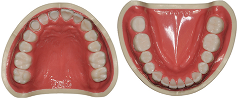
Fig. 1 Dental models with various preparation designs. Right maxillary incisor and canine (#11, 13); 3-unit fixed dental prosthesis, right maxillary second molar (#17); MO inlay, and right mandibular second molar (#47); crown.
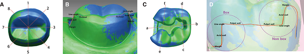
Fig. 2 Illustration of the sectioning of the superposed datasets and selected sites from preparation ouline form. (A) section lines of crown and bridge groups (sections 1, 3, 5, and 7 were investigated for the mesiodistal and buccolingual comparison). (B) measured sites for crown and bridge groups. (C) section line of inlay group (sections a and b were inspected for the box and non-box comparision). (D) sites from preparation outline form for inlay group.
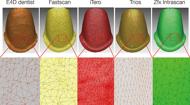
Fig. 3 Number of polygons for selected maxillary incisor for the comparison of the each IOS's resolution.
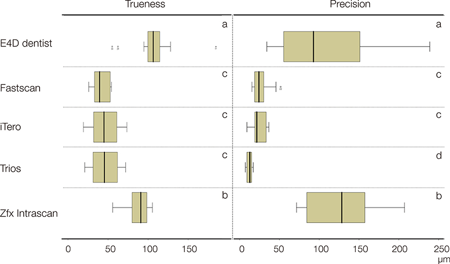
Fig. 4 Illustration of absolute mean trueness and precision values of intraoral scanners. Same letters denote significant differences in between the groups at the 5% significance level.
Cited by 2 articles
-
Comparative evaluation of marginal and internal fit of metal copings fabricated by various CAD/CAM methods
Seung-Jin Jeong, Hye-Won Cho, Ji-Hye Jung, Jeong-Mi Kim, Yu-Lee Kim
J Korean Acad Prosthodont. 2019;57(3):211-218. doi: 10.4047/jkap.2019.57.3.211.Digital evaluation of axial displacement by implant-abutment connection type: An in vitro study
Sung-Jun Kim, KeunBaDa Son, Kyu-Bok Lee
J Adv Prosthodont. 2018;10(5):388-394. doi: 10.4047/jap.2018.10.5.388.
Reference
-
1. Miyazaki T, Hotta Y, Kunii J, Kuriyama S, Tamaki Y. A review of dental CAD/CAM: current status and future perspectives from 20 years of experience. Dent Mater J. 2009; 28:44–56.2. Nedelcu RG, Persson AS. Scanning accuracy and precision in 4 intraoral scanners: an in vitro comparison based on 3-dimensional analysis. J Prosthet Dent. 2014; 112:1461–1471.3. Schaefer O, Decker M, Wittstock F, Kuepper H, Guentsch A. Impact of digital impression techniques on the adaption of ceramic partial crowns in vitro. J Dent. 2014; 42:677–683.4. Logozzo S, Zanetti EM, Franceschini G, Kilpelä A, Mäkynen A. Recent advances in dental optics – Part I: 3D intraoral scanners for restorative dentistry. Opt Laser Eng. 2014; 54:203–221.5. Seelbach P, Brueckel C, Wöstmann B. Accuracy of digital and conventional impression techniques and workflow. Clin Oral Investig. 2013; 17:1759–1764.6. Ender A, Mehl A. Full arch scans: conventional versus digital impressions-an in-vitro study. Int J Comput Dent. 2011; 14:11–21.7. Ender A, Mehl A. Influence of scanning strategies on the accuracy of digital intraoral scanning systems. Int J Comput Dent. 2013; 16:11–21.8. Ender A, Mehl A. In-vitro evaluation of the accuracy of conventional and digital methods of obtaining full-arch dental impressions. Quintessence Int. 2015; 46:9–17.9. Patzelt SB, Vonau S, Stampf S, Att W. Assessing the feasibility and accuracy of digitizing edentulous jaws. J Am Dent Assoc. 2013; 144:914–920.10. Patzelt SB, Emmanouilidi A, Stampf S, Strub JR, Att W. Accuracy of full-arch scans using intraoral scanners. Clin Oral Investig. 2014; 18:1687–1694.11. Papaspyridakos P, Gallucci GO, Chen CJ, Hanssen S, Naert I, Vandenberghe B. Digital versus conventional implant impressions for edentulous patients: accuracy outcomes. Clin Oral Implants Res. 2016; 27:465–472.12. Andriessen FS, Rijkens DR, van der Meer WJ, Wismeijer DW. Applicability and accuracy of an intraoral scanner for scanning multiple implants in edentulous mandibles: a pilot study. J Prosthet Dent. 2014; 111:186–194.13. van der Meer WJ, Andriessen FS, Wismeijer D, Ren Y. Application of intra-oral dental scanners in the digital workflow of implantology. PLoS One. 2012; 7:e43312.14. Kim SR, Lee WS, Kim WC, Kim HY, Kim JH. Digitization of dental alginate impression: Three-dimensional evaluation of point cloud. Dent Mater J. 2015; 34:835–840.15. Atzeni E, Iuliano L, Minetola P, Salmi A. Proposal of an innovative benchmark for accuracy evaluation of dental crown manufacturing. Comput Biol Med. 2012; 42:548–555.16. Paranhos LR, Lima CS, da Silva RH, Daruge Júnior E, Torres FC. Correlation between maxillary central incisor crown morphology and mandibular dental arch form in normal occlusion subjects. Braz Dent J. 2012; 23:149–153.17. Paranhos LR, Zaroni M, Carli JP, Okamoto R, Zogheib LV, Torres FC. Association between the facial type and morphology of the upper central incisor in normal occlusion subjects. J Contemp Dent Pract. 2014; 15:29–33.18. Martin CB, Chalmers EV, McIntyre GT, Cochrane H, Mossey PA. Orthodontic scanners: what's available? J Orthod. 2015; 42:136–143.19. Sannino G, Gloria F, Schiavetti R, Ottria L, Barlattani A. Dental Wings CAD/CAM system precision: an internal and marginal fit sperimental analisys. Oral Implantol (Rome). 2009; 2:11–20.20. Mehl A, Ender A, Mörmann W, Attin T. Accuracy testing of a new intraoral 3D camera. Int J Comput Dent. 2009; 12:11–28.21. Kim SY, Kim MJ, Han JS, Yeo IS, Lim YJ, Kwon HB. Accuracy of dies captured by an intraoral digital impression system using parallel confocal imaging. Int J Prosthodont. 2013; 26:161–163.22. Ender A, Mehl A. Accuracy of complete-arch dental impressions: a new method of measuring trueness and precision. J Prosthet Dent. 2013; 109:121–128.23. An S, Kim S, Choi H, Lee JH, Moon HS. Evaluating the marginal fit of zirconia copings with digital impressions with an intraoral digital scanner. J Prosthet Dent. 2014; 112:1171–1175.24. Keul C, Stawarczyk B, Erdelt KJ, Beuer F, Edelhoff D, Güth JF. Fit of 4-unit FDPs made of zirconia and CoCr-alloy after chairside and labside digitalization-a laboratory study. Dent Mater. 2014; 30:400–407.25. Brawek PK, Wolfart S, Endres L, Kirsten A, Reich S. The clinical accuracy of single crowns exclusively fabricated by digital workflow-the comparison of two systems. Clin Oral Investig. 2013; 17:2119–2125.26. Sturdevant JR, Bayne SC, Heymann HO. Margin gap size of ceramic inlays using second-generation CAD/CAM equipment. J Esthet Dent. 1999; 11:206–214.27. Rupf S, Berger H, Buchter A, Harth V, Ong MF, Hannig M. Exposure of patient and dental staff to fine and ultrafine particles from scanning spray. Clin Oral Investig. 2015; 19:823–830.
- Full Text Links
-
- Actions
-
Cited
- CITED
-
- Close
- Share
-
- Similar articles
-
- Full-arch accuracy of five intraoral scanners:In vivo analysis of trueness and precision
- A comparison of the accuracy of intraoral scanners using an intraoral environment simulator
- Effect of the volumetric dimensions of a complete arch on the accuracy of scanners
- Effect of scanning strategies on the accuracy of digital intraoral scanners: a meta-analysis of in vitro studies
- Accuracy of 14 intraoral scanners for the All-on-4 treatment concept: a comparative in vitro study
